Advertisement

Main menu

  • Home
  • Content
    • Current Issue
    • Accepted Manuscripts
    • Article Preview
    • Past Issue Archive
    • AJNR Case Collection
    • Case of the Week Archive
    • Classic Case Archive
    • Case of the Month Archive
  • Special Collections
    • Spinal CSF Leak Articles (Jan 2020-June 2024)
    • 2024 AJNR Journal Awards
    • Most Impactful AJNR Articles
  • Multimedia
    • AJNR Podcast
    • AJNR Scantastics
    • Video Articles
  • For Authors
    • Submit a Manuscript
    • Author Policies
    • Fast publishing of Accepted Manuscripts
    • Graphical Abstract Preparation
    • Manuscript Submission Guidelines
    • Imaging Protocol Submission
    • Submit a Case for the Case Collection
  • About Us
    • About AJNR
    • Editorial Board
  • More
    • Become a Reviewer/Academy of Reviewers
    • Subscribers
    • Permissions
    • Alerts
    • Feedback
    • Advertisers
    • ASNR Home
  • Other Publications
    • ajnr

User menu

  • Alerts
  • Log in

Search

  • Advanced search
American Journal of Neuroradiology

American Journal of Neuroradiology

ASHNR American Society of Functional Neuroradiology ASHNR American Society of Pediatric Neuroradiology ASSR
  • Alerts
  • Log in

Advanced Search

  • Home
  • Content
    • Current Issue
    • Accepted Manuscripts
    • Article Preview
    • Past Issue Archive
    • AJNR Case Collection
    • Case of the Week Archive
    • Classic Case Archive
    • Case of the Month Archive
  • Special Collections
    • Spinal CSF Leak Articles (Jan 2020-June 2024)
    • 2024 AJNR Journal Awards
    • Most Impactful AJNR Articles
  • Multimedia
    • AJNR Podcast
    • AJNR Scantastics
    • Video Articles
  • For Authors
    • Submit a Manuscript
    • Author Policies
    • Fast publishing of Accepted Manuscripts
    • Graphical Abstract Preparation
    • Manuscript Submission Guidelines
    • Imaging Protocol Submission
    • Submit a Case for the Case Collection
  • About Us
    • About AJNR
    • Editorial Board
  • More
    • Become a Reviewer/Academy of Reviewers
    • Subscribers
    • Permissions
    • Alerts
    • Feedback
    • Advertisers
    • ASNR Home
  • Follow AJNR on Twitter
  • Visit AJNR on Facebook
  • Follow AJNR on Instagram
  • Join AJNR on LinkedIn
  • RSS Feeds

Welcome to the new AJNR, Updated Hall of Fame, and more. Read the full announcements.


AJNR is seeking candidates for the position of Associate Section Editor, AJNR Case Collection. Read the full announcement.


Research ArticleBrain

MR Imaging Features of Amyloid-Related Imaging Abnormalities

J. Barakos, R. Sperling, S. Salloway, C. Jack, A. Gass, J.B. Fiebach, D. Tampieri, D. Melançon, Y. Miaux, G. Rippon, R. Black, Y. Lu, H.R. Brashear, H.M. Arrighi, K.A. Morris and M. Grundman
American Journal of Neuroradiology October 2013, 34 (10) 1958-1965; DOI: https://doi.org/10.3174/ajnr.A3500
J. Barakos
aFrom the California Pacific Medical Center (J.B.), San Francisco, California
bSynarc (J.B., Y.M.), Newark, California
  • Find this author on Google Scholar
  • Find this author on PubMed
  • Search for this author on this site
R. Sperling
cBrigham and Women's Hospital (R.S.), Massachusetts General Hospital, Harvard Medical School, Boston, Massachusetts
  • Find this author on Google Scholar
  • Find this author on PubMed
  • Search for this author on this site
S. Salloway
dWarren Alpert Medical School of Brown University (S.S.), Providence, Rhode Island
  • Find this author on Google Scholar
  • Find this author on PubMed
  • Search for this author on this site
C. Jack
eMayo Clinic (C.J.), Rochester, Minnesota
  • Find this author on Google Scholar
  • Find this author on PubMed
  • Search for this author on this site
A. Gass
fUniversity Hospital Mannheim (A.G.), University of Heidelberg, Mannheim, Germany
  • Find this author on Google Scholar
  • Find this author on PubMed
  • Search for this author on this site
J.B. Fiebach
gCenter for Stroke Research (J.B.F.), Charité, Berlin, Germany
  • Find this author on Google Scholar
  • Find this author on PubMed
  • Search for this author on this site
D. Tampieri
hNeuroRx Research (D.T., D.M.), Montreal, Quebec, Canada
  • Find this author on Google Scholar
  • Find this author on PubMed
  • Search for this author on this site
D. Melançon
hNeuroRx Research (D.T., D.M.), Montreal, Quebec, Canada
  • Find this author on Google Scholar
  • Find this author on PubMed
  • Search for this author on this site
Y. Miaux
bSynarc (J.B., Y.M.), Newark, California
  • Find this author on Google Scholar
  • Find this author on PubMed
  • Search for this author on this site
G. Rippon
iPfizer Inc (G.R., R.B.), Collegeville, Pennsylvania
  • Find this author on Google Scholar
  • Find this author on PubMed
  • Search for this author on this site
R. Black
iPfizer Inc (G.R., R.B.), Collegeville, Pennsylvania
  • Find this author on Google Scholar
  • Find this author on PubMed
  • Search for this author on this site
Y. Lu
aFrom the California Pacific Medical Center (J.B.), San Francisco, California
  • Find this author on Google Scholar
  • Find this author on PubMed
  • Search for this author on this site
H.R. Brashear
jJanssen Alzheimer Immunotherapy Research & Development, LLC (H.R.B., H.M.A., K.A.M., M.G.), South San Francisco, California
  • Find this author on Google Scholar
  • Find this author on PubMed
  • Search for this author on this site
H.M. Arrighi
jJanssen Alzheimer Immunotherapy Research & Development, LLC (H.R.B., H.M.A., K.A.M., M.G.), South San Francisco, California
  • Find this author on Google Scholar
  • Find this author on PubMed
  • Search for this author on this site
K.A. Morris
jJanssen Alzheimer Immunotherapy Research & Development, LLC (H.R.B., H.M.A., K.A.M., M.G.), South San Francisco, California
  • Find this author on Google Scholar
  • Find this author on PubMed
  • Search for this author on this site
M. Grundman
jJanssen Alzheimer Immunotherapy Research & Development, LLC (H.R.B., H.M.A., K.A.M., M.G.), South San Francisco, California
kGlobal Partners Inc (M.G.), Cambridge, Massachusetts.
  • Find this author on Google Scholar
  • Find this author on PubMed
  • Search for this author on this site
  • Article
  • Figures & Data
  • Info & Metrics
  • Responses
  • References
  • PDF
Loading

Article Figures & Data

Figures

  • Tables
  • Fig 1.
    • Download figure
    • Open in new tab
    • Download powerpoint
    Fig 1.

    ARIA-E (effusion); sulcal FLAIR hyperintensity. FLAIR MR imaging at baseline (A) and week 19 (B). On the follow-up surveillance scan (week 19), sulcal FLAIR hyperintensity developed in the right frontal region (arrow). No associated signal abnormality was identified on other imaging sequences, including T1 and gradient recalled-echo/T2* imaging (not shown). Because the imaging appearance mimicked that of a subarachnoid hemorrhage, CT and lumbar puncture were performed, both of which had normal findings, revealing no evidence of blood products.

  • Fig 2.
    • Download figure
    • Open in new tab
    • Download powerpoint
    Fig 2.

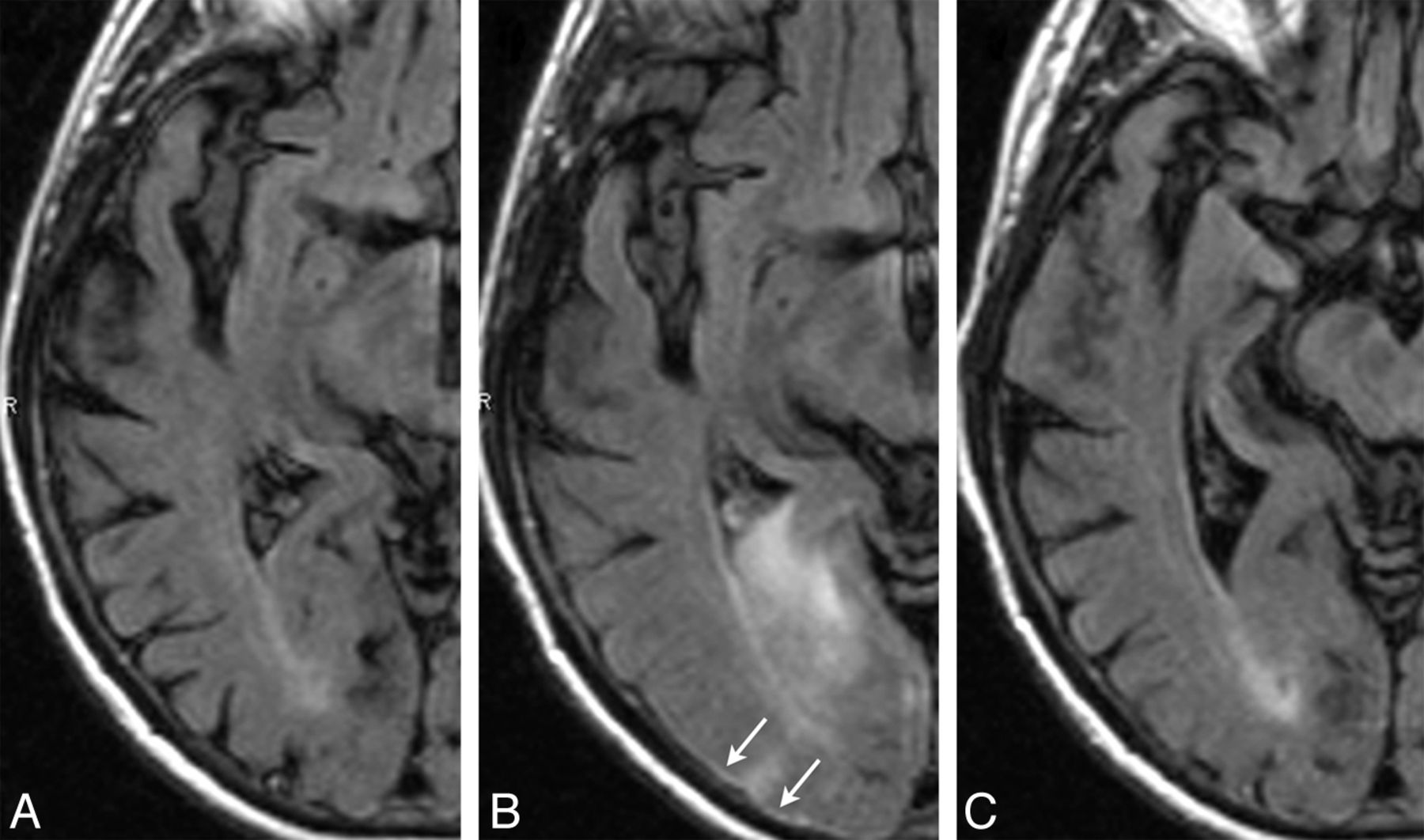
    ARIA-E (edema and effusion); parenchymal and sulcal FLAIR hyperintensity. FLAIR sequences at baseline (A), week 19 (B), and week 32 (C). By week 19 (6 weeks after the second dose of bapineuzumab), a zone of cortical swelling, sulcal effacement, and parenchymal hyperintensity has developed in the right temporal-occipital lobe. A small sulcal effusion and faint overlying linear leptomeningeal hyperintensity (arrows) help define the area of abnormality. By week 32, these findings have largely resolved. As is typically the case with bapineuzumab-related ARIA, the patient remained asymptomatic throughout all imaging time points.

  • Fig 3.
    • Download figure
    • Open in new tab
    • Download powerpoint
    Fig 3.

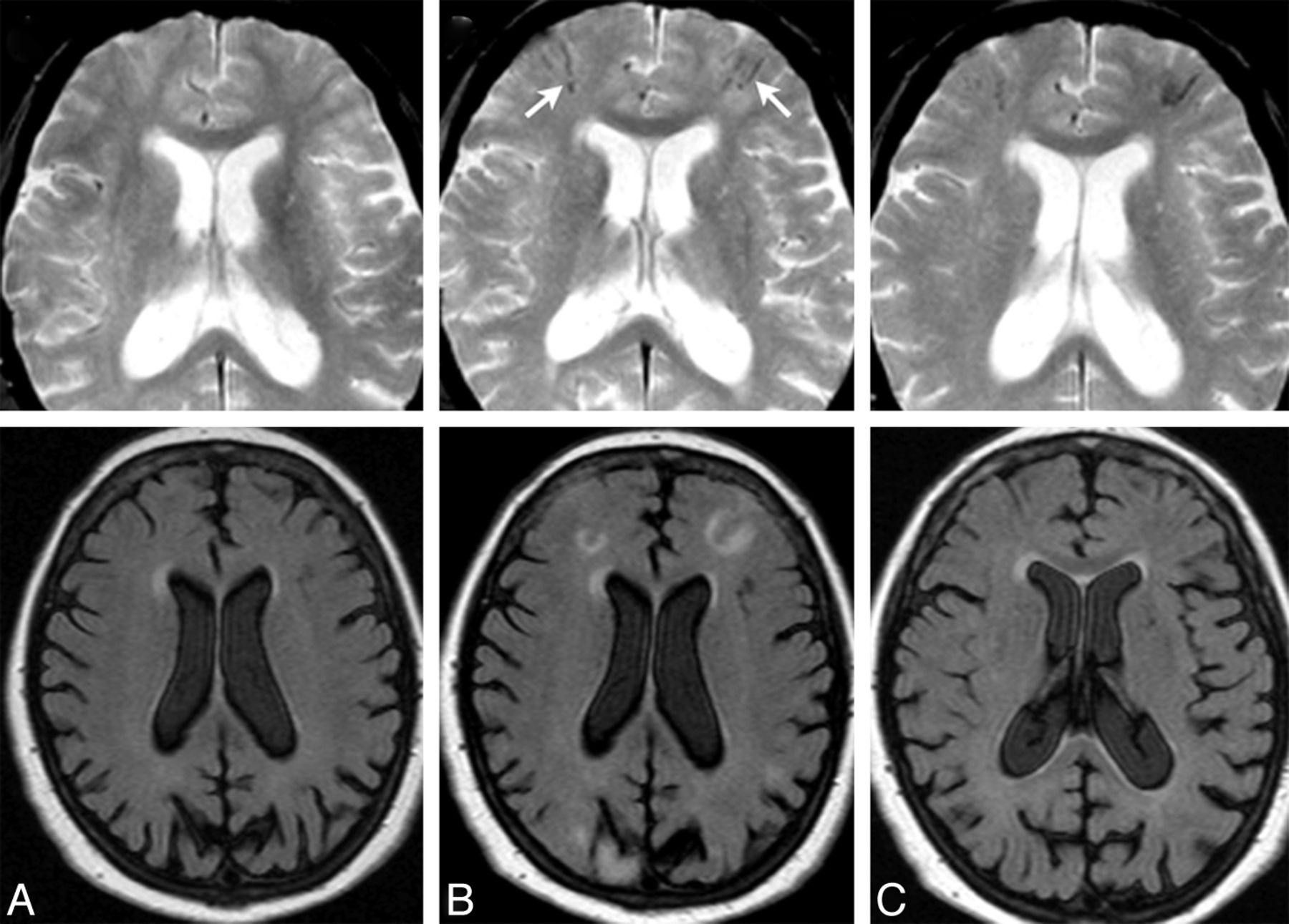
    ARIA-E (edema); week 19 parenchymal FLAIR hyperintensity. Week 19 FLAIR (A) and corresponding DWI (B) and week 19 gadolinium (C). On the surveillance scan for week 19 (A), several large regions of parenchymal edema develop, principally in the bifrontal and left temporal regions, which resolve on follow-up (not shown). Week 19 DWI reveals minimal T2 shinethrough (B), but no ADC abnormality (not shown). With gadolinium, there is no frank enhancement with only mild prominence of overlying cortical vasculature, in keeping with nonspecific cerebral swelling (C).

  • Fig 4.
    • Download figure
    • Open in new tab
    • Download powerpoint
    Fig 4.

    ARIA-E (edema) with incident ARIA-H (microhemorrhages). FLAIR and corresponding gradient recalled-echo/T2* sequences of the same patient, at baseline (A and B) and week 19 (C and D). By week 19 (6 weeks after the second dose of bapineuzumab), significant right-hemispheric edema has developed. As is characteristic of ARIA-E, despite the extensive parenchymal changes, DWI and ADC findings remained normal (not shown), confirming these findings as reflecting vasogenic as opposed to cytotoxic edema. On the corresponding gradient recalled-echo/T2* images, there is concomitant development of several punctate microhemorrhages in the right parietal region (circle). On subsequent imaging, the cerebral edema resolved while the microhemorrhages remained stable (not shown). In general, the severity of ARIA changes was associated with both Apolipoprotein ε4 allele status and drug dosage. As such, this patient was an Apolipoprotein ε4 homozygote and in the highest drug-dose arm.

  • Fig 5.
    • Download figure
    • Open in new tab
    • Download powerpoint
    Fig 5.

    ARIA-E (edema) with incident ARIA-H (superficial hemosiderin deposits). Gradient recalled-echo/T2* and corresponding FLAIR sequences from the same patient, at baseline (A), week 19 (B), and week 28 (C). By week 19 (B), several regions of parenchymal edema have developed, including bifrontal regions and the right parieto-occipital lobe. On the corresponding gradient recalled-echo/T2* images, by week 19, there is concomitant development of several superficial linear areas of blood-degradation products (arrows) in the bifrontal regions (≥10 mm). By week 28, the FLAIR changes relating to ARIA-E have resolved, with blood products re-demonstrated.

Tables

  • Figures
    • View popup
    Table 1:

    Clinical characteristics of ARIA

    ARIA ComponentPrimary Diagnostic Imaging SequenceNature of Leakage ProductsLocation of Increased Vascular Permeability
    ParenchymaLeptomeninges
    ARIA-EFLAIRProteinaceous fluidVasogenic edemaSulcal effusion/exudate
    ARIA-HGRE/T2*Heme productsMicrohemorrhage (hemosiderin deposits <10 mm or large hemosiderin deposits ≥10 mm)Superficial hemosiderin deposits
    • Note:—GRE indicates gradient recalled-echo.

    • View popup
    Table 2:

    Results

    ARIA Type: IncidenceLocation: Isolated LeptomeningealLocation: Isolated ParenchymalLocation: Combined
    ARIA-E: 36/210 (17%)ARIA-E: sulcal effusion: 15/36 (42%)ARIA-E: parenchymal edema: 8/36 (22%)ARIA-E: effusion and edema combined: 13/36 (36%)
    ARIA-H: 26/210 (12.4%)ARIA-H: superficial hemosiderin deposit: 2/26 (8%)ARIA-H: parenchymal microhemorrhage: 24/26 (92%)
PreviousNext
Back to top

In this issue

American Journal of Neuroradiology: 34 (10)
American Journal of Neuroradiology
Vol. 34, Issue 10
1 Oct 2013
  • Table of Contents
  • Index by author
  • Complete Issue (PDF)
Advertisement
Print
Download PDF
Email Article

Thank you for your interest in spreading the word on American Journal of Neuroradiology.

NOTE: We only request your email address so that the person you are recommending the page to knows that you wanted them to see it, and that it is not junk mail. We do not capture any email address.

Enter multiple addresses on separate lines or separate them with commas.
MR Imaging Features of Amyloid-Related Imaging Abnormalities
(Your Name) has sent you a message from American Journal of Neuroradiology
(Your Name) thought you would like to see the American Journal of Neuroradiology web site.
CAPTCHA
This question is for testing whether or not you are a human visitor and to prevent automated spam submissions.
Cite this article
J. Barakos, R. Sperling, S. Salloway, C. Jack, A. Gass, J.B. Fiebach, D. Tampieri, D. Melançon, Y. Miaux, G. Rippon, R. Black, Y. Lu, H.R. Brashear, H.M. Arrighi, K.A. Morris, M. Grundman
MR Imaging Features of Amyloid-Related Imaging Abnormalities
American Journal of Neuroradiology Oct 2013, 34 (10) 1958-1965; DOI: 10.3174/ajnr.A3500

Citation Manager Formats

  • BibTeX
  • Bookends
  • EasyBib
  • EndNote (tagged)
  • EndNote 8 (xml)
  • Medlars
  • Mendeley
  • Papers
  • RefWorks Tagged
  • Ref Manager
  • RIS
  • Zotero
0 Responses
Respond to this article
Share
Bookmark this article
MR Imaging Features of Amyloid-Related Imaging Abnormalities
J. Barakos, R. Sperling, S. Salloway, C. Jack, A. Gass, J.B. Fiebach, D. Tampieri, D. Melançon, Y. Miaux, G. Rippon, R. Black, Y. Lu, H.R. Brashear, H.M. Arrighi, K.A. Morris, M. Grundman
American Journal of Neuroradiology Oct 2013, 34 (10) 1958-1965; DOI: 10.3174/ajnr.A3500
del.icio.us logo Twitter logo Facebook logo Mendeley logo
  • Tweet Widget
  • Facebook Like
  • Google Plus One
Purchase

Jump to section

  • Article
    • Abstract
    • ABBREVIATIONS:
    • Materials and Methods
    • Results
    • Discussion
    • Conclusions
    • ACKNOWLEDGMENTS
    • Footnotes
    • REFERENCES
  • Figures & Data
  • Info & Metrics
  • Responses
  • References
  • PDF

Related Articles

  • No related articles found.
  • PubMed
  • Google Scholar

Cited By...

  • Alzheimer Disease Anti-Amyloid Immunotherapies: Imaging Recommendations and Practice Considerations for Monitoring of Amyloid-Related Imaging Abnormalities
  • Monoclonal Antibodies: What the Diagnostic Neuroradiologist Needs to Know
  • Incidence of Amyloid-Related Imaging Abnormalities in Patients With Alzheimer Disease Treated With Anti-{beta}-Amyloid Immunotherapy: A Meta-analysis
  • Amyloid-Related Imaging Abnormalities with Emerging Alzheimer Disease Therapeutics: Detection and Reporting Recommendations for Clinical Practice
  • Aducanumab Use in Symptomatic Alzheimer Disease Evidence in Focus: A Report of the AAN Guidelines Subcommittee
  • Tumefactive Primary Central Nervous System Vasculitis: Imaging Findings of a Rare and Underrecognized Neuroinflammatory Disease
  • Leptomeningeal contrast enhancement and blood-CSF barrier dysfunction in aseptic meningitis
  • Crossref (86)
  • Google Scholar

This article has been cited by the following articles in journals that are participating in Crossref Cited-by Linking.

  • The antibody aducanumab reduces Aβ plaques in Alzheimer’s disease
    Jeff Sevigny, Ping Chiao, Thierry Bussière, Paul H. Weinreb, Leslie Williams, Marcel Maier, Robert Dunstan, Stephen Salloway, Tianle Chen, Yan Ling, John O’Gorman, Fang Qian, Mahin Arastu, Mingwei Li, Sowmya Chollate, Melanie S. Brennan, Omar Quintero-Monzon, Robert H. Scannevin, H. Moore Arnold, Thomas Engber, Kenneth Rhodes, James Ferrero, Yaming Hang, Alvydas Mikulskis, Jan Grimm, Christoph Hock, Roger M. Nitsch, Alfred Sandrock
    Nature 2016 537 7618
  • Donanemab in Early Symptomatic Alzheimer Disease
    John R. Sims, Jennifer A. Zimmer, Cynthia D. Evans, Ming Lu, Paul Ardayfio, JonDavid Sparks, Alette M. Wessels, Sergey Shcherbinin, Hong Wang, Emel Serap Monkul Nery, Emily C. Collins, Paul Solomon, Stephen Salloway, Liana G. Apostolova, Oskar Hansson, Craig Ritchie, Dawn A. Brooks, Mark Mintun, Daniel M. Skovronsky, Rafael Abreu, Pinky Agarwal, Puja Aggarwal, Marc Agronin, Alison Allen, Dario Altamirano, Gustavo Alva, James Andersen, Allan Anderson, Donald Anderson, Jennifer Arnold, Takashi Asada, Yasuhiro Aso, Vikram Atit, Ricardo Ayala, Michael Badruddoja, Hanna Badzio-jagiello, Michal Bajacek, David Barton, David Bear, Sabrina Benjamin, Richard Bergeron, Perminder Bhatia, Sandra Black, Allan Block, Mohammad Bolouri, Wendy Bond, Jean Bouthillier, Sharon Brangman, Bruce Brew, Sarah Brisbin, Toby Brisken, Amy Brodtmann, Mark Brody, Jared Brosch, Celia Brown, Paul Brownstone, Sylwia Bukowczan, Jeffrey Burns, Alicia Cabrera, Horace Capote, Angel Carrasco, Jose Cevallos Yepez, Eric Chavez, Howard Chertkow, Urszula Chyrchel-paszkiewicz, Anthony Ciabarra, Edward Clemmons, Daniel Cohen, Robert Cohen, Ian Cohen, Mauricio Concha, Brian Costell, Denis Crimmins, Yvette Cruz-pagan, Adolfo Cueli, Robert Cupelo, Maciej Czarnecki, David Darby, P.l.j. Dautzenberg, Peter De Deyn, Jose De La Gandara, Kenneth Deck, David Dibenedetto, Mark Dibuono, Eric Dinnerstein, Ahmet Dirican, Shanker Dixit, Jacek Dobryniewski, Ryan Drake, Peter Drysdale, Ranjan Duara, John Duffy, Aaron Ellenbogen, Victor Faradji, Marc Feinberg, Robert Feldman, Simon Fishman, Stephen Flitman, Concetta Forchetti, Ivonne Fraga, Andrew Frank, Benjamin Frishberg, Hiroto Fujigasaki, Hiroyuki Fukase, Ileana Fumero, Kenichi Furihata, Christopher Galloway, Rekha Gandhi, Kristi George, Marcel Germain, Darren Gitelman, Nicholas Goetsch, Danielle Goldfarb, Mark Goldstein, Lawrence Goldstick, Yaneicy Gonzalez Rojas, Ira Goodman, David Greeley, Carl Griffin, Eric Grigsby, Daniel Grosz, Karl Hafner, David Hart, Sam Henein, Brad Herskowitz, Shinji Higashi, Yasuto Higashi, Gilbert Ho, Jonathan Hodgson, Mark Hohenberg, Larry Hollenbeck, Richard Holub, Tomokatsu Hori, Jakub Hort, Jan Ilkowski, K. Jennifer Ingram, Mitchell Isaac, Mitsunori Ishikawa, Lubos Janu, Mark Johnston, William Julio, William Justiz, Tomotsugu Kaga, Tatsuya Kakigi, Marvin Kalafer, Mikiko Kamijo, Jeffrey Kaplan, Michael Karathanos, Sadao Katayama, Siddharth Kaul, Andrew Keegan, Diana Kerwin, Uzma Khan, Arifulla Khan, Noriyuki Kimura, Gregory Kirk, Gabriela Klodowska, Hisatomo Kowa, Christen Kutz, Joseph Kwentus, Rosalyn Lai, Ayesha Lall, Mary Lawrence, Elly Lee, Ramon Leon, Gary Linker, Pawel Lisewski, Jonathan Liss, Collins Liu, Scott Losk, Ewelina Lukaszyk, Jennifer Lynch, Stephen Macfarlane, Josephine Macsweeney, Nicholas Mannering, Oto Markovic, Donald Marks, Joseph Masdeu, Yutaka Matsui, Kunitaka Matsuishi, Peter Mcallister, Brock Mcconnehey, Alvin Mcelveen, Lora Mcgill, Adam Mecca, Michael Mega, Jason Mensah, Anatol Mickielewicz, Artin Minaeian, Bharat Mocherla, Cynthia Murphy, Paul Murphy, Hirotaka Nagashima, Anil Nair, Malini Nair, John Nardandrea, Marshall Nash, Ziad Nasreddine, Yoshihiko Nishida, Jeffrey Norton, Lazaro Nunez, Jun Ochiai, Takuya Ohkubo, Yasuyuki Okamura, Eugene Okorie, Esteban Olivera, John O'mahony, Omid Omidvar, Desiree Ortiz-Cruz, Alexander Osowa, Michelle Papka, Alicia Parker, Paayal Patel, Ashok Patel, Meenakshi Patel, Claude Patry, Elizabeth Peckham, Michael Pfeffer, Alison Pietras, Michael Plopper, Anton Porsteinsson, Raphael Poulin Robitaille, Niels Prins, Orlando Puente, Marcin Ratajczak, Margaret Rhee, Angela Ritter, Ramon Rodriguez, Lilia Rodriguez Ables, Julio Rojas, Jeffrey Ross, Paule Royer, Jay Rubin, David Russell, Sterre Malou Rutgers, Stephanie Rutrick, Martin Sadowski, Beth Safirstein, Takafumi Sagisaka, Douglas Scharre, Lon Schneider, Curtis Schreiber, Michael Schrift, Paul Schulz, Harvey Schwartz, Julie Schwartzbard, John Scott, Lissette Selem, Pramod Sethi, Sharon Sha, Kenneth Sharlin, Sanjiv Sharma, Thomas Shiovitz, Rajinder Shiwach, Martin Sladek, Bart Sloan, Amanda Smith, Paul Solomon, Ehab Sorial, Evelio Sosa, Mary Stedman, Susan Steen, Lee Stein, Arkadiy Stolyar, John Stoukides, Shinji Sudoh, James Sutton, Junaid Syed, Kinga Szigeti, Hisatsugu Tachibana, Yuichi Takahashi, Amane Tateno, James Dale Taylor, Kelly Taylor, Oleg Tcheremissine, Adly Thebaud, Stephen Thein, Louise Thurman, Steven Toenjes, Hiromasa Toji, Misaki Toma, Duc Tran, Pilar Trueba, Masashi Tsujimoto, Raymond Turner, Akiyoshi Uchiyama, Dorota Ussorowska, Sanjeev Vaishnavi, Elena Valor, Joel Vandersluis, Alberto Vasquez, Juan Velez, Cherian Verghese, Klaudia Vodickova-borzova, David Watson, David Weidman, David Weisman, Alexander White, Katherine Willingham, Izabela Winkel, Paul Winner, Jaron Winston, Adam Wolff, Hideo Yagi, Hideki Yamamoto, Sanjay Yathiraj, Yasumasa Yoshiyama, Marzena Zboch
    JAMA 2023 330 6
  • Amyloid-Related Imaging Abnormalities in 2 Phase 3 Studies Evaluating Aducanumab in Patients With Early Alzheimer Disease
    Stephen Salloway, Spyros Chalkias, Frederik Barkhof, Patrick Burkett, Jerome Barakos, Derk Purcell, Joyce Suhy, Fiona Forrestal, Ying Tian, Kimberly Umans, Guanfang Wang, Priya Singhal, Samantha Budd Haeberlein, Karen Smirnakis
    JAMA Neurology 2022 79 1
  • Amyloid-related imaging abnormalities (ARIA): radiological, biological and clinical characteristics
    Harald Hampel, Aya Elhage, Min Cho, Liana G Apostolova, James A R Nicoll, Alireza Atri
    Brain 2023 146 11
  • Anti-amyloid antibody therapies in Alzheimer’s disease
    Robert Perneczky, Frank Jessen, Timo Grimmer, Johannes Levin, Agnes Flöel, Oliver Peters, Lutz Froelich
    Brain 2023 146 3
  • Amyloid-Related Imaging Abnormalities and β-Amyloid–Targeting Antibodies
    Massimo Filippi, Giordano Cecchetti, Edoardo Gioele Spinelli, Paolo Vezzulli, Andrea Falini, Federica Agosta
    JAMA Neurology 2022 79 3
  • Amyloid-Related Imaging Abnormalities With Anti-amyloid Antibodies for the Treatment of Dementia Due to Alzheimer&#039;s Disease
    Charles G. Withington, R. Scott Turner
    Frontiers in Neurology 2022 13
  • Safety, tolerability and immunogenicity of an active anti-Aβ40 vaccine (ABvac40) in patients with Alzheimer’s disease: a randomised, double-blind, placebo-controlled, phase I trial
    Ana-María Lacosta, María Pascual-Lucas, Pedro Pesini, Diego Casabona, Virginia Pérez-Grijalba, Iván Marcos-Campos, Leticia Sarasa, Jesus Canudas, Hassnae Badi, Inmaculada Monleón, Itziar San-José, Josep Munuera, Octavio Rodríguez-Gómez, Carla Abdelnour, Asunción Lafuente, Mar Buendía, Mercè Boada, Lluis Tárraga, Agustín Ruiz, Manuel Sarasa
    Alzheimer's Research & Therapy 2018 10 1
  • Amyloid-Related Imaging Abnormalities with Emerging Alzheimer Disease Therapeutics: Detection and Reporting Recommendations for Clinical Practice
    P.M. Cogswell, J.A. Barakos, F. Barkhof, T.S. Benzinger, C.R. Jack, T.Y. Poussaint, C.A. Raji, V.K. Ramanan, C.T. Whitlow
    American Journal of Neuroradiology 2022 43 9
  • Structural MR Imaging in the Diagnosis of Alzheimer&#039;s Disease and Other Neurodegenerative Dementia: Current Imaging Approach and Future Perspectives
    Mina Park, Won-Jin Moon
    Korean Journal of Radiology 2016 17 6

More in this TOC Section

  • Multimodal CT Provides Improved Performance for Lacunar Infarct Detection
  • Statin Therapy Does Not Affect the Radiographic and Clinical Profile of Patients with TIA and Minor Stroke
  • Optimal MRI Sequence for Identifying Occlusion Location in Acute Stroke: Which Value of Time-Resolved Contrast-Enhanced MRA?
Show more Brain

Similar Articles

Advertisement

Indexed Content

  • Current Issue
  • Accepted Manuscripts
  • Article Preview
  • Past Issues
  • Editorials
  • Editors Choice
  • Fellow Journal Club
  • Letters to the Editor

Cases

  • Case Collection
  • Archive - Case of the Week
  • Archive - Case of the Month
  • Archive - Classic Case

Special Collections

  • Special Collections

Resources

  • News and Updates
  • Turn around Times
  • Submit a Manuscript
  • Author Policies
  • Manuscript Submission Guidelines
  • Evidence-Based Medicine Level Guide
  • Publishing Checklists
  • Graphical Abstract Preparation
  • Imaging Protocol Submission
  • Submit a Case
  • Become a Reviewer/Academy of Reviewers
  • Get Peer Review Credit from Publons

Multimedia

  • AJNR Podcast
  • AJNR SCANtastic
  • Video Articles

About Us

  • About AJNR
  • Editorial Board
  • Not an AJNR Subscriber? Join Now
  • Alerts
  • Feedback
  • Advertise with us
  • Librarian Resources
  • Permissions
  • Terms and Conditions

American Society of Neuroradiology

  • Not an ASNR Member? Join Now

© 2025 by the American Society of Neuroradiology All rights, including for text and data mining, AI training, and similar technologies, are reserved.
Print ISSN: 0195-6108 Online ISSN: 1936-959X

Powered by HighWire