
Article Figures & Data
Figures
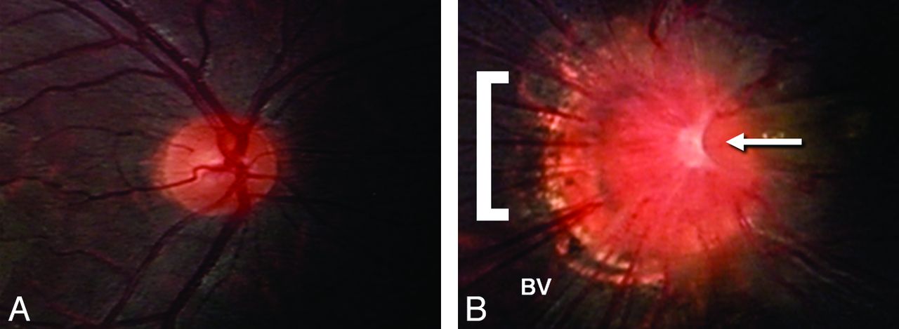
- Fig 1.
Fundus photos illustrating normal right optic nerve (A) and morning glory optic nerve anomaly (B). Note the larger overall size, central glial tuft (arrow), halo of pigmentary changes surrounding the nerve (bracket), and the radial orientation of the blood vessels (BV) emanating from the anomalous optic nerve.
- Fig 2.
A, Axial fat-saturated T2WI through the orbits of a patient with right MGDA demonstrates a funnel-shaped morphologic pattern of the optic disc (white arrow) with elevation of the adjacent retinal surface. This image also demonstrates abnormal tissue associated with the distal intraorbital segment of the ipsilateral optic nerve and effacement of the subarachnoid space at that level (white arrowhead). Also note discontinuity of the uveoscleral coat at the optic nerve insertion (compare with normal left globe where the curvilinear hypointensity of the uveoscleral coat is continuous across the lamina cribrosa; black arrowhead). B, Axial T1WI demonstrates hyperintensity within the elevated region of the retina (white arrow) as well as fat within the distal optic nerve sheath (white arrowheads).
- Fig 3.
A, Axial T1WI through the orbits of a different patient with right-sided MGDA again demonstrates fat within the distal optic nerve sheath (white arrowheads). B, Axial gadolinium-enhanced fat-saturated T1WI through the orbits in the same patient demonstrates enhancement in the region of the distal optic nerve (white arrowheads), which is thought to represent displaced choroidal tissue, perhaps in concert with glial, fibrous, and pigment epithelial proliferation. Careful inspection and cross-correlation established the locale of enhancement to be immediately internal to the fat in the optic nerve sheath. Of note, the fat within the nerve sheath is saturated on this sequence, confirming its identity. These images also demonstrate an associated retinal detachment; the patient is status post a scleral banding procedure.


Tables
Patient characteristics and imaging findings in MGDA
Pt No. Age (y) Sex Side Funnel-Shaped Optic Disc Retinal Elevation Optic Nerve Soft Tissue Uveoscleral Discontinuity Nerve Sheath Fat Enhancement Associated Abnormalities 1 2 M R Present Present Present Present Present Present Retinal detachment 2 1 F L Present Present Present Present Present Absent Small chiasm contralateral to MGDA 3 2 M R Present Present Present Present Absent Absent Persistent craniopharyngeal canal 4 0.67 F B Present Present Present Present Absent Present Moyamoya disease 5 1 M L Present Present Present Present Absent Absent N/A 6 1 F L Present Present Present Present Present Absent N/A Note:—N/A indicates not applicable; R, right; L, left; B, bilateral.


