
Article Figures & Data
Figures
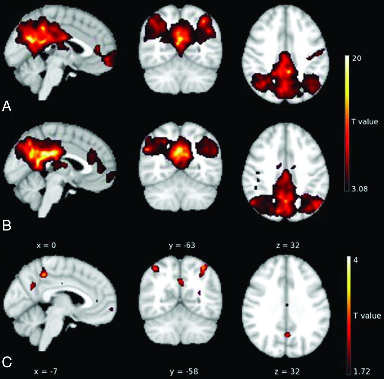
- Fig. 1.
Random-effects analysis of the DMN in controls (A) and (B) patients. C, The patients demonstrate decreased DMN connectivity in the PCC/PCUN, bilateral lateral parietal cortex, and anterior and midcingulate.
- Fig. 2.
Functional connectivity within the DMN in controls (A) and patients (B). Numbers in the rectangles correspond to the average pair-wise correlation coefficients. C, Comparison of functional connectivity within the DMN in controls versus patients shows significant reduction in mPFC–right lateral parietal cortex (LPC), PCC/PCUN–right LPC, and PCC/PCUN–left LPC. Numbers in rectangles represent the P value for the comparison of the mean correlation coefficients of control and patient groups. Thick lines represent significant changes in the pair-wise correlation between controls and patients (P < .05).


Tables
No./Age (yr)/Sex Location of Epilepsy Age at Seizure Onset (yr) Duration of Epilepsy (yr) Antiepileptic Medications Seizure Frequency/Week 1/15.2/F R frontal 7 8 Lamotrigine, levetiracetam 7 2/16.3/F R frontal paracentral 14 2.3 Carbamazepine 7 3/10.0/F R neocortical inferior temporal 7 3 Lamotrigine, topiramate 10 4/15.3/F R frontal paracentral 3 12.3 Carbamazepine, lamotrigine, divalproex 10 5/14.0/M L frontal 8 6 Valproic acid, lamotrigine 20 6/10.1/F L neocortical anterior temporal 8 2 Carbamazepine 10 7/16.2/M L frontal paracentral 8 8 Carbamazepine, levetiracetam 10 8/14.1/M L neocortical anterior temporal 12 2 Lamotrigine, levetiracetam 2 9/14.7/F R hemisphere 13 1 Levetiracetam, divalproex, valproic acid 2.5 10/15.0/F L frontal 4.5 10.5 Oxcarbazepine 40 11/14.1/F R lateral parietal occipital 10 4 Levetiracetam, topiramate 40 Note:—R indicates right; L, left.
Anatomic Regions Hemisphere MNI Coordinates (x, y, z) Peak T Value Posterior cingulate cortex/precuneus L/R −6, −54, 28 15.87 Medial prefrontal cortex L/R −4, 58, −10 13.89 Lateral parietal cortex L −44, −62, 33 9.91 R 42, −63, 33 10.51 Note:—R indicates right; L, left.
↵a P < .05 after correction for multiple comparison using false discovery rate.
Anatomic Regions MNI Coordinates (x, y, and z) Voxel Size (mm3) Peak T Value Right anterior cingulate 4, 40, 8 75 3.549 Right midcingulate 4, −16, 40 55 2.679 Right posterior cingulate cortex/precuneus 10, −68, 52 23 2.214 Right lateral parietal cortex 42, −52, 56 183 3.606 Left lateral parietal cortex −36, −62, 56 142 4.187 Left posterior cingulate cortex/precuneus 0, −60, 34 91 2.682


