
Article Figures & Data
Figures
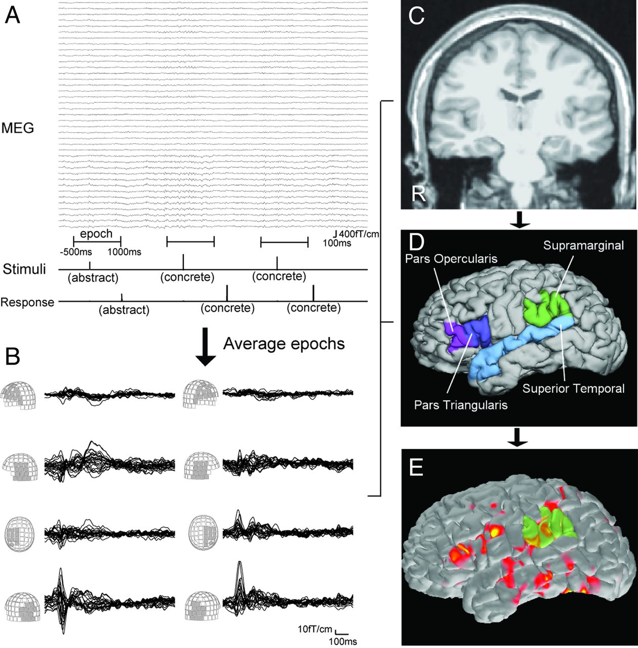
- Fig. 1.
A, Schematic representation of language MEG processing. Stimuli are visually presented as “abstract” or “concrete” words (Stimuli line). Patient responses are also recorded (Response line). Using stimuli as a trigger, epochs from −500 ms to 1000 ms are averaged. B, Waveforms of the averaged MEG from 0 ms to 1000 ms. Waveforms of each MEG sensor group are superimposed. Language activation is seen around 250–550 ms in the frontal, temporal, and parietal sensors. MPRAGE MR imaging (C) provides the cortical surface of each patient by reconstruction processing. This procedure also gives anatomic parcellation of the cortex (D). Four ROIs per each hemisphere are used for calculating LI. E, Spatiotemporal source distribution of the language MEG is mapped on the cortical surface by using dSPM. The value of activation in ROIs is extracted from dSPM.
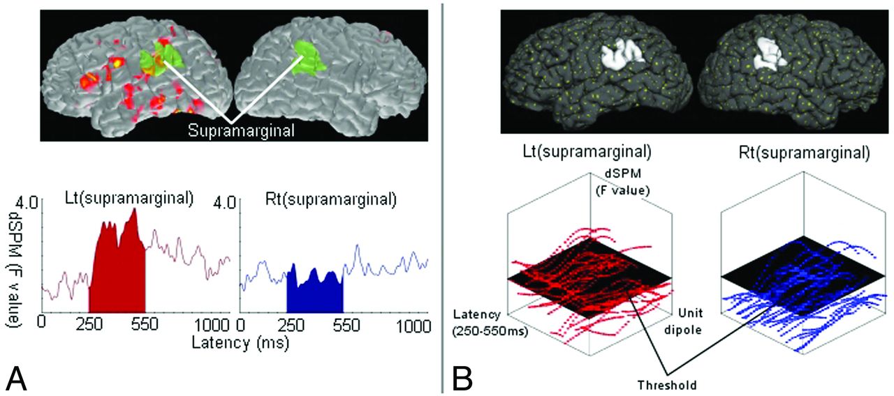
- Fig. 2.
Schematic representation for calculating the LI. LI is calculated by the formula (L − R)/(L + R), where L and R represent parameters obtained from the source distribution within a time window of 250–550 ms in the left and right ROIs, respectively. A, dSPM-amplitude method. The source waveform averaged in each region of interest is extracted from dSPM. L and R are the sum of these waveforms in left and right ROIs. The figure shows an example of processing in a pair of ROIs (supramarginal gyri). The dSPM (top) provides source waveforms averaged within the left and right supramarginal gyrus (bottom). B, dSPM-counting method. In dSPM, each unit dipole (top; yellow dots) has a value of activation. Within each region of interest (eg, the left and right supramarginal gyrus; top), the values of numerous unit dipoles are obtained at each time point (bottom). The number of unit dipoles with a value over a threshold is counted. L and R are the total number of unit dipoles counted in left and right ROIs.


Tables
Patient profile and laterality indices
Patient Age/Sex Diagnosis LI-Amplitude LI-Counting 1 16/F Lt TLE 0.27 0.88 2 13/F Lt FLE −0.03a 0.30 3 15/M Lt TLE 0.05a 0.58 4 15/F Rt TLE 0.02a 0.14 5 34/F Lt TLE 0.54 1.00 6 20/F Rt TLE 0.12 0.98 7 19/M Rt FLE 0.12 0.89 8 15/M Rt PLE 0.24 0.68 9 17/F Lt TLE 0.11 0.68 10 9/F Lt TLE 0.02a 0.28 11 14/M Rt FLE −0.06a −0.08a 12 14/F Lt TLE 0.15 0.62 13 16/F Lt TLE 0.16 0.63 14 28/M Lt FLE 0.08a 0.16 15 18/M Lt OLE 0.02a 0.05a 16 21/M Lt TLE 0.01a 0.33 17 38/M Rt TLE 0.04a 0.62 18 30/M Rt TLE −0.07a 0.15 19 28/F Rt TLE 0.26 0.99 20 42/F Rt TLE 0.10 0.15 21 50/M Lt TLE 0.10 −0.04a 22 18/M Rt TLE 0.12 0.22 23 59/M Lt TLE 0.17 0.88 24 26/M Lt TLE −0.08a 0.10 25 44/M Lt TLE 0.00a 0.23 26 15/M Lt TLE 0.07a 0.23 27 23/M Rt TLE 0.11 0.47 28 18/F Lt TLE 0.11 0.20 29 28/F Rt TLE 0.11 0.59 30 26/F Rt TLE 0.06a 0.13 31 30/F Lt TLE 0.11 0.56 32 14/F Rt OLE 0.24 1.00 33b 15/M Lt FLE −0.02a −0.17 34b 43/M Lt TLE −0.06a −0.13 35b 12/M Lt TLE −0.09a −0.33 Note:—LI indicates laterality index; F, female; M, male; Rt, right; Lt, left; TLE, temporal lobe epilepsy; FLE, frontal lobe epilepsy; PLE, parietal lobe epilepsy; OLE, occipital lobe epilepsy.
↵a MEG-derived language lateralization is not consistent with IAP.
↵b Right-hemisphere language dominance in IAP.


