Advertisement

Main menu

  • Home
  • Content
    • Current Issue
    • Accepted Manuscripts
    • Article Preview
    • Past Issue Archive
    • Video Articles
    • AJNR Case Collection
    • Case of the Week Archive
    • Case of the Month Archive
    • Classic Case Archive
  • Special Collections
    • AJNR Awards
    • Low-Field MRI
    • Alzheimer Disease
    • ASNR Foundation Special Collection
    • Photon-Counting CT
    • View All
  • Multimedia
    • AJNR Podcasts
    • AJNR SCANtastic
    • Trainee Corner
    • MRI Safety Corner
    • Imaging Protocols
  • For Authors
    • Submit a Manuscript
    • Submit a Video Article
    • Submit an eLetter to the Editor/Response
    • Manuscript Submission Guidelines
    • Statistical Tips
    • Fast Publishing of Accepted Manuscripts
    • Graphical Abstract Preparation
    • Imaging Protocol Submission
    • Author Policies
  • About Us
    • About AJNR
    • Editorial Board
    • Editorial Board Alumni
  • More
    • Become a Reviewer/Academy of Reviewers
    • Subscribers
    • Permissions
    • Alerts
    • Feedback
    • Advertisers
    • ASNR Home

User menu

  • Alerts
  • Log in

Search

  • Advanced search
American Journal of Neuroradiology

American Journal of Neuroradiology

ASHNR American Society of Functional Neuroradiology ASHNR American Society of Pediatric Neuroradiology ASSR
  • Alerts
  • Log in

Advanced Search

  • Home
  • Content
    • Current Issue
    • Accepted Manuscripts
    • Article Preview
    • Past Issue Archive
    • Video Articles
    • AJNR Case Collection
    • Case of the Week Archive
    • Case of the Month Archive
    • Classic Case Archive
  • Special Collections
    • AJNR Awards
    • Low-Field MRI
    • Alzheimer Disease
    • ASNR Foundation Special Collection
    • Photon-Counting CT
    • View All
  • Multimedia
    • AJNR Podcasts
    • AJNR SCANtastic
    • Trainee Corner
    • MRI Safety Corner
    • Imaging Protocols
  • For Authors
    • Submit a Manuscript
    • Submit a Video Article
    • Submit an eLetter to the Editor/Response
    • Manuscript Submission Guidelines
    • Statistical Tips
    • Fast Publishing of Accepted Manuscripts
    • Graphical Abstract Preparation
    • Imaging Protocol Submission
    • Author Policies
  • About Us
    • About AJNR
    • Editorial Board
    • Editorial Board Alumni
  • More
    • Become a Reviewer/Academy of Reviewers
    • Subscribers
    • Permissions
    • Alerts
    • Feedback
    • Advertisers
    • ASNR Home
  • Follow AJNR on Twitter
  • Visit AJNR on Facebook
  • Follow AJNR on Instagram
  • Join AJNR on LinkedIn
  • RSS Feeds

AJNR Awards, New Junior Editors, and more. Read the latest AJNR updates

Research ArticlePediatric Neuroimaging

Comparison of Spin-Echo T1- and T2-Weighted and Gradient-Echo T1-Weighted Images at 3T in Evaluating Very Preterm Neonates at Term-Equivalent Age

B. Sarikaya, A.M. McKinney, B. Spilseth and C.L. Truwit
American Journal of Neuroradiology May 2013, 34 (5) 1098-1103; DOI: https://doi.org/10.3174/ajnr.A3323
B. Sarikaya
aFrom the Department of Radiology/Neuroradiology, University of Minnesota and Hennepin County Medical Centers, Minneapolis, Minnesota.
  • Find this author on Google Scholar
  • Find this author on PubMed
  • Search for this author on this site
A.M. McKinney
aFrom the Department of Radiology/Neuroradiology, University of Minnesota and Hennepin County Medical Centers, Minneapolis, Minnesota.
  • Find this author on Google Scholar
  • Find this author on PubMed
  • Search for this author on this site
B. Spilseth
aFrom the Department of Radiology/Neuroradiology, University of Minnesota and Hennepin County Medical Centers, Minneapolis, Minnesota.
  • Find this author on Google Scholar
  • Find this author on PubMed
  • Search for this author on this site
C.L. Truwit
aFrom the Department of Radiology/Neuroradiology, University of Minnesota and Hennepin County Medical Centers, Minneapolis, Minnesota.
  • Find this author on Google Scholar
  • Find this author on PubMed
  • Search for this author on this site
  • Article
  • Figures & Data
  • Info & Metrics
  • Responses
  • References
  • PDF
Loading

Article Figures & Data

Figures

  • Tables
  • Fig 1.
    • Download figure
    • Open in new tab
    • Download powerpoint
    Fig 1.

    GRE-T1WI (A), SE-T1WI (B), and SE-T2WI (C) at the level of the nonmyelinated anterior commissure (arrows) and optic radiations (dashed arrows) in a very preterm infant (27 weeks +3 days) imaged at term-equivalent age (40 weeks +2 days). Note the myelinated PLIC (thin arrows) and habenular commissure (thin dashed arrows), both of which were myelinated in most infants on both GRE-T1WI and on SE-T2WI.

  • Fig 2.
    • Download figure
    • Open in new tab
    • Download powerpoint
    Fig 2.

    GRE-T1WI (A), SE-T1WI (B), and SE-T2WI (C) at the level of the pontomesencephalic junction in a very preterm infant (26 weeks +5 days at birth), imaged at term-equivalent age (39 weeks +5 days). The medial lemniscus (ML) (arrows) and the lateral lemniscus (dashed arrows) are well-depicted on both GRE-T1WI and SE-T2WI but not on SE-T1WI. These 2 structures were myelinated on both sequences in the large majority of term-equivalent infants. A level just caudal to that (D–F) demonstrates that the ML is well-visualized on each sequence, while the cranial nerve V fascicle (thin arrows) is well-visualized on both GRE-T1WI and SE-T2WI but not on SE-T1WI.

  • Fig 3.
    • Download figure
    • Open in new tab
    • Download powerpoint
    Fig 3.

    Comparison among GRE-T1WI (A), SE-T1WI (B), and SE-T2WI (C) at the level of the midbrain in a very preterm infant (26 weeks +0 days) imaged at term-equivalent age (40 weeks +1 day). The DSCP (arrows) and the brachium of the inferior colliculus (dashed arrows) are depicted. The DSCP was myelinated in all term-equivalent infants on both GRE-T1WI and SE-T2WI, while the brachium of the inferior colliculus was variably myelinated on these 2 sequences.

  • Fig 4.
    • Download figure
    • Open in new tab
    • Download powerpoint
    Fig 4.

    GRE-T1WI (A and C) and SE-T2WI (B and D) at the level of the medulla in a very preterm infant (26 weeks +0 days) imaged at term-equivalent age (40 weeks +1 day). Regarding A and B, the spinal tract and nucleus of cranial nerve V (STV) (arrows) are more easily delineated on SE-T2WI, while the pyramidal decussation (PD) (dashed arrows) is visible on both sequences. Note the cuneate fasciculi (arrowheads) just medial to the STV. At a level 1 section higher (C and D), the ICP (thin arrows) and the PD (dashed arrows) are visible on both sequences, but the ICP is more clearly visible on SE-T2WI due to a greater contrast with the remainder of the medulla.
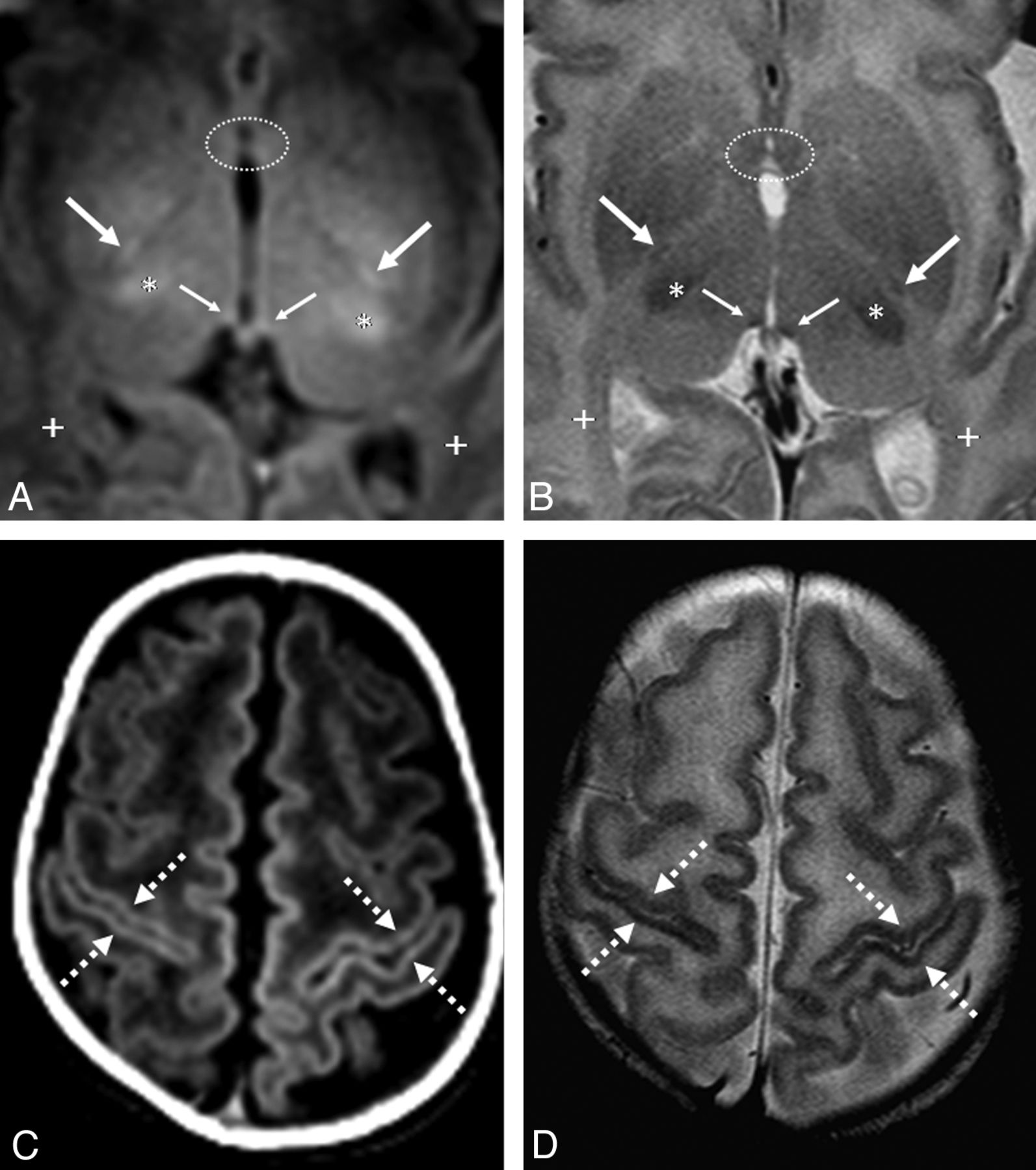
  • Fig 5.
    • Download figure
    • Open in new tab
    • Download powerpoint
    Fig 5.

    GRE-T1WI (A and C) and SE-T2WI (B and D) in a very preterm infant (26 weeks +4 days) imaged at term-equivalent age (40 weeks +2 days). A and B, The PLIC (arrows) is better delineated on GRE-T1WI, while the habenular commissure (thin arrows) is visible on both sequences. Note the myelinated ventrolateral thalami (asterisk) along with the nonmyelinated anterior commissure anteriorly (dotted circles) and optic radiations posteriorly (+). Near the vertex (C and D), the perirolandic cortex (dashed arrows) is visibly myelinated on both sequences. The PLIC has a slightly higher rate of myelination on GRE-T1WI than on SE-T2WI.

Tables

  • Figures
  • Percentage of patients myelinated in each region per the 2 observers

    SE T2WISE T1WIGRE T1WI
    Observer 1Observer 2Observer 1Observer 2Observer 1Observer 2
    AC36.4% (4/11)0% (0/11)0% (0/11)0% (0/11)0% (0/11)0% (0/11)
    ALIC0% (0/11)0% (0/11)0% (0/11)0% (0/11)0% (0/11)0% (0/11)
    BIC54.5% (6/11)100% (11/11)9% (1/11)45.5% (5/11)36.4% (4/11)100% (11/11)
    CCS90.9% (10/11)a90.9% (10/11)a9% (1/11)45.5% (5/11)18.2% (2/11)90.9% (10/11)
    CNV63.6% (7/11)100% (11/11)0% (0/11)45.5% (5/11)81.8% (9/11)90.9% (10/11)
    CST0% (0/11)36.4% (4/11)27.3% (3/11)45.5% (5/11)36.4% (4/11)81.8% (9/11)
    DSCP100% (11/11)a100% (11/11)a27.3% (3/11)81.8% (9/11)100% (11/11)a100% (11/11)a
    HC81.8% (9/11)72.7% (8/11)54.5% (6/11)63.6% (7/11)72.7% (8/11)72.7% (8/11)
    ICP90.9% (10/11)a100% (11/11)a72.7% (8/11)63.6% (7/11)90.9% (10/11)a100% (11/11)a
    LGN18.2% (2/11)63.6% (7/11)0% (0/11)27.3% (3/11)27.3% (3/11)72.7% (8/11)
    LL100% (11/11)a100% (11/11)a54.5% (6/11)81.8% (9/11)100% (11/11)a100% (11/11)a
    ML81.8% (9/11)100% (11/11)9.1% (1/11)81.8% (9/11)90.9% (10/11)a100% (11/11)a
    MLF27.3% (3/11)36.4% (4/11)0% (0/11)0% (0/11)0% (0/11)9% (1/11)
    ON45.5% (5/11)18.2% (2/11)9% (1/11)9% (1/11)72.7% (8/11)27.3% (3/11)
    OR0% (0/11)0% (0/11)0% (0/11)0% (0/11)0% (0/11)0% (0/11)
    OT81.8% (9/11)63.6% (7/11)27.3% (3/11)27.3% (3/11)45.5% (5/11)36.4% (4/11)
    PC18.2% (2/11)9.1% (1/11)0% (0/11)0% (0/11)0% (0/11)0% (0/11)
    PD63.6% (7/11)100% (11/11)18.2% (2/11)27.3% (3/11)90.9% (10/11)a100% (11/11)a
    PLIC81.8% (9/11)90.9% (10/11)81.8% (9/11)81.8% (9/11)100% (11/11)a100% (11/11)a
    PRC81.8% (9/11)100% (11/11)0% (0/11)45.5% (5/11)18.2% (2/11)72.7% (8/11)
    SCP81.8% (9/11)90.9% (10/11)36.4% (4/11)54.5%(6/11)90.9% (10/11)a100% (11/11)a
    STV90.9% (10/11)a100% (11/11)a18.2% (2/11)36.4% (4/11)72.7% (8/11)63.6% (7/11)
    • Note:—AC indicates anterior commissure; ALIC, anterior limb of the internal capsule; BIC, brachium of the inferior colliculus; CCS, corpus callosum splenium; CNV, cranial nerve V; CST, corticospinal tracts; HC, habenular commissure; LGN, lateral geniculate nucleus; LL, lateral lemniscus; ML, medial lemniscus; MLF, medial longitudinal fasciculus; ON, optic nerve; OR, optic radiations; OT, optic tracts; PC, posterior commissure; PRC, perirolandic cortex; PD, pyramidal decussation; SCP, superior cerebellar peduncle; STV, spiral tract/nucleus of cranial nerve V.

    • ↵a Structures that both reviewers and observers found to be myelinated in 90%–100% of term-equivalent patients.

PreviousNext
Back to top

In this issue

American Journal of Neuroradiology: 34 (5)
American Journal of Neuroradiology
Vol. 34, Issue 5
1 May 2013
  • Table of Contents
  • Index by author
Advertisement
Print
Download PDF
Email Article

Thank you for your interest in spreading the word on American Journal of Neuroradiology.

NOTE: We only request your email address so that the person you are recommending the page to knows that you wanted them to see it, and that it is not junk mail. We do not capture any email address.

Enter multiple addresses on separate lines or separate them with commas.
Comparison of Spin-Echo T1- and T2-Weighted and Gradient-Echo T1-Weighted Images at 3T in Evaluating Very Preterm Neonates at Term-Equivalent Age
(Your Name) has sent you a message from American Journal of Neuroradiology
(Your Name) thought you would like to see the American Journal of Neuroradiology web site.
CAPTCHA
This question is for testing whether or not you are a human visitor and to prevent automated spam submissions.
Cite this article
B. Sarikaya, A.M. McKinney, B. Spilseth, C.L. Truwit
Comparison of Spin-Echo T1- and T2-Weighted and Gradient-Echo T1-Weighted Images at 3T in Evaluating Very Preterm Neonates at Term-Equivalent Age
American Journal of Neuroradiology May 2013, 34 (5) 1098-1103; DOI: 10.3174/ajnr.A3323

Citation Manager Formats

  • BibTeX
  • Bookends
  • EasyBib
  • EndNote (tagged)
  • EndNote 8 (xml)
  • Medlars
  • Mendeley
  • Papers
  • RefWorks Tagged
  • Ref Manager
  • RIS
  • Zotero
0 Responses
Respond to this article
Share
Bookmark this article
Comparison of Spin-Echo T1- and T2-Weighted and Gradient-Echo T1-Weighted Images at 3T in Evaluating Very Preterm Neonates at Term-Equivalent Age
B. Sarikaya, A.M. McKinney, B. Spilseth, C.L. Truwit
American Journal of Neuroradiology May 2013, 34 (5) 1098-1103; DOI: 10.3174/ajnr.A3323
del.icio.us logo Twitter logo Facebook logo Mendeley logo
  • Tweet Widget
  • Facebook Like
  • Google Plus One
Purchase

Jump to section

  • Article
    • Abstract
    • ABBREVIATIONS:
    • Materials and Methods
    • Results
    • Discussion
    • Conclusions
    • Footnotes
    • References
  • Figures & Data
  • Info & Metrics
  • Responses
  • References
  • PDF

Related Articles

  • No related articles found.
  • PubMed
  • Google Scholar

Cited By...

  • Quiet T1-Weighted Pointwise Encoding Time Reduction with Radial Acquisition for Assessing Myelination in the Pediatric Brain
  • Comparison of Spin-Echo and Gradient-Echo T1-Weighted and Spin-Echo T2-Weighted Images at 3T in Evaluating Term-Neonatal Myelination
  • Crossref
  • Google Scholar

This article has not yet been cited by articles in journals that are participating in Crossref Cited-by Linking.

More in this TOC Section

  • FRACTURE MR in Congenital Vertebral Anomalies
  • Comparing MRI Perfusion in Pediatric Brain Tumors
  • Sodium MRI in Pediatric Brain Tumors
Show more Pediatric Neuroimaging

Similar Articles

Advertisement

Indexed Content

  • Current Issue
  • Accepted Manuscripts
  • Article Preview
  • Past Issues
  • Editorials
  • Editor's Choice
  • Fellows' Journal Club
  • Letters to the Editor
  • Video Articles

Cases

  • Case Collection
  • Archive - Case of the Week
  • Archive - Case of the Month
  • Archive - Classic Case

More from AJNR

  • Trainee Corner
  • Imaging Protocols
  • MRI Safety Corner
  • Book Reviews

Multimedia

  • AJNR Podcasts
  • AJNR Scantastics

Resources

  • Turnaround Time
  • Submit a Manuscript
  • Submit a Video Article
  • Submit an eLetter to the Editor/Response
  • Manuscript Submission Guidelines
  • Statistical Tips
  • Fast Publishing of Accepted Manuscripts
  • Graphical Abstract Preparation
  • Imaging Protocol Submission
  • Evidence-Based Medicine Level Guide
  • Publishing Checklists
  • Author Policies
  • Become a Reviewer/Academy of Reviewers
  • News and Updates

About Us

  • About AJNR
  • Editorial Board
  • Editorial Board Alumni
  • Alerts
  • Permissions
  • Not an AJNR Subscriber? Join Now
  • Advertise with Us
  • Librarian Resources
  • Feedback
  • Terms and Conditions
  • AJNR Editorial Board Alumni

American Society of Neuroradiology

  • Not an ASNR Member? Join Now

© 2025 by the American Society of Neuroradiology All rights, including for text and data mining, AI training, and similar technologies, are reserved.
Print ISSN: 0195-6108 Online ISSN: 1936-959X

Powered by HighWire