
Article Figures & Data
Figures
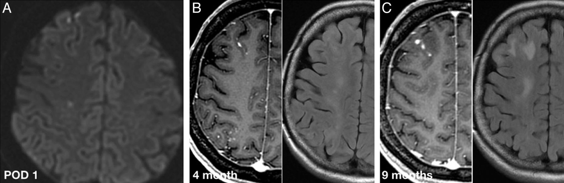
- Fig 1.
A 54-year-old woman with a right supraclinoid ICA unruptured aneurysm treated with a flow diverter. Axial DWI on postoperative day (POD) 1 (A), contrast-enhanced T1-weighted and T2-FLAIR at 4 months (B), and 9 months after procedure (C). POD 1 MR image shows a few asymptomatic DWI lesions in the frontal lobe. Four-month follow-up MR shows subcortical enhancing lesions in both the frontal and parietal lobes, far more numerous than the DWI lesions seen in POD 1. The enhancing lesions and the perilesional edema persist in the 9-month follow-up MR imaging, which is not the expected evolution for ischemic lesions. Foreign body reaction was the presumed diagnosis after all clinical and laboratory work-up was negative for infection.
- Fig 2.
A 32-year-old woman with an unruptured left MCA aneurysm treated with balloon-assisted coiling. Axial FLAIR and axial T1-weighted contrast-enhanced MR images at 6 months (A) and 11 months (B) follow-up show multiple enhancing cortico-subcortical lesions in the distal MCA territory surrounded by vasogenic edema. Combined antibiotic therapy was initiated after (A) with only partial improvement in the next follow-up. Note how the middle temporal gyrus lesion disappears over time while the middle frontal lesions only decrease in number.
- Fig 3.
A 51-year-old woman with an asymptomatic intradural left cavernous ICA aneurysm treated with a flow diverter. Axial contrast-enhanced T1-weighted, T2-FLAIR, and DWI brain MR images at 3 months (A) and 6 months (B). A, Representation of 1 of the rim-enhancing lesions in the frontal operculum, with significant perilesional edema and restricted diffusion. An infectious cause was ruled out on clinical grounds and a foreign body reaction was considered as the most possible cause. The patient received 6 weeks of combined oral antibiotic therapy and remained asymptomatic. B, At 6-month follow-up, the lesions show a solid pattern of enhancement, with no restricted diffusion and only partial improvement of the edema. This was considered to represent the evolution of an aseptic abscess into a foreign body granuloma. The lesions and the surrounding edema decrease slowly in size but at 2-year follow up still exist (not shown).


Jump to section
Related Articles
Cited By...
- Non-ischemic cerebral enhancing (NICE) lesions after flow diversion for intracranial aneurysms: a multicenter study
- Longitudinal radiological follow-up of individual level non-ischemic cerebral enhancing lesions following endovascular aneurysm treatment
- Non-ischemic cerebral enhancing lesions after intracranial aneurysm endovascular repair: a retrospective French national registry
- Effect of the Shelving Technique on the Outcome of Embolization in Intracranial Bifurcation Aneurysms
- Medical treatment of polymeric cerebral granulomatous reactions following endovascular procedures
- Microcatheter Originating Debris during Neuroendovascular Procedures: Mechanism of Dislodgement and Its Prevention
- Delayed Leukoencephalopathy: A Rare Complication after Coiling of Cerebral Aneurysms
- Intrathrombus polymer coating deposition: a pilot study of 91 patients undergoing endovascular therapy for acute large vessel stroke. Part I: Histologic frequency
- Delayed leucoencephalopathy after coil embolisation of unruptured cerebral aneurysm
- Delayed neurological deficits after endovascular placement of a pipeline embolisation device: clinical manifestation and treatment
- Teaching NeuroImages: Intracranial foreign body reaction after endovascular procedures
- Delayed enhancing lesions after coil embolization of aneurysms: clinical experience and benchtop analyses
- Treatment of Middle Cerebral Artery Aneurysms with Flow-Diverter Stents: A Systematic Review and Meta-Analysis
- Multicenter Experience with FRED Jr Flow Re-Direction Endoluminal Device for Intracranial Aneurysms in Small Arteries
- FRED Flow Diverter: A Study on Safety and Efficacy in a Consecutive Group of 50 Patients
- Suspected Metallic Embolism following Endovascular Treatment of Intracranial Aneurysms
- Risk Factors for Ischemic Complications following Pipeline Embolization Device Treatment of Intracranial Aneurysms: Results from the IntrePED Study
- Retrospective Analysis of Delayed Intraparenchymal Hemorrhage after Flow-Diverter Treatment: Presentation of a Retrospective Multicenter Trial
- Foreign Body Emboli following Cerebrovascular Interventions: Clinical, Radiographic, and Histopathologic Features