Advertisement

Main menu

  • Home
  • Content
    • Current Issue
    • Accepted Manuscripts
    • Article Preview
    • Past Issue Archive
    • AJNR Case Collection
    • Case of the Week Archive
    • Classic Case Archive
    • Case of the Month Archive
  • Special Collections
    • Spinal CSF Leak Articles (Jan 2020-June 2024)
    • 2024 AJNR Journal Awards
    • Most Impactful AJNR Articles
  • Multimedia
    • AJNR Podcast
    • AJNR Scantastics
    • Video Articles
  • For Authors
    • Submit a Manuscript
    • Author Policies
    • Fast publishing of Accepted Manuscripts
    • Graphical Abstract Preparation
    • Manuscript Submission Guidelines
    • Imaging Protocol Submission
    • Submit a Case for the Case Collection
  • About Us
    • About AJNR
    • Editorial Board
  • More
    • Become a Reviewer/Academy of Reviewers
    • Subscribers
    • Permissions
    • Alerts
    • Feedback
    • Advertisers
    • ASNR Home
  • Other Publications
    • ajnr

User menu

  • Alerts
  • Log in

Search

  • Advanced search
American Journal of Neuroradiology

American Journal of Neuroradiology

ASHNR American Society of Functional Neuroradiology ASHNR American Society of Pediatric Neuroradiology ASSR
  • Alerts
  • Log in

Advanced Search

  • Home
  • Content
    • Current Issue
    • Accepted Manuscripts
    • Article Preview
    • Past Issue Archive
    • AJNR Case Collection
    • Case of the Week Archive
    • Classic Case Archive
    • Case of the Month Archive
  • Special Collections
    • Spinal CSF Leak Articles (Jan 2020-June 2024)
    • 2024 AJNR Journal Awards
    • Most Impactful AJNR Articles
  • Multimedia
    • AJNR Podcast
    • AJNR Scantastics
    • Video Articles
  • For Authors
    • Submit a Manuscript
    • Author Policies
    • Fast publishing of Accepted Manuscripts
    • Graphical Abstract Preparation
    • Manuscript Submission Guidelines
    • Imaging Protocol Submission
    • Submit a Case for the Case Collection
  • About Us
    • About AJNR
    • Editorial Board
  • More
    • Become a Reviewer/Academy of Reviewers
    • Subscribers
    • Permissions
    • Alerts
    • Feedback
    • Advertisers
    • ASNR Home
  • Follow AJNR on Twitter
  • Visit AJNR on Facebook
  • Follow AJNR on Instagram
  • Join AJNR on LinkedIn
  • RSS Feeds

Welcome to the new AJNR, Updated Hall of Fame, and more. Read the full announcements.


AJNR is seeking candidates for the position of Associate Section Editor, AJNR Case Collection. Read the full announcement.


Research ArticleHead & Neck

Imaging Findings of Head and Neck Dermatofibrosarcoma Protuberans

G.G. Millare, N. Guha-Thakurta, E.M. Sturgis, A.K. El-Naggar and J.M. Debnam
American Journal of Neuroradiology February 2014, 35 (2) 373-378; DOI: https://doi.org/10.3174/ajnr.A3650
G.G. Millare
aFrom the Department of Diagnostic Radiology (G.G.M.), Baylor College of Medicine, Houston, Texas
  • Find this author on Google Scholar
  • Find this author on PubMed
  • Search for this author on this site
N. Guha-Thakurta
bDepartment of Diagnostic Radiology (N.G.-T., J.M.D.), Section of Neuroradiology
  • Find this author on Google Scholar
  • Find this author on PubMed
  • Search for this author on this site
E.M. Sturgis
cDepartments of Head and Neck Surgery (E.M.S.)
dEpidemiology (E.M.S.)
  • Find this author on Google Scholar
  • Find this author on PubMed
  • Search for this author on this site
A.K. El-Naggar
ePathology (A.K.E.-N.), The University of Texas MD Anderson Cancer Center, Houston, Texas.
  • Find this author on Google Scholar
  • Find this author on PubMed
  • Search for this author on this site
J.M. Debnam
bDepartment of Diagnostic Radiology (N.G.-T., J.M.D.), Section of Neuroradiology
  • Find this author on Google Scholar
  • Find this author on PubMed
  • Search for this author on this site
  • Article
  • Figures & Data
  • Info & Metrics
  • Responses
  • References
  • PDF
Loading

Abstract

BACKGROUND AND PURPOSE: Dermatofibrosarcoma protuberans is a rare, locally aggressive sarcoma of the skin in children and adults, usually involving the trunk and extremities and less commonly the head and neck. Despite clinical reports in the literature on the management of dermatofibrosarcoma protuberans, there are limited articles describing its imaging features.

MATERIALS AND METHODS: We retrospectively reviewed the demographics and imaging findings in all 24 patients with pathologically proven dermatofibrosarcoma protuberans of the head and neck seen at a tertiary cancer center between 2001 and 2010.

RESULTS: Twenty-two of the 24 lesions were nodular and well circumscribed; 19 of the 24 were located on the scalp. On imaging, all 24 lesions involved subcutaneous tissues. The lesions ranged in size from 0.6–9.5 cm (mean, 3.7 cm; standard deviation, 2.3 cm). Twelve lesions involved the soft tissues either at or extending directly to the midline. Thirteen lesions were associated with bulging of the skin surface. Fourteen lesions were imaged with CT and 14 with MR imaging. Whereas variable enhancement patterns were noted on CT and MR imaging, dermatofibrosarcoma protuberans was usually T2-hyperintense and demonstrated marked enhancement. None of the lesions was associated with bone invasion, perineural spread, or nodal/distant metastasis.

CONCLUSIONS: Knowledge of the imaging characteristics of dermatofibrosarcoma protuberans may alert neuroradiologists to include dermatofibrosarcoma protuberans in the differential diagnosis of lesions about the head and neck with similar imaging characteristics.

ABBREVIATIONS:

DFSP
dermatofibrosarcoma protuberans
PDGFB
platelet-derived growth factor β-chain
PDGF-R
platelet-derived growth factor receptor tyrosine kinase

Dermatofibrosarcoma protuberans (DFSP) is a rare, locally aggressive sarcoma of the dermis with a high cure rate.1,2 DFSP often presents in the third or fourth decade of life, most commonly involving the trunk (50–60%) or proximal extremities (20–30%) and less commonly involving the head and neck (10–15%).3,4 DFSP grows in an infiltrative manner and has the capacity for local recurrence, though it rarely metastasizes.5,6

The imaging features of DFSP of the trunk and extremities are nonspecific, including low signal on T1 and high on T2 on MR imaging, with uniform enhancement after contrast administration.7,8 CT findings include isoattenuated to hypoattenuated lesions without calcifications and uniform (small lesions) to heterogeneous (large lesions) enhancement after contrast administration.9 Previously published reports on DFSP have highlighted its clinical features, but only a few small series have outlined its imaging features.7,8,10 The purpose of this study was to more comprehensively define the CT and MR imaging appearance of DFSP of the head and neck.

Materials and Methods

This study was approved by the Institutional Review Board of The University of Texas MD Anderson Cancer Center, which waived the requirement for informed consent. We retrospectively reviewed the demographics and radiologic images of 24 patients with pathologically proven DFSP of the head and neck seen at our institution between 2001–2010.

Fourteen patients underwent CT of the head and neck, and 14 patients underwent MR imaging of the head and neck. The CT examinations were performed after intravenous administration of contrast material. Soft tissue and bone windows were evaluated in all patients. MR imaging sequences included axial T1 precontrast, orthogonal T1 postcontrast, and T2-weighted sequences (Table).

View this table:
  • View inline
  • View popup

Patient demographics and image appearance

The following imaging features were documented: lesion size, location, enhancement pattern, calcification, and perineural spread; associated osseous abnormality; and presence of nodal or distant metastasis.

Results

Clinical Features

The patients ranged in age from 15–72 years (mean, 42.5 years; median, 40 years) and included 12 women and 12 men. Seventeen patients presented with a primary, untreated tumor, and 7 patients presented with recurrent disease. The most common presenting symptom was a slowly enlarging mass (n = 18). Six patients described a history of trauma before development of a slowly growing mass at the same site as the trauma. The trauma occurred 2 years before presentation in 2 patients, 6–11 years before presentation in 3 patients, and as a child in 1 patient. DFSP was diagnosed through biopsy in all 24 patients.

Imaging Findings

On imaging, 19 lesions involved the scalp; other locations included the posterior neck (n = 1), left face to midline (n = 1), pre-auricular region (n = 1), chin (n = 1), and cheek (n = 1). All 24 lesions involved the subcutaneous soft tissues. Eighteen lesions extended up to the skin surface, and, of these, 13 were associated with bulging of the skin, 2 were associated with an exophytic fungating component, and 2 were associated with skin retraction; 1 was associated with none of these findings. Twelve of the lesions involved the soft tissues either at (n = 6) or near the midline (n = 6). The lesions ranged in size from 0.6–9.5 cm (mean, 3.7 cm; standard deviation, 2.3 cm). Twenty-two of the 24 lesions were nodular and well-circumscribed; the nodules were singular in 13 cases, coalescent in 6 cases, and multiple but separated in 3 cases. Two lesions had an infiltrative pattern; both manifested as recurrences in the operative field. None of the patients had evidence of osseous destruction, cervical lymph node, or distant metastasis.

CT Findings

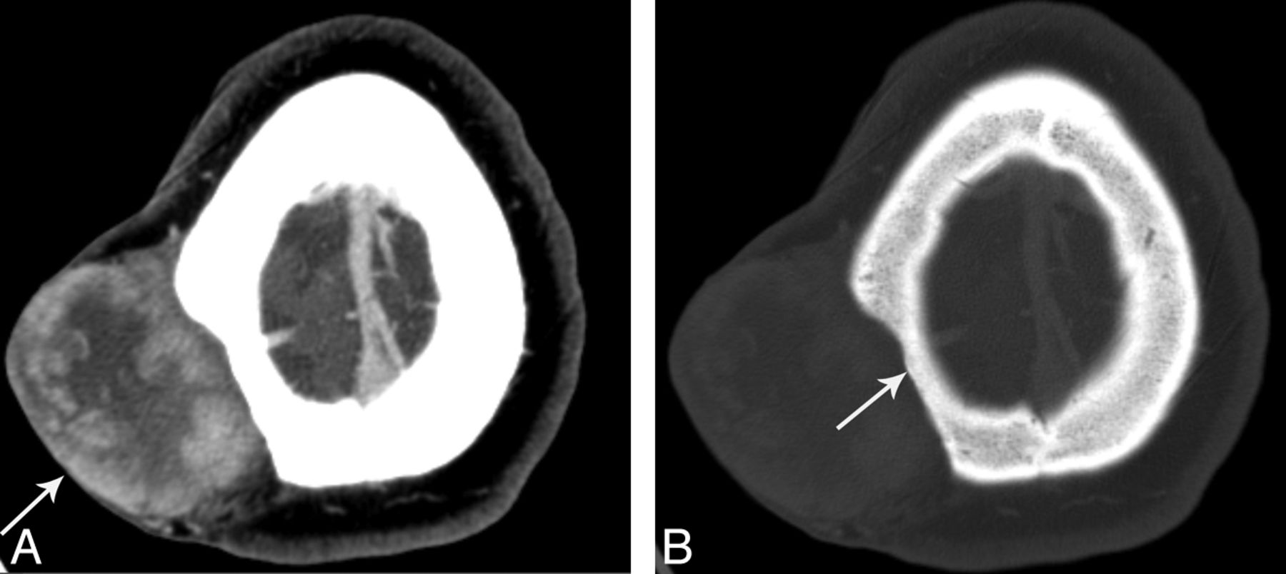
The 14 lesions imaged with CT demonstrated enhancement that was either minimal (n = 3), heterogeneous (n = 3) (Fig 1A), or homogeneous (n = 8). There was no evidence of calcification, and none of the lesions demonstrated CT evidence of perineural spread. Osseous destruction was absent; however, thinning of the underlying calvaria (Fig 1B) was seen in 2 patients (patients 10 and 16).

Fig 1.
  • Download figure
  • Open in new tab
  • Download powerpoint
Fig 1.

A 31-year-old woman (patient 10) with DFSP of the right parietal scalp. Contrast-enhanced axial CT in soft tissue (A) and bone window (B) shows a 6.1 × 4.8-cm heterogeneously enhancing mass (A, arrow) that bulges the skin surface outward and thins underlying calvaria (B, arrow).

MR Imaging Findings

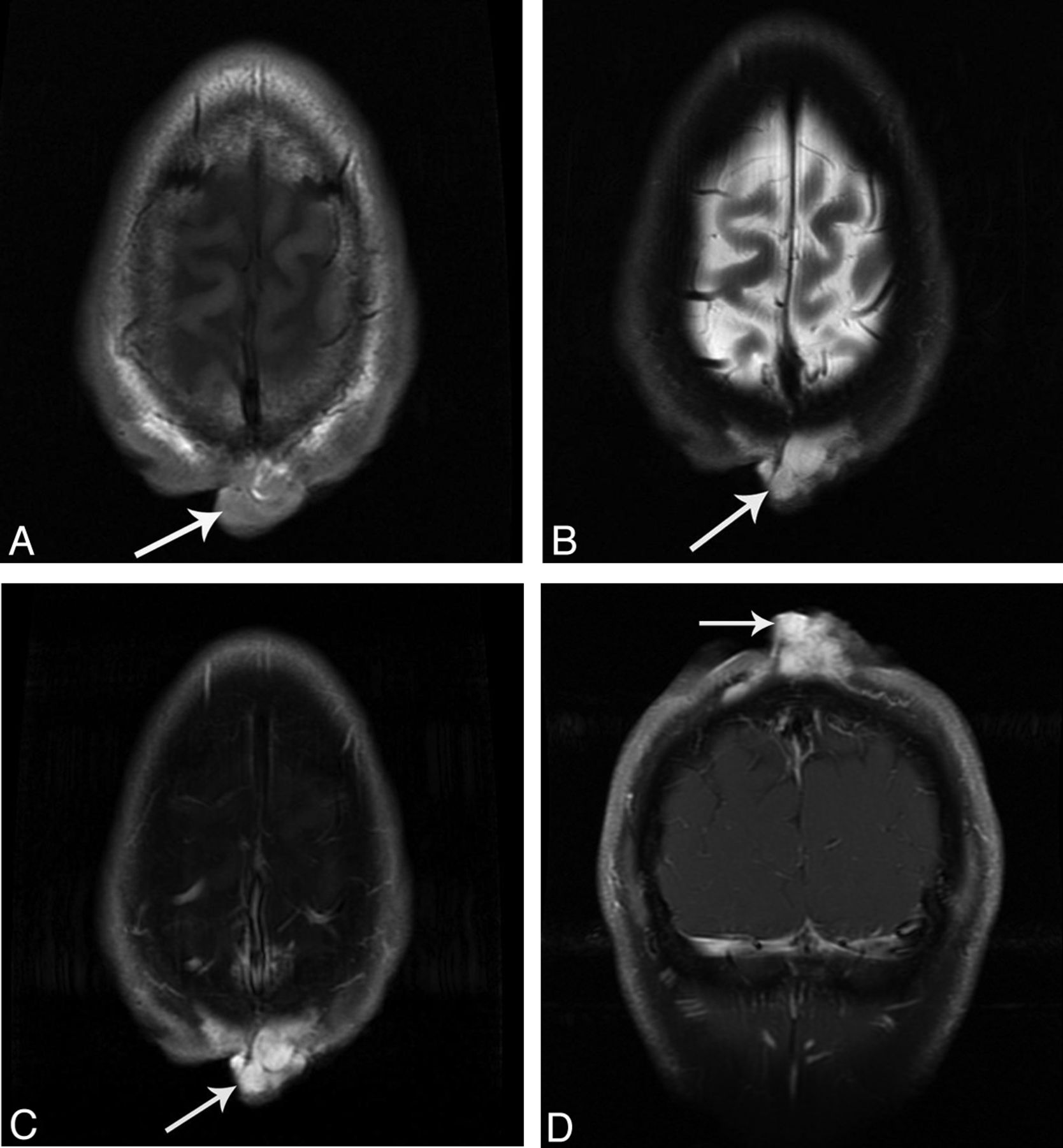
All 14 lesions imaged with MR were T1-isointense to muscle (Fig 2A); 10 lesions were T2-hyperintense (Fig 2B) and 4 were iso-intense to hypointense to muscle. T1 postcontrast enhancement patterns ranged from mild (n = 1) to markedly heterogeneous (n = 4) or markedly homogeneous (n = 9) (Fig 2C,-D). Neither perineural spread nor bone involvement was present in any case.

Fig 2.
  • Download figure
  • Open in new tab
  • Download powerpoint
Fig 2.

A 31-year-old man (patient 1) with DFSP of the parietal midline scalp. MR imaging shows a 3.5 × 1.8-cm T1-isointense, T2-hyperintense, coalescing nodular mass with marked homogeneous enhancement in the parietal midline scalp (arrows). A, Axial noncontrast T1. B, Fast spin-echo T2 with fat saturation. C, T1 postcontrast axial with fat saturation. D, T1 postcontrast coronal with fat saturation.

Pathologic Findings

Most the lesions were found in the lower dermis and the subcutaneous tissue. All tumors manifested spindle cell proliferation arranged in light storiform patterns. Although infiltrative with ill-defined boundaries, the cells were bland and lacking mitotic activity and nuclear pleomorphism.

Seven patients presented to our institution with recurrent disease after having undergone resection at other centers. In 6 of the 7 patients, the pathologic specimens from the initial resection were submitted and reviewed by our pathology department. In all 6 patients, DFSP was present at the surgical margins.

Treatment

The lesions were treated with surgical excision (n = 9), excision and radiation (n = 8), excision and chemotherapy (n = 3), chemotherapy (n = 3), or all 3 treatments (n = 1). All of the patients were still alive at the time of this report.

Discussion

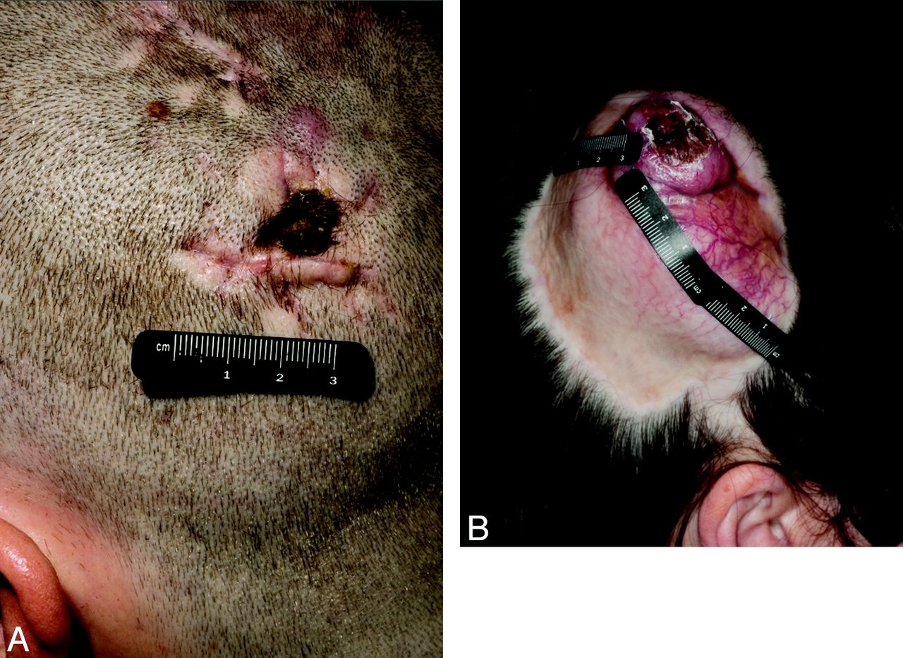
DFSP typically presents as a slow-growing, painless, and firm subcutaneous nodule.11 The lesions can be multiple and may coalesce to form an indurated plaque (Fig 3), which on clinical examination is often violaceous.12,13 The growth rate is variable; some lesions remain stable in size for many years, whereas others demonstrate slow progressive growth.14 DFSP can arise in healthy skin or in areas of repeated trauma, vaccination sites, irradiated skin, or scars.15 Six patients in our series had a history of local trauma before the development of DFSP, and the interval between the trauma and development of DFSP varied. Tattoo sites also show a predilection for benign and malignant changes and should be evaluated during regular skin examinations; DFSP should be considered in the differential diagnosis of neoplasms arising within areas of tattoos.16

Fig 3.
  • Download figure
  • Open in new tab
  • Download powerpoint
Fig 3.

Photographs of 2 patients with DFSP. A, Patient 16, a 25-year-old man with a large DFSP of the scalp extending from the midline near the vertex down to the left lower parietal region with subcutaneous infiltration. B, Patient 10, a 31-year-old woman with a large DFSP of the right paramedian posterior parietal scalp.

In our series, DFSP lesions ranged in size from 0.6–9.5 cm, and half were located at or extended to the midline. Variable enhancement was noted on CT (Fig 1A), similar to prior reports.8,9 On MR imaging, almost all of the cases showed marked enhancement (Fig 2C,-D), which has not previously been reported.7,8 None of the lesions was associated with perineural spread, nodal, or distant metastases. No osseous destruction was demonstrated; however, calvarial thinning was seen in 2 patients. These 2 patients (patients 10 and 16) had the largest lesions, which suggests that the thinning could be related to a pressure effect. Although none of the lesions in our series showed intracranial extension, this has been described in 2 case reports.10,17

None of the patients in our series had local or nodal metastasis, perineural spread, or bone destruction. In contrast, 2–3% of all patients with squamous cell carcinoma of the skin have metastasis, which occurs almost exclusively through lymphatic channels and only rarely through hematogenous spread.18 Perineural spread occurs in basal cell carcinoma, squamous cell carcinoma, and melanoma (desmoplastic), usually involving cranial nerves V and VII.19,20

Bony destruction is uncommon with skin tumors, but when bone destruction is noted on imaging, malignant melanoma and squamous cell carcinoma should be considered.21⇓–23

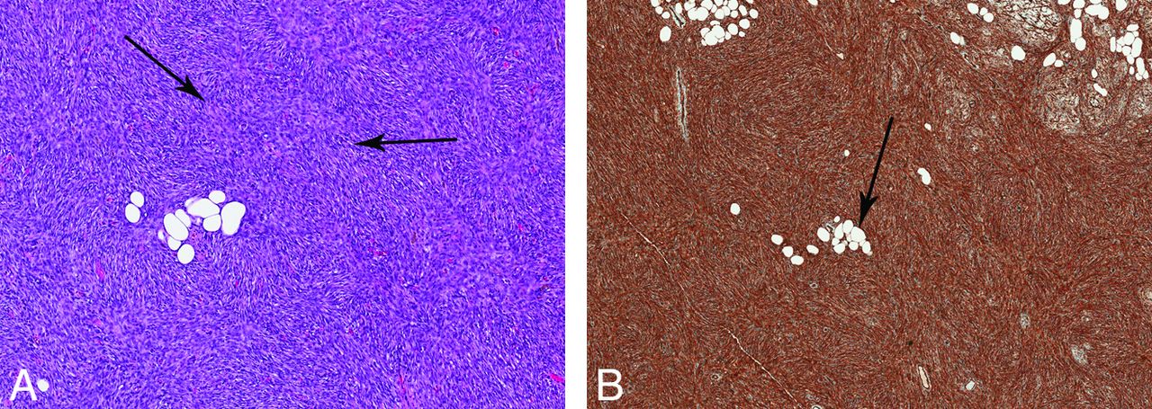
Histologically, DFSP is an infiltrative soft tissue neoplasm of the dermis and subcutaneous tissues. DFSP is composed of bland, elongated spindle cells with a storiform, or whorled, pattern arranged around foci of collagen or vascular spaces, radiating outward like the spokes of a wheel (Fig 4A).24 Infiltration around fat cells may produce a honeycomb appearance of the fat (Fig 4B).25 Immunohistochemical analysis may assist if the diagnosis as DFSP is positive for CD34 staining (Fig 4B), with a reported sensitivity of 84–100%.26⇓–28 Because other skin lesions are also positive for CD34 staining, CD34 is not specific for DFSP; however, CD34 positivity in combination with the histologic features makes the diagnosis.

Fig 4.
  • Download figure
  • Open in new tab
  • Download powerpoint
Fig 4.

Pathologic specimen of DFSP. A, Hematoxylin and eosin stain. Elongated spindle cells with a storiform (whorled) pattern arranged around foci of collagen or vascular spaces, radiating outward (arrows). B, CD34 stain. The specimen is positive for CD34 staining. Fat entrapment is noted with a honeycomb pattern (arrow).

At the genetic level, the critical initiating event in DFSP development is rearrangement of chromosomes 17 and 22 in the form of a supernumerary ring chromosome [r (17; 22)] or a reciprocal translocation [t (17; 22)]. Either of these rearrangements results in chimeric fusion of the genes encoding the platelet-derived growth factor β-chain (PDGFB) and the collagen type 1 α1-chain, which in turn results in autocrine activation of the platelet-derived growth factor receptor tyrosine kinase (PDGF-R), which triggers the proliferation of DFSP tumor cells.29,30

Treatment options include complete surgical excision, including wide excision and Mohs micrographic surgery, radiation, and imatinib mesylate.2,11,31 Imatinib, a competitive tyrosine kinase inhibitor, is currently approved by the Food and Drug Administration for the treatment of adult patients with unresectable, recurrent, and/or metastatic DFSP, and this may in turn facilitate tumor resection and or decrease disfigurement.32 After local resection, the 5-year survival rate is 93–100%.33,34 In agreement with these previously reported findings, none of the patients in our series had died of disease at the time of this report. Local recurrence occurs in 20–55% of cases.12⇓–14,35 Most local recurrences occur within 3 years11,12,35; only 30% occur after 5 years.32 The lungs are the most common site of distant metastasis,12,30,36,37 and metastasis has been reported to occur in 5–6% of cases.13,14,34,35,38,39

Differential diagnoses of other cutaneous neoplasms that could be confused with DFSP include dermatofibroma, epidermal cyst, peripheral nerve sheath tumor, nodular fasciitis, cavernous hemangioma, liposarcoma, nodular type of melanoma, and mycosis fungoides.21,40,41 Dermatofibroma can have imaging findings similar to DFSP on CT and MR imaging but is most frequently seen in the extremities, whereas DFSP is most commonly seen in the trunk.42,43 Epidermal cyst, most commonly an incidental finding, does not enhance on both CT and MR, unless infected, whereas DFSP shows variable enhancement on CT and MR imaging.44,45 Peripheral nerve sheath tumors are usually close to a major nerve, with certain signs such as split fat or fascicular or target sign, which are absent in DFSP.45,46 Plexiform neurofibromas tend to be asymmetric and diffuse, whereas DFSP most commonly is nodular.47,48 Clinically, patients with plexiform neurofibromas have neurofibromatosis type 1. Nodular fasciitis can be indistinguishable from DFSP by imaging. However, on MRI, one may see fascial tail sign, which is the linear extension of the lesion along the fascia, which also may appear to be enhanced. Nodular fasciitis favors the upper extremity, whereas DFSP favors the trunk.40,41,49 Cavernous hemangioma has low attenuation on CT, and calcifications are common, features not seen in DFSP.49,50 Liposarcoma contains fat, which is not present on DFSP.45 The nodular type of melanoma is mostly clinically blue to black, and DFSP is often violaceous. Melanotic melanoma is hyperintense on T1-weighted images and hypointense on T2-weighted images. Melanomas can have associated lymphadenopathy and osseous destruction not usually found in DFSP.44,51,52 Mycosis fungoides is the most common type of cutaneous T-cell lymphoma. CT findings include skin thickening, edema, soft tissue infiltration, lymphadenopathy, and systemic manifestations, not typically found in DFSP.45,53 The imaging findings of DFSP are nonspecific, but the combination of imaging findings, location of the lesion, and clinical features of the lesion help narrow the differential diagnosis of these cutaneous lesions mimicking DFSP.

Limitations of the study include the retrospective nature of the review. Similar imaging studies were not available for each patient, and, for the 7 patients presenting to our center with recurrent disease, the initial imaging of the primary lesion was not submitted for interpretation. However, the pathologic specimens for 6 of the 7 patients with recurrent DFSP were reviewed by our pathology department. DFSP was present at the surgical margins in all 6 specimens, which suggests that recurrent disease may be associated with incomplete surgical resection. This is in keeping with published reports of high recurrence rates in patients with positive surgical margins.54 The most significant prognostic factor in patients with DFSP has been the extent of surgical resection.2

Conclusions

DFSP of the head and neck region typically involves the dermis of the scalp, is often well-circumscribed with a nodular appearance, presents as a subcutaneous mass of variable size, often occurs near the midline, and is associated with bulging of the skin surface. Variable enhancement patterns are noted on CT, but, on MR imaging, DFSP is usually T2-hyperintense with marked enhancement. There is a high propensity for local recurrence but a low incidence of bone invasion, perineural spread, and nodal or distant metastasis. Neuroradiologists should be familiar with the imaging appearance of DFSP and include DFSP in the differential diagnosis of head and neck dermal tumors with similar imaging characteristics.

Acknowledgments

We wish to thank Stephanie Deming for editorial assistance.

Footnotes

  • Disclosures: Adel K. El-Naggar—UNRELATED: Grants/Grants Pending: NIH,* NCI,* NIDCR*; Other: Salivary Gland Tissue Biorepository,* Comments: Contract with NIH to collect extremely rare cancers of the head and neck and distribute to researchers conducting relevant research (*money paid to institution).

  • Paper previously presented as a poster at: Annual Meeting of the American Society of Head and Neck Radiology, October 3–7, 2012; Miami Beach, Florida.

  • The University of Texas MD Anderson Cancer Center is supported in part by the National Institutes of Health through Cancer Center Support Grant CA016672.

References

  1. 1.↵
    1. Snow SN,
    2. Gordon EM,
    3. Larson PO,
    4. et al
    . Dermatofibrosarcoma protuberans: a report on 29 patients treated by Mohs micrographic surgery with long-term follow-up and review of the literature. Cancer 2004;101:28–38
    CrossRefPubMed
  2. 2.↵
    1. Doreen L,
    2. Mügge O,
    3. Mentzel T,
    4. et al
    . Current treatment options in dermatofibrosarcoma. J Cancer Res Clin Oncol 2009;135:653–65
    CrossRefPubMed
  3. 3.↵
    1. Gloster HM
    . Dermatofibrosarcoma protuberans. J Am Acad Dermatol 1996;35:355–74
    CrossRefPubMed
  4. 4.↵
    1. Maggoudi D,
    2. Vahtsevanos K,
    3. Psomaderi K,
    4. et al
    . Dermatofibrosarcoma protuberans of the face: report of 2 cases and an overview of the recent literature. J Oral Maxillofac Surg 2006;64:140–44
    CrossRefPubMed
  5. 5.↵
    1. Gatlin JL,
    2. Hosch R,
    3. Khan M
    . Dermatofibrosarcoma protuberans of the scalp with fibrosarcomatous degeneration and pulmonary metastasis. J Clin Imaging Sci 2011;1:55
    CrossRefPubMed
  6. 6.↵
    1. Lal P,
    2. Goel A,
    3. Mandal AK
    . Dermatofibrosarcoma of the scalp with cervical lymph node metastasis. Sarcoma 2004;8:43–45
    CrossRefPubMed
  7. 7.↵
    1. Torreggiani WC,
    2. Al-Ismail K,
    3. Munk PL,
    4. et al
    . Dermatofibrosarcoma protuberans: MR imaging features. AJR Am J Roentgenol 2002;178:989–93
    PubMed
  8. 8.↵
    1. Kransdorf MJ,
    2. Meis-Kindblom JM
    . Dermatofibrosarcoma protuberans: radiologic appearance. AJR Am J Roentgenol 1994;163:391–94
    PubMed
  9. 9.↵
    1. Li X,
    2. Zhang W,
    3. Xiao L,
    4. et al
    . Computed tomographic and pathological findings of dermatofibrosarcoma protuberans. J Comput Assist Tomogr 2012;36:462–68
    CrossRefPubMed
  10. 10.↵
    1. Das L,
    2. Grover SB,
    3. Chand K
    . Intracranial extension of a dermatofibrosarcoma protuberans of the scalp: a case report with brief review of literature. Surg Neurol 2000;54:452–54
    CrossRefPubMed
  11. 11.↵
    1. Stojadinovic A,
    2. Karpoff HM,
    3. Antonescu CR,
    4. et al
    . Dermatofibrosarcoma protuberans of the head and neck. Ann Surg Oncol 2000;7:696–704
    CrossRefPubMed
  12. 12.↵
    1. Brenner W,
    2. Schaefler K,
    3. Chhabra H,
    4. et al
    . Dermatofibrosarcoma protuberans metastatic to a regional lymph node: report of a case and review. Cancer 1975;36:1897–902
    CrossRefPubMed
  13. 13.↵
    1. Simstein NL,
    2. Tuthill RJ,
    3. Sperber EE,
    4. et al
    . Dermatofibrosarcoma protuberans: case reports and review of literature. South Med J 1977;70:487–89
    CrossRefPubMed
  14. 14.↵
    1. Phelan JT,
    2. Juardo J
    Dermatofibrosarcoma protuberans. Am J Surg 1963;106:943–48
    CrossRefPubMed
  15. 15.↵
    1. McGuire JF,
    2. Ge NN,
    3. Dyson S,
    4. et al
    . Nonmelanoma skin cancer of the head and neck: histopathology and clinical behavior. Am J Otolaryngology 2009;30:121–33
    CrossRef
  16. 16.↵
    1. Baker PA,
    2. Dowd GJ,
    3. Khan IU
    . Dermatofibrosarcoma protuberans arising in a decorative tattoo. Sarcoma 2005;9:37–41
    CrossRefPubMed
  17. 17.↵
    1. Marakovic J,
    2. Vilendecic M,
    3. Marinovic T,
    4. et al
    . Intracranial recurrence and distant metastasis of scalp dermatofibrosarcoma protuberans. J Neurooncol 2008;88:305–08
    CrossRefPubMed
  18. 18.↵
    1. Vargo NL
    . Basal and squamous cell carcinomas: an overview. Semin Oncol Nurs 1991;7:13–25
    CrossRefPubMed
  19. 19.↵
    1. Williams LS,
    2. Mancuso AA,
    3. Mendenhall WM
    . Perineural spread of cutaneous squamous and basal cell carcinoma: CT and MR detection and its impact on patient management and prognosis. Int J Radiat Oncol Biol Phys 2001;49:1061–69
    CrossRefPubMed
  20. 20.↵
    1. Chang PC,
    2. Fischbein NJ,
    3. McCalmont TH
    . Perineural spread of malignant melanoma of the head and neck: clinical and imaging features. AJNR Am J Neuroradiol 2004;25:5–11
    Abstract/FREE Full Text
  21. 21.↵
    1. Kim JH,
    2. Kim JY,
    3. Chun KA,
    4. et al
    . MR imaging manifestations of skin tumors. Eur Radiol 2008;18:2652–61
    CrossRefPubMed
  22. 22.↵
    1. Tsao SW,
    2. Burman JF,
    3. Pittam MR
    . Further observations on mechanisms of bone destruction by squamous carcinomas of the head and neck: the role of host stroma. Br J Cancer 1983;48:697–704
    CrossRefPubMed
  23. 23.↵
    1. Schwarze HP,
    2. Loche F,
    3. Gorguet MC
    . Invasive cutaneous squamous cell carcinoma associated with actinic keratosis: a case with orbital invasion and meningeal infiltration. Dermatol Surg 1999;25:587–89
    CrossRefPubMed
  24. 24.↵
    1. Taylor HB,
    2. Hewlwig EB
    . Dermatofibrosarcoma protuberans: a study of 115 cases. Cancer 1962;15:717–25
    CrossRefPubMed
  25. 25.↵
    1. Reimann JD,
    2. Fletcher CD
    . Myxoid dermatofibrosarcoma protuberans: a rare variant analyzed in a series of 23 cases. Am J Surg Pathol 2007;31:1371–77
    CrossRefPubMed
  26. 26.↵
    1. Haycox CL,
    2. Odland PB,
    3. Olbricht SM,
    4. et al
    . Immunohistochemical characterization of dermatofibrosarcoma protuberans with practical applications for diagnosis and treatment. J Am Acad Dermatol 1997;37:438–44
    CrossRefPubMed
  27. 27.↵
    1. Abenoza P,
    2. Lillemoe T
    . CD34 and factor XIIIa in the differential diagnosis of dermatofibroma and dermatofibrosarcoma protuberans. Am J Dermatopathol 1993;15:429–34
    CrossRefPubMed
  28. 28.↵
    1. McArthur GA,
    2. Demetri GD,
    3. van Oosterom A,
    4. et al
    . Molecular and clinical analysis of locally advanced dermatofibrosarcoma protuberans treated with imatinib: Imatinib Target Exploration Consortium Study B2225. J Clin Oncol 2005;23:866–73
    Abstract/FREE Full Text
  29. 29.↵
    1. Nikolaos A,
    2. Panagiotis K,
    3. Waseem J,
    4. et al
    . Dermatofibrosarcoma protuberans with fibrosarcomatous transformation of the head and neck. Head Neck Oncology 2011;3:5
    CrossRefPubMed
  30. 30.↵
    1. Shimizu A,
    2. O'Brien KP,
    3. Sjöblom T,
    4. et al
    . The dermatofibrosarcoma protuberans-associated collagen type Ialpha1/platelet-derived growth factor (PDGF) B-chain fusion gene generates a transforming protein that is processed to functional PDGF-BB. Cancer Res 1999;59:3719–23
    Abstract/FREE Full Text
  31. 31.↵
    1. DuBay D,
    2. Cimmino V,
    3. Lowe L,
    4. et al
    . Low recurrence rate after surgery for dermatofibrosarcoma protuberans: a multidisciplinary approach from a single institution. Cancer 2004;100:1008–16
    CrossRefPubMed
  32. 32.↵
    1. Rutkowski P,
    2. Debiec-Rychter M,
    3. Nowecki ZI,
    4. et al
    . Treatment of advanced dermatofibrosarcoma protuberans with imatinib mesylate with or without surgical resection. J Eur Acad Dermatol Venereol 2011;25:264–70
    CrossRefPubMed
  33. 33.↵
    1. Mark RJ,
    2. Bailet JW,
    3. Tran LM,
    4. et al
    . Dermatofibrosarcoma protuberans of the head and neck: a report of 16 cases. Arch Otolaryngol Head Neck Surg 1993;119:891–96
    CrossRefPubMed
  34. 34.↵
    1. Hashimoto K,
    2. Brownstein MH,
    3. Jakobiec FA
    . Dermatofibrosarcoma protuberans. Arch Dermatol 1974;110:874–85
    CrossRefPubMed
  35. 35.↵
    1. Batsakis JG,
    2. Manning JT
    . Soft tissue tumors: unusual forms. Otolaryngology Clin North Am 1986;19:659–83
  36. 36.↵
    1. Rockley PF,
    2. Robinson JK,
    3. Magid M,
    4. et al
    . Dermatofibrosarcoma protuberans of the scalp: a series of cases. J Am Acad Dermatol 1989;21:278–82
    CrossRefPubMed
  37. 37.↵
    1. Voth H,
    2. Landsberg J,
    3. Hinz T,
    4. et al
    . Management of dermatofibrosarcoma protuberans with fibrosarcomatous transformation: an evidence-based review of the literature. J Eur Acad Dermatol Venereol 2011;25:1385–91
    CrossRefPubMed
  38. 38.↵
    1. Mendenhall WM,
    2. Zlotecki RA,
    3. Scarborough MT
    . Dermatofibrosarcoma protuberans. Cancer 2004;101:2503–08
    CrossRefPubMed
  39. 39.↵
    1. Nakra T,
    2. Cook T,
    3. Douglas RS,
    4. et al
    . Dermatofibrosarcoma protuberans metastatic to the orbit. Arch Ophthalmol 2004;122:1240–01
    CrossRefPubMed
  40. 40.↵
    1. Dinauer PA,
    2. Brixey CJ,
    3. Moncur JT,
    4. et al
    . Pathologic and MR imaging features of benign fibrous soft-tissue tumors in adults. RadioGraphics 2007;27:173–87
    CrossRefPubMed
  41. 41.↵
    1. Beaman FD,
    2. Kransdorf MJ,
    3. Andrews TR,
    4. et al
    . Superficial soft-tissue masses: analysis, diagnosis, and differential considerations. RadioGraphics 2007;27:509–23
    CrossRefPubMed
  42. 42.↵
    1. Skoulakis CE,
    2. Papadakis CE,
    3. Datseris GE,
    4. et al
    . Subcutaneous benign fibrous histiocytoma of the cheek: case report and review of the literature. Acta Otorhinolaryngol Ital 2007;27:90–93
    PubMed
  43. 43.↵
    1. Ito T,
    2. Yoshida Y,
    3. Masutaka F,
    4. et al
    . Subcutaneous benign fibrous histiocytoma showing nerve involvement on the eyebrow region. Acta Derm Venereol 2013;93:1–2
  44. 44.↵
    1. Kim HK,
    2. Kim SM,
    3. Lee SH,
    4. et al
    . Subcutaneous epidermal inclusion cysts: ultrasound (US) and MR imaging findings. Skelet Radiol 2011;40:1415–19
    CrossRef
  45. 45.↵
    1. Beaman FD,
    2. Kransdorf MJ,
    3. Menke DM
    . Schwannoma: radiologic-pathologic correlation. RadioGraphics 2004;24:1477–81
    CrossRefPubMed
  46. 46.↵
    1. Lin J,
    2. Martel W
    . Cross-sectional imaging of peripheral nerve sheath tumors: characteristic signs on CT, MR imaging, and sonography. AJR 2001;176:75–82
    PubMed
  47. 47.↵
    1. Lim R,
    2. Jaramillo D,
    3. Poussaint TY,
    4. et al
    . Superficial neurofibroma: a lesion with unique MRI characteristics in patients with neurofibromatosis type 1. AJR 2005;184:962–68
    CrossRefPubMed
  48. 48.↵
    1. Hassell DS,
    2. Bancroft LW,
    3. Kransdorf MJ,
    4. et al
    . Imaging appearance of diffuse neurofibroma. AJR 2008;190:582–88
    CrossRefPubMed
  49. 49.↵
    1. Walker EA,
    2. Fenton ME,
    3. Salesky JS,
    4. et al
    . Magnetic resonance imaging of benign soft tissue neoplasms in adults. Radiol Clin North Am 2011: 1197–217
  50. 50.↵
    1. Dubois J,
    2. Soulez G,
    3. Oliva VL,
    4. et al
    . Soft-tissue venous malformations in adult patients: imaging and therapeutic issues. RadioGraphics 2001;21:1519–31
    CrossRefPubMed
  51. 51.↵
    1. Bolognia JL,
    2. Jorizzo JL,
    3. Schaffer JV,
    4. et al
    . Dermatology. Tampa, Florida: Elsevier, Saunders; 2012:1892
  52. 52.↵
    1. Kienstra MA,
    2. Padhya TA
    . Head and neck melanoma. Cancer Control 2005;12:242–47
    PubMed
  53. 53.↵
    1. Shapeero LG,
    2. Young SW
    . Mycosis fungoides: manifestations on computed tomography. Radiology 1983:148:202
    PubMed
  54. 54.↵
    1. Bowne WB,
    2. Antonescu CR,
    3. Leung DH,
    4. et al
    . Dermatofibrosarcoma protuberans a clinicopathologic analysis of patients treated and followed at a single institution. Cancer 2000;88:2711–20
    CrossRefPubMed
  • Received February 10, 2013.
  • Accepted after revision April 19, 2013.
  • © 2014 by American Journal of Neuroradiology
View Abstract
PreviousNext
Back to top

In this issue

American Journal of Neuroradiology: 35 (2)
American Journal of Neuroradiology
Vol. 35, Issue 2
1 Feb 2014
  • Table of Contents
  • Index by author
  • Complete Issue (PDF)
Advertisement
Print
Download PDF
Email Article

Thank you for your interest in spreading the word on American Journal of Neuroradiology.

NOTE: We only request your email address so that the person you are recommending the page to knows that you wanted them to see it, and that it is not junk mail. We do not capture any email address.

Enter multiple addresses on separate lines or separate them with commas.
Imaging Findings of Head and Neck Dermatofibrosarcoma Protuberans
(Your Name) has sent you a message from American Journal of Neuroradiology
(Your Name) thought you would like to see the American Journal of Neuroradiology web site.
CAPTCHA
This question is for testing whether or not you are a human visitor and to prevent automated spam submissions.
Cite this article
G.G. Millare, N. Guha-Thakurta, E.M. Sturgis, A.K. El-Naggar, J.M. Debnam
Imaging Findings of Head and Neck Dermatofibrosarcoma Protuberans
American Journal of Neuroradiology Feb 2014, 35 (2) 373-378; DOI: 10.3174/ajnr.A3650

Citation Manager Formats

  • BibTeX
  • Bookends
  • EasyBib
  • EndNote (tagged)
  • EndNote 8 (xml)
  • Medlars
  • Mendeley
  • Papers
  • RefWorks Tagged
  • Ref Manager
  • RIS
  • Zotero
0 Responses
Respond to this article
Share
Bookmark this article
Imaging Findings of Head and Neck Dermatofibrosarcoma Protuberans
G.G. Millare, N. Guha-Thakurta, E.M. Sturgis, A.K. El-Naggar, J.M. Debnam
American Journal of Neuroradiology Feb 2014, 35 (2) 373-378; DOI: 10.3174/ajnr.A3650
del.icio.us logo Twitter logo Facebook logo Mendeley logo
  • Tweet Widget
  • Facebook Like
  • Google Plus One
Purchase

Jump to section

  • Article
    • Abstract
    • ABBREVIATIONS:
    • Materials and Methods
    • Results
    • Discussion
    • Conclusions
    • Acknowledgments
    • Footnotes
    • References
  • Figures & Data
  • Info & Metrics
  • Responses
  • References
  • PDF

Related Articles

  • No related articles found.
  • PubMed
  • Google Scholar

Cited By...

  • No citing articles found.
  • Crossref (34)
  • Google Scholar

This article has been cited by the following articles in journals that are participating in Crossref Cited-by Linking.

  • The role of imaging in the management of patients with nonmelanoma skin cancer
    Tatyana R. Humphreys, Komal Shah, Ashley Wysong, Frank Lexa, Deborah MacFarlane
    Journal of the American Academy of Dermatology 2017 76 4
  • Dermatofibrosarcoma Protuberans
    Liang Zhang, Qing-yu Liu, Yun Cao, Jin-shuang Zhong, Wei-dong Zhang
    Medicine 2015 94 24
  • Dermatofibrosarcoma Protuberans: Pathological and Imaging Review
    Bilal Mujtaba, Felicia Wang, Ahmed Taher, Rizwan Aslam, John E. Madewell, Rosario Spear, Sameh Nassar
    Current Problems in Diagnostic Radiology 2021 50 2
  • Wide Local Excision for Dermatofibrosarcoma Protuberans: A Single-Center Series of 90 Patients
    Byung Jun Kim, Hyeonwoo Kim, Ung Sik Jin, Kyung Won Minn, Hak Chang
    BioMed Research International 2015 2015
  • Head and Neck Sarcomas: A Review of Clinical and Imaging Findings Based on the 2013 World Health Organization Classification
    Chris L. Scelsi, Allan Wang, Corey M. Garvin, Manish Bajaj, Scott E. Forseen, Bruce C. Gilbert
    American Journal of Roentgenology 2019 212 3
  • Appearance of recurrent dermatofibrosarcoma protuberans in postoperative MRI follow-up
    Sam Sedaghat, Frederick Schmitz, Maya Sedaghat, Volkmar Nicolas
    Journal of Plastic, Reconstructive & Aesthetic Surgery 2020 73 11
  • Vascular anomaly imaging mimics and differential diagnoses
    Mark D. Mamlouk, Christina Danial, William P. McCullough
    Pediatric Radiology 2019 49 8
  • Dermatofibrosarcoma protuberans of the breast: A case report
    Kamil Pohlodek, Iveta Mečiarová, Petr Grossmann, Zdeněk Kinkor
    Oncology Letters 2017 14 1
  • Perineural Spread of Nonmelanoma Skin Cancer to the Brachial Plexus: Identifying Anatomic Pathway(s)
    Tomas Marek, B. Matthew Howe, Kimberly K. Amrami, Robert J. Spinner
    World Neurosurgery 2018 114
  • Soft Tissue Special Issue: Imaging of Bone and Soft Tissue Sarcomas in the Head and Neck
    Ngoc-Anh Tran, Jeffrey P. Guenette, Jyothi Jagannathan
    Head and Neck Pathology 2020 14 1

More in this TOC Section

  • WHO Classification Update: Nasal&Skull Base Tumors
  • Peritumoral Signal in Vestibular Schwannomas
  • Chondrosarcoma vs Synovial Chondromatosis: Imaging
Show more HEAD & NECK

Similar Articles

Advertisement

Indexed Content

  • Current Issue
  • Accepted Manuscripts
  • Article Preview
  • Past Issues
  • Editorials
  • Editors Choice
  • Fellow Journal Club
  • Letters to the Editor

Cases

  • Case Collection
  • Archive - Case of the Week
  • Archive - Case of the Month
  • Archive - Classic Case

Special Collections

  • Special Collections

Resources

  • News and Updates
  • Turn around Times
  • Submit a Manuscript
  • Author Policies
  • Manuscript Submission Guidelines
  • Evidence-Based Medicine Level Guide
  • Publishing Checklists
  • Graphical Abstract Preparation
  • Imaging Protocol Submission
  • Submit a Case
  • Become a Reviewer/Academy of Reviewers
  • Get Peer Review Credit from Publons

Multimedia

  • AJNR Podcast
  • AJNR SCANtastic
  • Video Articles

About Us

  • About AJNR
  • Editorial Board
  • Not an AJNR Subscriber? Join Now
  • Alerts
  • Feedback
  • Advertise with us
  • Librarian Resources
  • Permissions
  • Terms and Conditions

American Society of Neuroradiology

  • Not an ASNR Member? Join Now

© 2025 by the American Society of Neuroradiology All rights, including for text and data mining, AI training, and similar technologies, are reserved.
Print ISSN: 0195-6108 Online ISSN: 1936-959X

Powered by HighWire