Advertisement

Main menu

  • Home
  • Content
    • Current Issue
    • Accepted Manuscripts
    • Article Preview
    • Past Issue Archive
    • Video Articles
    • AJNR Case Collection
    • Case of the Week Archive
    • Case of the Month Archive
    • Classic Case Archive
  • Special Collections
    • AJNR Awards
    • Low-Field MRI
    • Alzheimer Disease
    • ASNR Foundation Special Collection
    • Photon-Counting CT
    • View All
  • Multimedia
    • AJNR Podcasts
    • AJNR SCANtastic
    • Trainee Corner
    • MRI Safety Corner
    • Imaging Protocols
  • For Authors
    • Submit a Manuscript
    • Submit a Video Article
    • Submit an eLetter to the Editor/Response
    • Manuscript Submission Guidelines
    • Statistical Tips
    • Fast Publishing of Accepted Manuscripts
    • Graphical Abstract Preparation
    • Imaging Protocol Submission
    • Author Policies
  • About Us
    • About AJNR
    • Editorial Board
    • Editorial Board Alumni
  • More
    • Become a Reviewer/Academy of Reviewers
    • Subscribers
    • Permissions
    • Alerts
    • Feedback
    • Advertisers
    • ASNR Home

User menu

  • Alerts
  • Log in

Search

  • Advanced search
American Journal of Neuroradiology

American Journal of Neuroradiology

ASHNR American Society of Functional Neuroradiology ASHNR American Society of Pediatric Neuroradiology ASSR
  • Alerts
  • Log in

Advanced Search

  • Home
  • Content
    • Current Issue
    • Accepted Manuscripts
    • Article Preview
    • Past Issue Archive
    • Video Articles
    • AJNR Case Collection
    • Case of the Week Archive
    • Case of the Month Archive
    • Classic Case Archive
  • Special Collections
    • AJNR Awards
    • Low-Field MRI
    • Alzheimer Disease
    • ASNR Foundation Special Collection
    • Photon-Counting CT
    • View All
  • Multimedia
    • AJNR Podcasts
    • AJNR SCANtastic
    • Trainee Corner
    • MRI Safety Corner
    • Imaging Protocols
  • For Authors
    • Submit a Manuscript
    • Submit a Video Article
    • Submit an eLetter to the Editor/Response
    • Manuscript Submission Guidelines
    • Statistical Tips
    • Fast Publishing of Accepted Manuscripts
    • Graphical Abstract Preparation
    • Imaging Protocol Submission
    • Author Policies
  • About Us
    • About AJNR
    • Editorial Board
    • Editorial Board Alumni
  • More
    • Become a Reviewer/Academy of Reviewers
    • Subscribers
    • Permissions
    • Alerts
    • Feedback
    • Advertisers
    • ASNR Home
  • Follow AJNR on Twitter
  • Visit AJNR on Facebook
  • Follow AJNR on Instagram
  • Join AJNR on LinkedIn
  • RSS Feeds

AJNR Awards, New Junior Editors, and more. Read the latest AJNR updates

Getting new auth cookie, if you see this message a lot, tell someone!
Research ArticleBrain

Role of EPI-FLAIR in Patients with Acute Stroke: A Comparative Analysis with FLAIR

A. Meshksar, J.P. Villablanca, R. Khan, R. Carmody, B. Coull and K. Nael
American Journal of Neuroradiology May 2014, 35 (5) 878-883; DOI: https://doi.org/10.3174/ajnr.A3786
A. Meshksar
aFrom the Departments of Medical Imaging (A.M., R.K., R.C., K.N.)
  • Find this author on Google Scholar
  • Find this author on PubMed
  • Search for this author on this site
J.P. Villablanca
cDepartment of Radiological Sciences (J.P.V.), University of California at Los Angeles, Los Angeles, California.
  • Find this author on Google Scholar
  • Find this author on PubMed
  • Search for this author on this site
R. Khan
aFrom the Departments of Medical Imaging (A.M., R.K., R.C., K.N.)
  • Find this author on Google Scholar
  • Find this author on PubMed
  • Search for this author on this site
R. Carmody
aFrom the Departments of Medical Imaging (A.M., R.K., R.C., K.N.)
  • Find this author on Google Scholar
  • Find this author on PubMed
  • Search for this author on this site
B. Coull
bNeurology (B.C.), University of Arizona; Tucson, Arizona
  • Find this author on Google Scholar
  • Find this author on PubMed
  • Search for this author on this site
K. Nael
aFrom the Departments of Medical Imaging (A.M., R.K., R.C., K.N.)
  • Find this author on Google Scholar
  • Find this author on PubMed
  • Search for this author on this site
  • Article
  • Figures & Data
  • Info & Metrics
  • Responses
  • References
  • PDF
Loading

Article Figures & Data

Figures

  • Tables
  • Fig 1.
    • Download figure
    • Open in new tab
    • Download powerpoint
    Fig 1.

    An 86-year-old man with acute onset left-sided weakness after elective cardiac surgery, NIHSS:8. Axial DWI (4500/90, b = 1000 s/mm2) (A), FLAIR (9000/88/2500) (B), and EPI-FLAIR (10,000/106/2500) (C) images obtained 4.5 hours after the onset of symptoms. Watershed infarctions are noted along the right cerebral hemisphere deep white matter zones. Note the comparable image quality between FLAIR and EPI-FLAIR, both demonstrating increased signal intensity in the region of DWI abnormality, indicative of completed infarctions. Acquisition time for FLAIR was 3 minutes; for EPI-FLAIR, it was 52 seconds.

  • Fig 2.
    • Download figure
    • Open in new tab
    • Download powerpoint
    Fig 2.

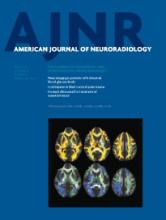
    A 60-year-old man with sudden-onset left-sided weakness after an aneurysm coil was placed for a proximal supraclinoid ICA aneurysm. Axial DWI (4500/90, b = 1000 s/mm2) (A), FLAIR (9000/88/2500) (B), and EPI-FLAIR (10,000/106/2500) (C) obtained approximately 60 minutes after onset of symptoms. Acute infarction along the right MCA territory is noted. The signal intensity ratio of the lesion to the contralateral hemisphere was elevated and measured 1.19 for FLAIR and 1.15 for EPI-FLAIR. Qualitatively, however, very subtle increased hyperintensity corresponding to the region of DWI abnormality is evident on FLAIR but is not clearly seen on EPI-FLAIR. Note the susceptibility artifacts related to the dislodged coil in the MCA bifurcation, which is more pronounced on EPI-FLAIR (arrow) and likely contributed to field inhomogeneity and may have explained the qualitative discrepancy with FLAIR. Note the hyperintense vessel sign (arrowheads) on both FLAIR and EPI-FLAIR caused by sluggish flow or clot in the sylvian MCA branches.

  • Fig 3.
    • Download figure
    • Open in new tab
    • Download powerpoint
    Fig 3.

    Bland-Altman plots and scatterplot show significant correlation (r = 0.899; z value = 8.677; P < .0001) for the SIR values between FLAIR and EPI-FLAIR in patients with acute infarction (DWI-positive lesions [n = 38]).

Tables

  • Figures
    • View popup
    Table 1:

    Correlation of FLAIR and EPI-FLAIR compared with DWI (n = 50)

    NDWIFLAIR (Signal)EPI-FLAIR (Signal)Conclusion
    12−−−No ischemic infarction
    10+−−Concordant FLAIR–EPI-FLAIR, early stage of ischemic infarction
    26+++Concordant FLAIR–EPI-FLAIR, completed infarction
    2++−EPI-FLAIR discordant with FLAIR
    • View popup
    Table 2:

    Comparison of SIR values between FLAIR and EPI-FLAIR studies (n = 50)

    FLAIR SIREPI-FLAIR SIRT Test (P Value)
    DWI negative
        (−) FLAIR, (−) EPI-FLAIR (n = 12)1.02 ± 0.0051.02 ± 0.006.2
    DWI positive
        <4.5 hours of time from onset to MR imaging (n = 12)1.14 ± 0.081.10 ± 0.06.22
        >4.5 hours of time from onset to MR imaging (n = 22)1.36 ± 0.161.33 ± 0.17.83
        (+) FLAIR, (−) EPI-FLAIR (n = 2)1.15 ± 0.031.13 ± 0.05/Aa
    • Note:—Data are mean ± standard deviation.

    • ↵a In 2 discrepant cases, the SIR values were comparable. The sample was too small for evaluation with the t test.

    • In 4 patients with unknown time of presentation, the SIR values were concordant between FLAIR and EPI-FLAIR (SIR > 1.3, n = 3; SIR < 1.3, n = 1).

PreviousNext
Back to top

In this issue

American Journal of Neuroradiology: 35 (5)
American Journal of Neuroradiology
Vol. 35, Issue 5
1 May 2014
  • Table of Contents
  • Index by author
  • Complete Issue (PDF)
Advertisement
Print
Download PDF
Email Article

Thank you for your interest in spreading the word on American Journal of Neuroradiology.

NOTE: We only request your email address so that the person you are recommending the page to knows that you wanted them to see it, and that it is not junk mail. We do not capture any email address.

Enter multiple addresses on separate lines or separate them with commas.
Role of EPI-FLAIR in Patients with Acute Stroke: A Comparative Analysis with FLAIR
(Your Name) has sent you a message from American Journal of Neuroradiology
(Your Name) thought you would like to see the American Journal of Neuroradiology web site.
CAPTCHA
This question is for testing whether or not you are a human visitor and to prevent automated spam submissions.
Cite this article
A. Meshksar, J.P. Villablanca, R. Khan, R. Carmody, B. Coull, K. Nael
Role of EPI-FLAIR in Patients with Acute Stroke: A Comparative Analysis with FLAIR
American Journal of Neuroradiology May 2014, 35 (5) 878-883; DOI: 10.3174/ajnr.A3786

Citation Manager Formats

  • BibTeX
  • Bookends
  • EasyBib
  • EndNote (tagged)
  • EndNote 8 (xml)
  • Medlars
  • Mendeley
  • Papers
  • RefWorks Tagged
  • Ref Manager
  • RIS
  • Zotero
0 Responses
Respond to this article
Share
Bookmark this article
Role of EPI-FLAIR in Patients with Acute Stroke: A Comparative Analysis with FLAIR
A. Meshksar, J.P. Villablanca, R. Khan, R. Carmody, B. Coull, K. Nael
American Journal of Neuroradiology May 2014, 35 (5) 878-883; DOI: 10.3174/ajnr.A3786
del.icio.us logo Twitter logo Facebook logo Mendeley logo
  • Tweet Widget
  • Facebook Like
  • Google Plus One
Purchase

Jump to section

  • Article
    • Abstract
    • ABBREVIATIONS:
    • Materials and Methods
    • Results
    • Discussion
    • Conclusions
    • Footnotes
    • REFERENCES
  • Figures & Data
  • Info & Metrics
  • Responses
  • References
  • PDF

Related Articles

  • No related articles found.
  • PubMed
  • Google Scholar

Cited By...

  • Effects of minocycline on patients with acute anterior circulation ischaemic stroke undergoing intravenous thrombectomy (MIST-A): the study protocol for a multicentre, prospective, randomised, open-label, blinded-endpoint trial
  • Clinical Experience of 1-Minute Brain MRI Using a Multicontrast EPI Sequence in a Different Scan Environment
  • Six-Minute Magnetic Resonance Imaging Protocol for Evaluation of Acute Ischemic Stroke: Pushing the Boundaries
  • Crossref
  • Google Scholar

This article has not yet been cited by articles in journals that are participating in Crossref Cited-by Linking.

More in this TOC Section

  • Enhanced Axonal Metabolism during Early Natalizumab Treatment in Relapsing-Remitting Multiple Sclerosis
  • SWI or T2*: Which MRI Sequence to Use in the Detection of Cerebral Microbleeds? The Karolinska Imaging Dementia Study
  • Progression of Microstructural Damage in Spinocerebellar Ataxia Type 2: A Longitudinal DTI Study
Show more Brain

Similar Articles

Advertisement

Indexed Content

  • Current Issue
  • Accepted Manuscripts
  • Article Preview
  • Past Issues
  • Editorials
  • Editor's Choice
  • Fellows' Journal Club
  • Letters to the Editor
  • Video Articles

Cases

  • Case Collection
  • Archive - Case of the Week
  • Archive - Case of the Month
  • Archive - Classic Case

More from AJNR

  • Trainee Corner
  • Imaging Protocols
  • MRI Safety Corner
  • Book Reviews

Multimedia

  • AJNR Podcasts
  • AJNR Scantastics

Resources

  • Turnaround Time
  • Submit a Manuscript
  • Submit a Video Article
  • Submit an eLetter to the Editor/Response
  • Manuscript Submission Guidelines
  • Statistical Tips
  • Fast Publishing of Accepted Manuscripts
  • Graphical Abstract Preparation
  • Imaging Protocol Submission
  • Evidence-Based Medicine Level Guide
  • Publishing Checklists
  • Author Policies
  • Become a Reviewer/Academy of Reviewers
  • News and Updates

About Us

  • About AJNR
  • Editorial Board
  • Editorial Board Alumni
  • Alerts
  • Permissions
  • Not an AJNR Subscriber? Join Now
  • Advertise with Us
  • Librarian Resources
  • Feedback
  • Terms and Conditions
  • AJNR Editorial Board Alumni

American Society of Neuroradiology

  • Not an ASNR Member? Join Now

© 2025 by the American Society of Neuroradiology All rights, including for text and data mining, AI training, and similar technologies, are reserved.
Print ISSN: 0195-6108 Online ISSN: 1936-959X

Powered by HighWire