
Article Figures & Data
Figures
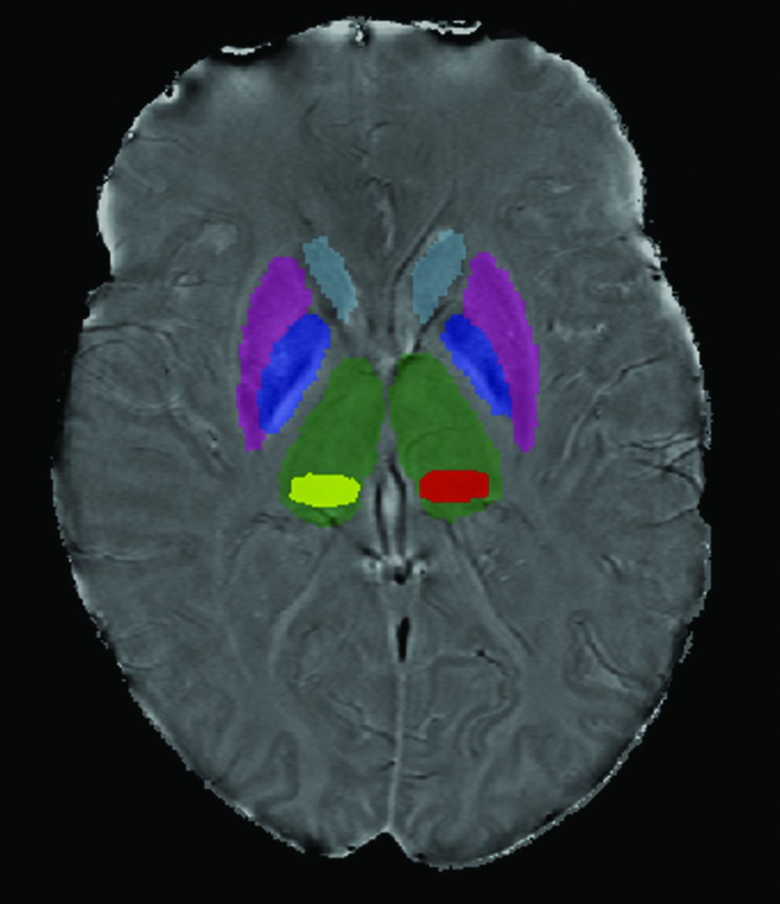
- Fig 1.
Scan demonstrating deep gray matter structure segmentation. FMRIB's Integrated Registration and Segmentation Tool was applied to the 3D high-resolution T1WI to segment the DGM. Pulvinar (yellow and red) was segmented using a semiautomated contouring technique.

Tables
- Table 1:
Demographic characteristics, neuropsychological test scores, and mean phase of the low-phase voxels across subcortical deep gray matter structures
MS (n = 85) NC (n = 27) P Value Characteristics Age 46.0 ± 9.2 41.9 ± 10.7 NSa Education 14.4 ± 2.2 15.3 ± 2.4 NSa Female, n (%) 59 (69.4) 17 (63.0) NSa Caucasian, n (%) 73 (85.9) 24 (88.9) NSa Right-handed, n (%) 77 (90.6) 25 (92.6) NSa Impaired, n (%) 52 (61.2) 1 (3.7) <.001a Disease duration 10.9 ± 7.6 DMT durationb 3.8 ± 3.6 EDSSc 3.6 ± 1.9 T2 lesion volume 18.5 ± 20.3 T1 lesion volume 3.4 ± 7.1 NP test scores SDMT 50.8 ± 15.8 66.0 ± 10.5 <.001d PASAT 42.0 ± 15.7 49.8 ± 8.7 .017d DKEFS-CS 9.5 ± 3.0 11.7 ± 2.4 <.001d CVLT2 52.1 ± 13.1 64.4 ± 7.4 <.001d BVMT-R 7.8 ± 2.8 10.4 ± 1.5 <.001d MP-LPV Thalamus −0.099 ± 0.016 −0.092 ± 0.011 .027a Caudate nucleus −0.178 ± 0.021 −0.166 ± 0.011 .007a Putamen −0.184 ± 0.033 −0.172 ± 0.028 NSa Globus pallidus −0.195 ± 0.036 −0.179 ± 0.029 NSa Pulvinar −0.157 ± 0.036 −0.139 ± 0.012 .014a Note:—NC indicates healthy control; DMT, disease-modifying therapy; EDSS, Expanded Disability Status Scale; NS, not significant.
↵a P value is based on 1-way ANOVA.
↵b n = 68 for calculation of DMT duration as only 68 were on therapy.
↵c n = 83 for calculation of mean EDSS as 2 patients were not assessed during their clinical visit.
↵d P value is based on 1-way ANCOVA, controlling for age and years of education. Age, education, disease duration, and DMT duration data are given as mean years ± SD. Lesion volume is given as mean milliliters ± SD. NP test data for the SDMT, PASAT, DKEFS-CS, CVLT2, and BVMT-R are given as mean test score ± standard deviation. MP-LPV data in the thalamus, caudate nucleus, putamen, globus pallidus, and pulvinar structures are given as radians ± SD.
- Table 2:
Partial correlation of structure volume and mean phase of the low-phase voxels to neuropsychological tests in patients with multiple sclerosis
SDMT PASAT DKEFS-CS CVLT2 BVMT-R r P r P r P r P r P Volumea Thalamus 0.548 <.001 0.354 .001 0.422 <.001 0.313 .004 0.415 <.001 Caudate nucleus 0.470 <.001 0.357 .001 0.409 <.001 0.257 .019 0.348 .001 Putamen 0.516 <.001 0.405 <.001 0.429 <.001 0.382 <.001 0.518 <.001 Globus pallidus 0.449 <.001 0.296 .007 0.348 .001 NS 0.282 .010 Pulvinar 0.279 .011 0.275 .012 0.309 .005 0.221 .044 0.327 .003 MP-LPVb Caudate nucleus 0.240 .030 0.232 .036 NS NS NS Putamen 0.368 .001 NS 0.252 .022 NS 0.238 .031 Globus pallidus NS NS NS NS 0.235 .033 Pulvinar 0.244 .027 NS 0.255 .021 NS 0.251 .023 - Table 3:
Mean phase of the low-phase voxels correlated with structure volume and with low-phase voxel volume
Volume LPV Volume r P r P Caudate nucleus 0.268 .013 NS Putamen NS −0.502 <.001 Globus pallidus −0.452 <.001 −0.483 <.001 Pulvinar 0.266 .014 −0.659 <.001 Note:—NS indicates not significant.
- Table 4:
Summary of hierarchical linear regressions predicting neuropsychological test scores
NP Test Independent Variable (Final Standardized Beta, P Value) R2 R2 Change Model P Value F Change F Change P Value SDMT Block 1 Age (−0.115, .238), Ed (0.146, .125) 0.064 0.064 .068 2.780 NS Block 2 Putamen LPV volume (0.012, .929) 0.082 0.018 .074 1.609 NS Block 3 Putamen MP-LPV (0.154, .241) 0.206 0.124 .001 12.515 .001 Block 4 Putamen volume (0.468, <.001) 0.333 0.127 <.001 15.028 <.001 PASAT Block 1 Age (0.112, .318), Ed (0.175, .096) 0.033 0.033 .255 1.388 NS Block 2 Caudate n. LPV volume (0.009, .941) 0.042 0.010 .318 0.806 NS Block 3 Caudate n. MP-LPV (0.120, .304) 0.094 0.052 .092 4.564 .036 Block 4 Caudate n. volume (0.319, .009) 0.169 0.075 .011 7.093 .009 DKEFS-CS Block 1 Age (−0.081, .444), Ed (0.191, .078) 0.058 0.058 .085 2.544 NS Block 2 Pulvinar LPV volume (0.161, .365) 0.072 0.013 .109 1.142 NS Block 3 Pulvinar MP-LPV (0.160, .373) 0.132 0.061 .022 5.577 .021 Block 4 Pulvinar volume (0.214, .123) 0.158 0.026 .017 2.43 NS CVLT2 Block 1 Age (−0.207, .049), Ed (0.158, .122) 0.093 0.093 .018 4.212 .018 Block 2 Putamen LPV volume (0.066, .661) 0.133 0.040 .009 3.710 NS Block 3 Putamen MP-LPV (0.019, .894) 0.164 0.031 .006 2.942 NS Block 4 Putamen volume (0.333, .012) 0.228 0.064 .001 6.589 .012 BVMT-R Block 1 Age (−0.144, .175), Ed (0.083, .437) 0.042 0.042 .172 1.798 NS Block 2 Pulvinar LPV volume (0.221, .213) 0.076 0.034 .092 2.981 NS Block 3 Pulvinar MP-LPV (0.146, .413) 0.134 0.058 .02 5.365 .023 Block 4 Pulvinar volume (0.222, .109) 0.162 0.028 .014 2.625 NS Note:—Ed indicates education; n, nucleus; NS, not significant.


Jump to section
Related Articles
Cited By...
- Gray Matter alterations in MS and CIS: a Coordinate based Meta-analysis and regression
- Quantitative Susceptibility Mapping of the Thalamus: Relationships with Thalamic Volume, Total Gray Matter Volume, and T2 Lesion Burden
- Thalamic Iron Differentiates Primary-Progressive and Relapsing-Remitting Multiple Sclerosis
- Cognitive Implications of Deep Gray Matter Iron in Multiple Sclerosis