Advertisement

Main menu

  • Home
  • Content
    • Current Issue
    • Accepted Manuscripts
    • Article Preview
    • Past Issue Archive
    • AJNR Case Collection
    • Case of the Week Archive
    • Classic Case Archive
    • Case of the Month Archive
  • Special Collections
    • Spinal CSF Leak Articles (Jan 2020-June 2024)
    • 2024 AJNR Journal Awards
    • Most Impactful AJNR Articles
  • Multimedia
    • AJNR Podcast
    • AJNR Scantastics
    • Video Articles
  • For Authors
    • Submit a Manuscript
    • Author Policies
    • Fast publishing of Accepted Manuscripts
    • Graphical Abstract Preparation
    • Manuscript Submission Guidelines
    • Imaging Protocol Submission
    • Submit a Case for the Case Collection
  • About Us
    • About AJNR
    • Editorial Board
  • More
    • Become a Reviewer/Academy of Reviewers
    • Subscribers
    • Permissions
    • Alerts
    • Feedback
    • Advertisers
    • ASNR Home
  • Other Publications
    • ajnr

User menu

  • Alerts
  • Log in

Search

  • Advanced search
American Journal of Neuroradiology

American Journal of Neuroradiology

ASHNR American Society of Functional Neuroradiology ASHNR American Society of Pediatric Neuroradiology ASSR
  • Alerts
  • Log in

Advanced Search

  • Home
  • Content
    • Current Issue
    • Accepted Manuscripts
    • Article Preview
    • Past Issue Archive
    • AJNR Case Collection
    • Case of the Week Archive
    • Classic Case Archive
    • Case of the Month Archive
  • Special Collections
    • Spinal CSF Leak Articles (Jan 2020-June 2024)
    • 2024 AJNR Journal Awards
    • Most Impactful AJNR Articles
  • Multimedia
    • AJNR Podcast
    • AJNR Scantastics
    • Video Articles
  • For Authors
    • Submit a Manuscript
    • Author Policies
    • Fast publishing of Accepted Manuscripts
    • Graphical Abstract Preparation
    • Manuscript Submission Guidelines
    • Imaging Protocol Submission
    • Submit a Case for the Case Collection
  • About Us
    • About AJNR
    • Editorial Board
  • More
    • Become a Reviewer/Academy of Reviewers
    • Subscribers
    • Permissions
    • Alerts
    • Feedback
    • Advertisers
    • ASNR Home
  • Follow AJNR on Twitter
  • Visit AJNR on Facebook
  • Follow AJNR on Instagram
  • Join AJNR on LinkedIn
  • RSS Feeds

Welcome to the new AJNR, Updated Hall of Fame, and more. Read the full announcements.


AJNR is seeking candidates for the position of Associate Section Editor, AJNR Case Collection. Read the full announcement.


Research ArticleADULT BRAIN
Open Access

Patterns of Tumor Contrast Enhancement Predict the Prognosis of Anaplastic Gliomas with IDH1 Mutation

Y.Y. Wang, K. Wang, S.W. Li, J.F. Wang, J. Ma, T. Jiang and J.P. Dai
American Journal of Neuroradiology November 2015, 36 (11) 2023-2029; DOI: https://doi.org/10.3174/ajnr.A4407
Y.Y. Wang
aFrom the Departments of Neurosurgery (Y.Y.W., J.F.W., T.J.)
cBeijing Neurosurgical Institute (Y.Y.W., T.J., J.P.D.), Capital Medical University, Beijing, China
  • Find this author on Google Scholar
  • Find this author on PubMed
  • Search for this author on this site
K. Wang
bNeuroradiology (K.W., S.W.L., J.M., J.P.D.), Beijing Tian Tan Hospital
  • Find this author on Google Scholar
  • Find this author on PubMed
  • Search for this author on this site
S.W. Li
bNeuroradiology (K.W., S.W.L., J.M., J.P.D.), Beijing Tian Tan Hospital
  • Find this author on Google Scholar
  • Find this author on PubMed
  • Search for this author on this site
J.F. Wang
aFrom the Departments of Neurosurgery (Y.Y.W., J.F.W., T.J.)
  • Find this author on Google Scholar
  • Find this author on PubMed
  • Search for this author on this site
J. Ma
bNeuroradiology (K.W., S.W.L., J.M., J.P.D.), Beijing Tian Tan Hospital
  • Find this author on Google Scholar
  • Find this author on PubMed
  • Search for this author on this site
T. Jiang
aFrom the Departments of Neurosurgery (Y.Y.W., J.F.W., T.J.)
cBeijing Neurosurgical Institute (Y.Y.W., T.J., J.P.D.), Capital Medical University, Beijing, China
dCenter for Brain Tumor (T.J.), Beijing Institute for Brain Disorders, Beijing, China.
  • Find this author on Google Scholar
  • Find this author on PubMed
  • Search for this author on this site
J.P. Dai
bNeuroradiology (K.W., S.W.L., J.M., J.P.D.), Beijing Tian Tan Hospital
cBeijing Neurosurgical Institute (Y.Y.W., T.J., J.P.D.), Capital Medical University, Beijing, China
  • Find this author on Google Scholar
  • Find this author on PubMed
  • Search for this author on this site
  • Article
  • Figures & Data
  • Info & Metrics
  • Responses
  • References
  • PDF
Loading

Article Figures & Data

Figures

  • Tables
  • Fig 1.
    • Download figure
    • Open in new tab
    • Download powerpoint
    Fig 1.

    Tumor contrast-enhancement patterns in AG. Postcontrast T1-weighted images depict the nodular (largest focal diameter of ≤1.5 cm), patchy (largest focal diameter of >1.5 cm), and ringlike (cystic necrosis with peripheral enhancement) enhancement patterns.
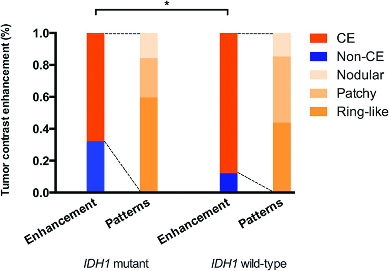
  • Fig 2.
    • Download figure
    • Open in new tab
    • Download powerpoint
    Fig 2.

    Constitution of tumor contrast enhancements between AG accompanied by mutant or wild type IDH1. The difference in contrast-enhancement rate (asterisk) between tumors from patients with mutant and wild type IDH1 was significant (P < .001). There was no significant difference in enhancement-pattern distribution between tumors from patients with mutant and wild type IDH1 (P = .135). CE indicates contrast enhancement.
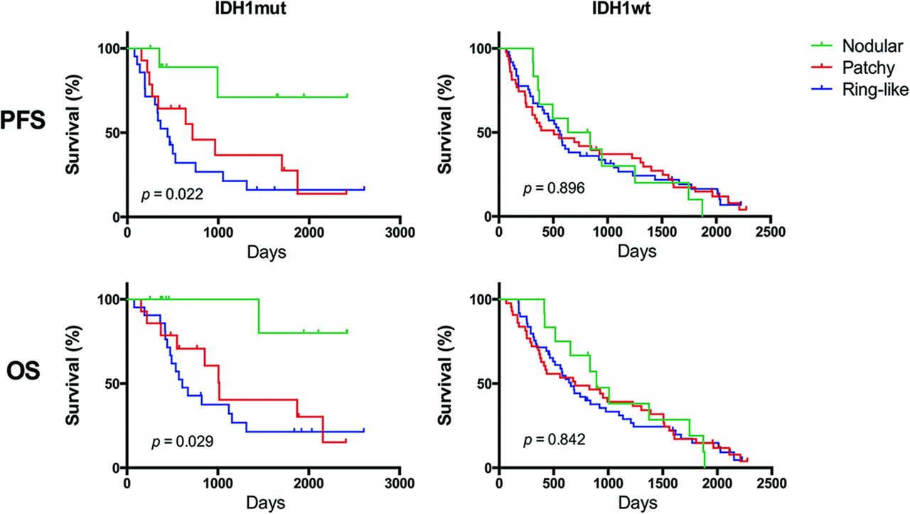
  • Fig 4.
    • Download figure
    • Open in new tab
    • Download powerpoint
    Fig 4.

    Kaplan-Meier plots showing that the tumor contrast enhancement pattern enabled stratification of the PFS and OS of patients with mutant IDH1 (PFS, P = .022; OS, P = .029). Meanwhile, Kaplan-Meier plots show that the tumor contrast-enhancement pattern did not enable stratification of the PFS and OS of patients with wild type IDH1 (PFS, P = .896; OS, P = .842).

Tables

  • Figures
    • View popup
    Table 1:

    IDH1 mutation status of patients with AG

    CharacteristicsIDH1 StatusP Valuea
    Total (n = 216)Mutant (n = 84)Wild Type (n = 132)
    Age (yr)
        Median (range)44 (18–87)43 (18–71)45 (18–87)
        50 or older/50 or younger77:13923:6154:78.043
    Sex
        Male/female135:8149:3586/46.313
    KPS
        ≥80/<80181/3578/6103/29.004
    Contrast enhancement
        Yes/no173/4357/27116/16<.001
    Pattern of enhancement
        Nodular/patchy/ringlike26/62/859/14/3417/48/51.084
    Extent of resection
        GTR/<GTR123/9356/2867/65.021
    Histopathology
        AA/AO/AOA57/44/11516/20/4841/24/67.135
    • Note:—AA indicates anaplastic astrocytomas; AO, anaplastic oligodendrogliomas; AOA, anaplastic oligoastrocytomas.

    • ↵a Results of the χ2 test.

    • View popup
    Table 2:

    Univariate analysis of survival outcomes for patients with AG

    CharacteristicPFSOS
    P ValueHR95% CIP ValueHR95% CI
    Age 50 yr or older<.0011.8131.318–2.494.0071.7291.134–2.421
    Sex (male).2150.8730.695–1.064.4150.9140.612–1.283
    Preoperative KPS < 80.0042.6031.154–3.548.0022.8721.270–3.341
    Enhancement.5701.2000.641–2.247.6251.1870.596–2.365
    Enhancement pattern.1501.1070.964–1.271.2471.9020.941–1.266
    <GTR/GTR.0011.7341.237–2.429<.0011.9261.346–2.758
    Histopathology.0860.8550.715–1.022.0540.7890.654–1.002
    IDH1 wild type.0022.3641.362–4.098.0042.6881.231–4.717
    • View popup
    Table 3:

    Multivariate analysis of survival outcomes

    PredictorP ValueaHR95% CI
    PFS
        Age 50 yr or older.0181.8571.111–3.106
        Preoperative KPS < 80.0152.1581.179–3.471
        <GTR.0281.5981.053–2.597
        IDH1 wild type.0042.2771.303–3.968
    OS
         Age 50 yr or older.0161.4311.342–2.434
        Preoperative KPS < 80.0261.8361.087–3.402
        <GTR.0231.4881.210–2.432
        IDH1 wild-type.0022.4631.389–4.386
    • ↵a Cox proportional hazard regression analyses. A P value of .05 denoted significance.

PreviousNext
Back to top

In this issue

American Journal of Neuroradiology: 36 (11)
American Journal of Neuroradiology
Vol. 36, Issue 11
1 Nov 2015
  • Table of Contents
  • Index by author
  • Complete Issue (PDF)
Advertisement
Print
Download PDF
Email Article

Thank you for your interest in spreading the word on American Journal of Neuroradiology.

NOTE: We only request your email address so that the person you are recommending the page to knows that you wanted them to see it, and that it is not junk mail. We do not capture any email address.

Enter multiple addresses on separate lines or separate them with commas.
Patterns of Tumor Contrast Enhancement Predict the Prognosis of Anaplastic Gliomas with IDH1 Mutation
(Your Name) has sent you a message from American Journal of Neuroradiology
(Your Name) thought you would like to see the American Journal of Neuroradiology web site.
CAPTCHA
This question is for testing whether or not you are a human visitor and to prevent automated spam submissions.
Cite this article
Y.Y. Wang, K. Wang, S.W. Li, J.F. Wang, J. Ma, T. Jiang, J.P. Dai
Patterns of Tumor Contrast Enhancement Predict the Prognosis of Anaplastic Gliomas with IDH1 Mutation
American Journal of Neuroradiology Nov 2015, 36 (11) 2023-2029; DOI: 10.3174/ajnr.A4407

Citation Manager Formats

  • BibTeX
  • Bookends
  • EasyBib
  • EndNote (tagged)
  • EndNote 8 (xml)
  • Medlars
  • Mendeley
  • Papers
  • RefWorks Tagged
  • Ref Manager
  • RIS
  • Zotero
0 Responses
Respond to this article
Share
Bookmark this article
Patterns of Tumor Contrast Enhancement Predict the Prognosis of Anaplastic Gliomas with IDH1 Mutation
Y.Y. Wang, K. Wang, S.W. Li, J.F. Wang, J. Ma, T. Jiang, J.P. Dai
American Journal of Neuroradiology Nov 2015, 36 (11) 2023-2029; DOI: 10.3174/ajnr.A4407
del.icio.us logo Twitter logo Facebook logo Mendeley logo
  • Tweet Widget
  • Facebook Like
  • Google Plus One
Purchase

Jump to section

  • Article
    • Abstract
    • ABBREVIATIONS:
    • Materials and Methods
    • Results
    • Discussion
    • Conclusions
    • Acknowledgments
    • Footnotes
    • REFERENCES
  • Figures & Data
  • Info & Metrics
  • Responses
  • References
  • PDF

Related Articles

  • No related articles found.
  • PubMed
  • Google Scholar

Cited By...

  • Does Blood-Brain Barrier Disruption Define the Glioma Extracellular Metabolome?
  • Systemic Immune Bias Delineates Malignant Astrocytoma Survival Cohorts
  • Predicting Genotype and Survival in Glioma Using Standard Clinical MR Imaging Apparent Diffusion Coefficient Images: A Pilot Study from The Cancer Genome Atlas
  • MR Imaging Characteristics Associate with Tumor-Associated Macrophages in Glioblastoma and Provide an Improved Signature for Survival Prognostication
  • Crossref (42)
  • Google Scholar

This article has been cited by the following articles in journals that are participating in Crossref Cited-by Linking.

  • Multi-Channel 3D Deep Feature Learning for Survival Time Prediction of Brain Tumor Patients Using Multi-Modal Neuroimages
    Dong Nie, Junfeng Lu, Han Zhang, Ehsan Adeli, Jun Wang, Zhengda Yu, LuYan Liu, Qian Wang, Jinsong Wu, Dinggang Shen
    Scientific Reports 2019 9 1
  • Machine learning and radiomic phenotyping of lower grade gliomas: improving survival prediction
    Yoon Seong Choi, Sung Soo Ahn, Jong Hee Chang, Seok-Gu Kang, Eui Hyun Kim, Se Hoon Kim, Rajan Jain, Seung-Koo Lee
    European Radiology 2020 30 7
  • Predicting Genotype and Survival in Glioma Using Standard Clinical MR Imaging Apparent Diffusion Coefficient Images: A Pilot Study from The Cancer Genome Atlas
    C.-C. Wu, R. Jain, A. Radmanesh, L.M. Poisson, W.-Y. Guo, D. Zagzag, M. Snuderl, D.G. Placantonakis, J. Golfinos, A.S. Chi
    American Journal of Neuroradiology 2018 39 10
  • IDH mutation-specific radiomic signature in lower-grade gliomas
    Xing Liu, Yiming Li, Shaowu Li, Xing Fan, Zhiyan Sun, Zhengyi Yang, Kai Wang, Zhong Zhang, Tao Jiang, Yong Liu, Lei Wang, Yinyan Wang
    Aging 2019 11 2
  • Conventional MRI features of adult diffuse glioma molecular subtypes: a systematic review
    Arian Lasocki, Mustafa Anjari, Suna Ӧrs Kokurcan, Stefanie C. Thust
    Neuroradiology 2021 63 3
  • Overall survival time prediction for high-grade glioma patients based on large-scale brain functional networks
    Luyan Liu, Han Zhang, Jinsong Wu, Zhengda Yu, Xiaobo Chen, Islem Rekik, Qian Wang, Junfeng Lu, Dinggang Shen
    Brain Imaging and Behavior 2019 13 5
  • Prediction of Molecular Mutations in Diffuse Low-Grade Gliomas using MR Imaging Features
    Zeina A. Shboul, James Chen, Khan M. Iftekharuddin
    Scientific Reports 2020 10 1
  • Contrast enhancement is a prognostic factor in IDH1/2 mutant, but not in wild-type WHO grade II/III glioma as confirmed by machine learning
    Bogdana Suchorska, Ulrich Schüller, Annamaria Biczok, Markus Lenski, Nathalie L. Albert, Armin Giese, Friedrich-Wilhelm Kreth, Birgit Ertl-Wagner, Jörg-Christian Tonn, Michael Ingrisch
    European Journal of Cancer 2019 107
  • MRI texture analysis based on 3D tumor measurement reflects the IDH1 mutations in gliomas – A preliminary study
    Liang Han, Siyu Wang, Yanwei Miao, Huicong Shen, Yan Guo, Lizhi Xie, Yuqing Shang, Junyi Dong, Xiaoxin Li, Weiwei Wang, Qingwei Song
    European Journal of Radiology 2019 112
  • MRI-Based Radiomics and Radiogenomics in the Management of Low-Grade Gliomas: Evaluating the Evidence for a Paradigm Shift
    Ahmed Habib, Nicolina Jovanovich, Meagan Hoppe, Murat Ak, Priyadarshini Mamindla, Rivka R. Colen, Pascal O. Zinn
    Journal of Clinical Medicine 2021 10 7

More in this TOC Section

  • Diagnostic Neuroradiology of Monoclonal Antibodies
  • NCCT vs. MRI for Brain Atrophy in Acute Stroke
  • Clinical Outcomes After Chiari I Decompression
Show more Adult Brain

Similar Articles

Advertisement

Indexed Content

  • Current Issue
  • Accepted Manuscripts
  • Article Preview
  • Past Issues
  • Editorials
  • Editors Choice
  • Fellow Journal Club
  • Letters to the Editor

Cases

  • Case Collection
  • Archive - Case of the Week
  • Archive - Case of the Month
  • Archive - Classic Case

Special Collections

  • Special Collections

Resources

  • News and Updates
  • Turn around Times
  • Submit a Manuscript
  • Author Policies
  • Manuscript Submission Guidelines
  • Evidence-Based Medicine Level Guide
  • Publishing Checklists
  • Graphical Abstract Preparation
  • Imaging Protocol Submission
  • Submit a Case
  • Become a Reviewer/Academy of Reviewers
  • Get Peer Review Credit from Publons

Multimedia

  • AJNR Podcast
  • AJNR SCANtastic
  • Video Articles

About Us

  • About AJNR
  • Editorial Board
  • Not an AJNR Subscriber? Join Now
  • Alerts
  • Feedback
  • Advertise with us
  • Librarian Resources
  • Permissions
  • Terms and Conditions

American Society of Neuroradiology

  • Not an ASNR Member? Join Now

© 2025 by the American Society of Neuroradiology All rights, including for text and data mining, AI training, and similar technologies, are reserved.
Print ISSN: 0195-6108 Online ISSN: 1936-959X

Powered by HighWire