
Article Figures & Data
Figures
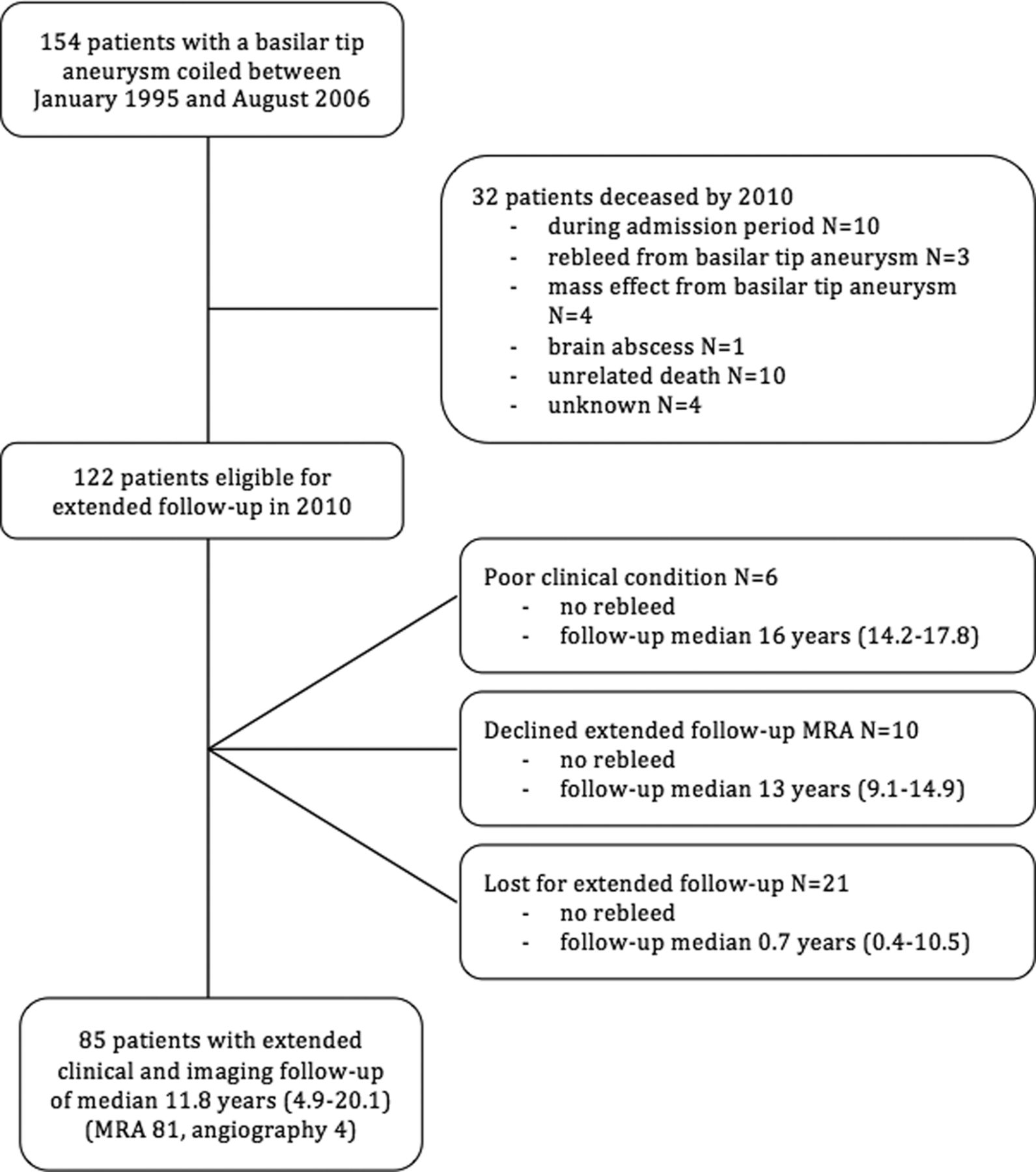
- Fig 1.
Follow-up scheme of 154 patients with a basilar tip aneurysm.
- Fig 2.
A 41-year-old man with a coiled ruptured basilar tip aneurysm in 1997 and a rebleed 15 years later. A, Angiography in 2006, 9 years after coiling in 1997, shows an adequately occluded basilar tip aneurysm. B, CT in 2012 demonstrates a rebleed from the basilar tip aneurysm. C, Angiography reveals regrowth of the aneurysm (arrow). D, After additional coiling, the aneurysm is completely occluded (arrow).
- Fig 3.
Timing of first retreatments in 37 patients with reopened basilar tip aneurysms.
- Fig 4.
Timing of retreatments in 15 patients with coiled basilar tip aneurysms and >1 recurrence.
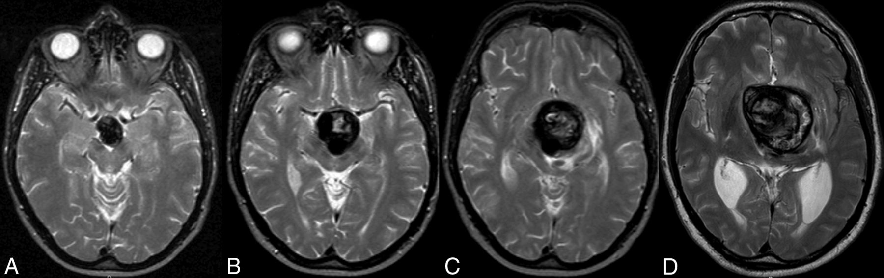
- Fig 5.
Serial MR images of a 40-year-old man with a coiled ruptured basilar tip aneurysm in 2003. A, Transversal T2-weighted MR image from December 2003 shows a basilar tip aneurysm 6 months after coiling. B, MR imaging in May 2008 shows enlargement of the aneurysm and compression of the brain stem. C, MR imaging in March 2009 shows further growth, now with edema in the brain stem. D, MR imaging in December 2009 shows a rapid increase in size with enormous compression of the brain stem. The patient died 1 month later.


Tables
- Table 1:
Univariate analysis of risk factors for retreatment of coiled basilar tip aneurysms in 144 patients
No Recurrence (n = 107) Recurrence (n = 37) OR (95% CI) P Value Median age at first treatment (yr) 54 46 0.97 (0.9–1.0) .13 Male sex 25 (23%) 16 (43%) 2.6 (1.2–5.6) .02 Median aneurysm size (mm) 9 15 1.1 (1.1–1.2) <.001 Size of aneurysm in quintilesa Quintile 2 versus 1 0.9 (0.2–3.3) Quintile 3 versus 1 0.9 (0.2–3.3) Quintile 4 versus 1 1.6 (0.4–6.3) Quintile 5 versus 1 8.5 (2.5–29.3) Ruptured aneurysm 88 (82%) 14 (38%) 0.8 (0.3–1.9) .62 Median follow-up (yr) 10.5 10 1.0 (0.98–1.14) .20 ↵a Size range in quintiles: quintile 1 = 2–5 mm; quintile 2 = 6–8 mm; quintile 3 = 9–12 mm; quintile 4 = 13–15 mm; quintile 5 = 16–30 mm.
- Table 2:
Characteristics of 6 patients with progressive mass effect by continuous growth of the basilar tip aneurysm despite (repeat) coiling
Date of First Treatment Sex, Age (yr) SAH First Signs of Mass Effect Compression Organ No.of Coilings Death Follow-Up (yr) Jan 10, 1996 M, 52 Yes January 1996 Brain stem 1 Yes 0.5 June 5, 1996 M, 39 Yes May 2008 Brain stem 5 No 11.8 June 18, 2001 F, 69 Yes September 2004 Brain stem 1 Yes 3.3 July 27, 2001 M, 39 Yes March 2009 Brain stem 5 Yes 8.4 November 5, 2001 M, 58 Yes July 2012 Brain stem 5 Yes 12.5 May 30, 2002 M, 49 No October 2002 Optic chiasm 2 Yes 0.5


