
Article Figures & Data
Figures
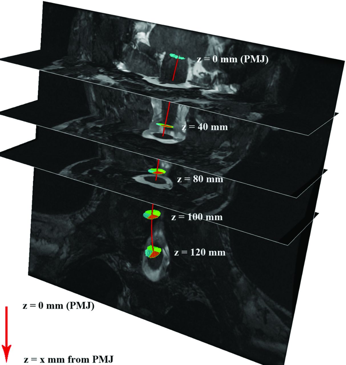
- Fig 1.
Results of spinal cord centerline extraction in 1 subject. The red line represents the spinal cord centerline. Distance from the PMJ is calculated along this centerline (z, in millimeters), for measuring the absolute location of the vertebral and spinal levels.
- Fig 2.
From left to right: T2-weighted MR image with the PMJ and superior (Sup.) and inferior (inf.) endplates of the C3 vertebral body marked with dashed white lines. Immediately adjacent to this, an artist's illustration demonstrates how person-specific markings are positioned relative to the individualized arc-length axis of the spinal cord (red line). Thus, distances can be compared across individuals along their personalized spinal cord axes. The T2-weighted coronal image depicts spinal rootlets (A) and the gap between adjacent segmental rootlets (B), delineated by dashed white lines. The far right artist's illustration depicts segmental rootlets and the formation of a spinal nerve root that emerges from the intervertebral foramen. To accurately localize segmental nerve rootlets, we followed the spinal nerve root that emerges from the intervertebral foramen of the respective vertebral body.
- Fig 3.
Nerve rootlet (NR, solid lines) and vertebral body (VB, dashed lines) distributions across our cohort of 20 individuals. The x-axis of this line graph represents the distance from the PMJ along the spinal cord axis, where zero is the PMJ and the increasing numbers are millimeter distances down the curved axis of the spinal cord to the midpoint of either the vertebral body or spinal cord segment as demarcated by nerve rootlets. The y-axis of the line graph represents the probability of finding either NR (solid line) or VB (dashed line) at a given distance down the curved spinal cord. The spinal cord segments and vertebral bodies represented by the colored lines are shown in the graph inset as distances in millimeters to the midpoint of either the vertebral bodies or spinal cord segments (as represented by nerve rootlets), projected orthogonally onto the spinal cord centerline, SDs, and range. The rostral-caudal extent of nerve rootlets corresponding to each spinal segment was relatively consistent across all spinal cord segments. The average length of spinal cord segments are the following: C3, 10.5 mm; C4, 9.9 mm; C5, 10.5 mm; C6, 9.7 mm; C7, 9.4 mm; and C8 9.6 mm.
- Fig 4.
Vertebral body and spinal cord segment location across 10 subjects enrolled in this study. Vertebral bodies are represented for each subject by light-shaded bars, whereas spinal cord segments are represented by colored bars (see graph inset).
- Fig 5.
Left: scaled relative distance of the C7 spinal cord segments (red bars) from the C6 (upper light-brown-shaded area) and C7 (lower light-brown-shaded area) vertebral bodies. When we visualize an individual's cervical spine MR imaging, we tend to hold the vertebral bodies constant. This figure illustrates that the position of the seventh cervical spinal cord segment varies relative to the position of the vertebral body across a cohort of individuals. If one were to assume that the C7 spinal segments are immediately adjacent to the C7 vertebral body, then 0% of the actual segments would be captured in such an analysis. Similarly, if one were to assume that the C7 spinal cord segments are 1 vertebral body length rostral to the C7 body, then one would capture 33% of the corresponding spinal segments for the 10 subjects shown, or 44% of the corresponding spinal segments across all 20 subjects. Depending on the goals of the imaging experiment, one should pay careful attention to the relative position of spinal segments and vertebral bodies across a cohort of individuals. Assuming that spinal segments are constant may lead to false-positive or false-negative results. Right: an artist's depiction shows the range in spinal cord segments relative to the vertebral bodies; light-shaded areas represent areas of population level overlap.
- Fig 6.
Automated analysis of the degree of neck flexion/extension of a subject. Top: the angle between the tangent of the spinal cord at any point and tangent at the C7 vertebral body for 1 subject with the neck in flexion (red) and extension (blue). Bottom: the difference between the flexion and extension curves or the maximum extent of curvature of the spinal cord at various locations down the longitudinal axis of the spinal cord (x-axis). VB indicates vertebral body; deg, degree; deg diff, degree difference.


Tables
Participant No. Age (yr) Sex Height (cm) 1 32 F 163 2 44 M 181 3 28 F 168 4 47 M 170 5 23 F 171 6 52 F 170 7 31 F 157 8 29 M 182 9 21 M 179 10 34 F 164 11 25 F 175 12 22 F 186 13 19 M 180 14 27 F 173 15 22 M 162 16 28 F 159 17 34 F 169 18 30 F 168 19 25 F 176 20 36 M 174 - Table 2:
Vertebral-spinal discrepancy reported in neutral position across 2 independent observers, between neutral and flexion positions and neutral and extension positionsa
C3 C4 C5 C6 C7 Neutral position across 2 independent reviewers (mm) 1.0 1.3 1.3 0.9 1.1 Neutral flexion (mm) 1.3 0.7 1.2 0.8 1.9 Neutral extension (mm) 1.4 0.7 1.0 1.0 1.0 ↵a Results are reported in millimeters. These results suggest that flexion or extension that is possible within the confines of the MR imaging environment does not affect the relative positions of vertebral and spinal cord segments.


Jump to section
Related Articles
Cited By...
- Mapping Hand Function with Simultaneous Brain-Spinal Cord Functional MRI
- A multi-channel electrophysiology approach to non-invasively and precisely record human spinal cord activity
- Reliability of task-based fMRI in the dorsal horn of the human spinal cord
- Normalizing Spinal Cord Compression Morphometric Measures: Application in Degenerative Cervical Myelopathy
- Thermal Stimulus Task fMRI in the Cervical Spinal Cord at 7 Tesla
- Spatial distribution of hand-grasp motor task activity in spinal cord functional magnetic resonance imaging
- Spatial distribution of hand-grasp motor task activity in spinal cord functional magnetic resonance imaging
- Reliability of resting-state functional connectivity in the human spinal cord: assessing the impact of distinct noise sources
- Reliability of resting-state functional connectivity in the human spinal cord: assessing the impact of distinct noise sources
- Normalizing automatic spinal cord cross-sectional area measures
- T1 Mapping for Microstructural Assessment of the Cervical Spinal Cord in the Evaluation of Patients with Degenerative Cervical Myelopathy
- Investigating resting-state functional connectivity in the cervical spinal cord at 3T
- Clinically Feasible Microstructural MRI to Quantify Cervical Spinal Cord Tissue Injury Using DTI, MT, and T2*-Weighted Imaging: Assessment of Normative Data and Reliability