Advertisement

Main menu

  • Home
  • Content
    • Current Issue
    • Accepted Manuscripts
    • Article Preview
    • Past Issue Archive
    • Video Articles
    • AJNR Case Collection
    • Case of the Week Archive
    • Case of the Month Archive
    • Classic Case Archive
  • Special Collections
    • AJNR Awards
    • Low-Field MRI
    • Alzheimer Disease
    • ASNR Foundation Special Collection
    • Photon-Counting CT
    • View All
  • Multimedia
    • AJNR Podcasts
    • AJNR SCANtastic
    • Trainee Corner
    • MRI Safety Corner
    • Imaging Protocols
  • For Authors
    • Submit a Manuscript
    • Submit a Video Article
    • Submit an eLetter to the Editor/Response
    • Manuscript Submission Guidelines
    • Statistical Tips
    • Fast Publishing of Accepted Manuscripts
    • Graphical Abstract Preparation
    • Imaging Protocol Submission
    • Author Policies
  • About Us
    • About AJNR
    • Editorial Board
    • Editorial Board Alumni
  • More
    • Become a Reviewer/Academy of Reviewers
    • Subscribers
    • Permissions
    • Alerts
    • Feedback
    • Advertisers
    • ASNR Home

User menu

  • Alerts
  • Log in

Search

  • Advanced search
American Journal of Neuroradiology

American Journal of Neuroradiology

ASHNR American Society of Functional Neuroradiology ASHNR American Society of Pediatric Neuroradiology ASSR
  • Alerts
  • Log in

Advanced Search

  • Home
  • Content
    • Current Issue
    • Accepted Manuscripts
    • Article Preview
    • Past Issue Archive
    • Video Articles
    • AJNR Case Collection
    • Case of the Week Archive
    • Case of the Month Archive
    • Classic Case Archive
  • Special Collections
    • AJNR Awards
    • Low-Field MRI
    • Alzheimer Disease
    • ASNR Foundation Special Collection
    • Photon-Counting CT
    • View All
  • Multimedia
    • AJNR Podcasts
    • AJNR SCANtastic
    • Trainee Corner
    • MRI Safety Corner
    • Imaging Protocols
  • For Authors
    • Submit a Manuscript
    • Submit a Video Article
    • Submit an eLetter to the Editor/Response
    • Manuscript Submission Guidelines
    • Statistical Tips
    • Fast Publishing of Accepted Manuscripts
    • Graphical Abstract Preparation
    • Imaging Protocol Submission
    • Author Policies
  • About Us
    • About AJNR
    • Editorial Board
    • Editorial Board Alumni
  • More
    • Become a Reviewer/Academy of Reviewers
    • Subscribers
    • Permissions
    • Alerts
    • Feedback
    • Advertisers
    • ASNR Home
  • Follow AJNR on Twitter
  • Visit AJNR on Facebook
  • Follow AJNR on Instagram
  • Join AJNR on LinkedIn
  • RSS Feeds

AJNR Awards, New Junior Editors, and more. Read the latest AJNR updates

Research ArticleNeurointervention
Open Access

A New Aneurysm Occlusion Classification after the Impact of Flow Modification

H.S. Cekirge and I. Saatci
American Journal of Neuroradiology January 2016, 37 (1) 19-24; DOI: https://doi.org/10.3174/ajnr.A4489
H.S. Cekirge
aFrom the Department of Radiology (H.S.C), Bayindir Hospital, Ankara, Turkey
  • Find this author on Google Scholar
  • Find this author on PubMed
  • Search for this author on this site
I. Saatci
bDepartment of Radiology (I.S.), Yüksek Ihtisas University, Koru Hospital, Ankara, Turkey.
  • Find this author on Google Scholar
  • Find this author on PubMed
  • Search for this author on this site
  • Article
  • Figures & Data
  • Supplemental
  • Info & Metrics
  • Responses
  • References
  • PDF
Loading

Article Figures & Data

Figures

  • Tables
  • Fig 1.
    • Download figure
    • Open in new tab
    • Download powerpoint
    Fig 1.

    Schematic drawing of class 1 subgroups and class 5 in different case settings. The last column shows the control angiographic appearance. The first example of class 5 represents the control result after intrasaccular FM placement, given that the control appearance remains unchanged, as required. The second and third examples of class 5 represent the remodeling after extrasaccular flow diverter treatment.

  • Fig 2.
    • Download figure
    • Open in new tab
    • Download powerpoint
    Fig 2.

    Class 1A. A and B, Preoperative images show the ICA aneurysm in which the anterior choroidal artery (arrow) is originating from the aneurysm at the neck. C, Six-month control angiography after single Pipeline device (Covidien, Irvine, California) placement demonstrates total occlusion of the aneurysm with the anterior choroidal artery preserved (arrow). Reprinted from Saatci et al.18

  • Fig 3.
    • Download figure
    • Open in new tab
    • Download powerpoint
    Fig 3.

    Class 1C. A, Preoperative angiography shows right vertebral artery aneurysm with the posterior inferior cerebellar artery originating from the sac. B, Single Pipeline device was placed, and a 6-month control angiography demonstrates complete occlusion of the aneurysm sac, along with the posterior inferior cerebellar artery. The patient was asymptomatic.
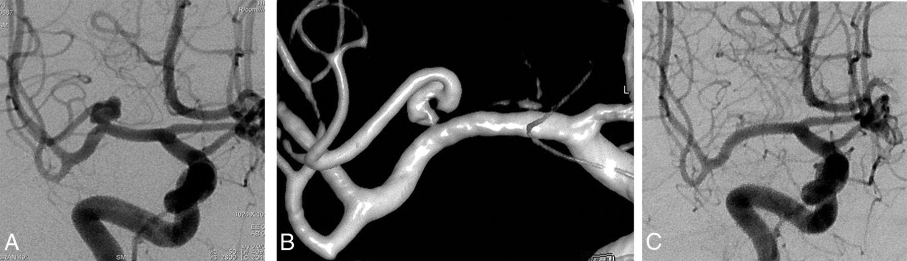
  • Fig 4.
    • Download figure
    • Open in new tab
    • Download powerpoint
    Fig 4.

    Class 5 evolving into class 1B eventually. A, Preoperative angiography shows right MCA aneurysm with a branch coming off from the sac. B, A 3D image from a 6-month control angiography after treatment with single Pipeline device shows remodeling of the flow with a tortuous appearance of the branch proximally at its direct continuation with the parent artery, and no sac filling. This appearance is referred to as class 5. C, An 18-month control angiography shows complete occlusion of the aneurysm with the originating branch in reduced caliber, that is, class 1B. Reprinted from Yavuz et al.19

  • Fig 5.
    • Download figure
    • Open in new tab
    • Download powerpoint
    Fig 5.

    Class 5. A, Right internal carotid angiogram shows a posterior communicating artery aneurysm (the ipsilateral P1 is aplastic, not shown). B, A 2-year angiography after a single Pipeline device placement shows that the aneurysm sac is not filling and the origin of the posterior communicating artery is remodeled. Reprinted from Saatci et al.18

  • Fig 6.
    • Download figure
    • Open in new tab
    • Download powerpoint
    Fig 6.

    Class 5. A, Preoperative angiography shows a large left MCA aneurysm at the trifurcation, with the branches incorporated in the sac. B, Six months after the treatment with a WEB device (Sequent Medical, Aliso Viejo, California), control angiography shows neck filling at the trifurcation, with the branches patent, and this result is classified as class 2. C, An 18-month control angiography shows an unchanged appearance of the MCA trifurcation, and this result is classified as class 5, with the apparently “stable” remodeling.

  • Fig 7.
    • Download figure
    • Open in new tab
    • Download powerpoint
    Fig 7.

    Class 5. A, Preoperative angiography shows a large, irregular shaped, right MCA bifurcation aneurysm with both bifurcation branches coming off the sac. A Pipeline device was placed, extending from the inferior trunk to the M1 in addition to a WEB device within the sac. B, A 6-month control angiography shows a patent inferior trunk, a tortuous origin of the superior trunk, no sac filling; this result is referred to as class 5.

Tables

  • Figures
  • Classification of angiographic results after endovascular treatment with any technique

    Classification
    Class 1: Complete occlusion of the aneurysm sac. When there is a branch integrated with the aneurysm sac, ie, coming off the aneurysm, at any point of the sac, further analysis is carried out with subgroups
        1A: Complete occlusion with the full patency of the integrated branch
        1B: Complete occlusion with the branch reduced in caliber
        1C: Complete occlusion with no antegrade filling of the branch
    Class 2: Neck filling
    Class 3: Incomplete occlusion with aneurysm filling
    Class 4: Aneurysm filling. This class is reserved for an immediate postoperative result based on end-of-treatment DSA; after extra- and/or intrasaccular flow modification treatment
        4A: With contrast stagnation—contrast stagnation is referred to when there happens to be any change in the duration of the contrast stay within the aneurysm sac after treatment
        4B: Without contrast stagnation
    Class 5: Stable remodeling with flow modification. Filling in the neck region, which stays unchanged or reduced; to be included in this group, there have to be at least 2 consecutive control angiographies, by definition, at least 6 months apart, and expanding for a period of not <1 year; exceptionally, 1 control angiography could be sufficient for definition of class 5, only in selected cases of contrast filling the branch coming off the sac, with an appearance of a different vessel course than the original, eg, tortuous or dilated, given that it is in continuation with the parent artery with no sac filling
PreviousNext
Back to top

In this issue

American Journal of Neuroradiology: 37 (1)
American Journal of Neuroradiology
Vol. 37, Issue 1
1 Jan 2016
  • Table of Contents
  • Index by author
  • Complete Issue (PDF)
Advertisement
Print
Download PDF
Email Article

Thank you for your interest in spreading the word on American Journal of Neuroradiology.

NOTE: We only request your email address so that the person you are recommending the page to knows that you wanted them to see it, and that it is not junk mail. We do not capture any email address.

Enter multiple addresses on separate lines or separate them with commas.
A New Aneurysm Occlusion Classification after the Impact of Flow Modification
(Your Name) has sent you a message from American Journal of Neuroradiology
(Your Name) thought you would like to see the American Journal of Neuroradiology web site.
CAPTCHA
This question is for testing whether or not you are a human visitor and to prevent automated spam submissions.
Cite this article
H.S. Cekirge, I. Saatci
A New Aneurysm Occlusion Classification after the Impact of Flow Modification
American Journal of Neuroradiology Jan 2016, 37 (1) 19-24; DOI: 10.3174/ajnr.A4489

Citation Manager Formats

  • BibTeX
  • Bookends
  • EasyBib
  • EndNote (tagged)
  • EndNote 8 (xml)
  • Medlars
  • Mendeley
  • Papers
  • RefWorks Tagged
  • Ref Manager
  • RIS
  • Zotero
0 Responses
Respond to this article
Share
Bookmark this article
A New Aneurysm Occlusion Classification after the Impact of Flow Modification
H.S. Cekirge, I. Saatci
American Journal of Neuroradiology Jan 2016, 37 (1) 19-24; DOI: 10.3174/ajnr.A4489
del.icio.us logo Twitter logo Facebook logo Mendeley logo
  • Tweet Widget
  • Facebook Like
  • Google Plus One
Purchase

Jump to section

  • Article
    • Abstract
    • ABBREVIATION:
    • Materials and Methods
    • Discussion
    • Conclusions
    • Footnotes
    • References
  • Figures & Data
  • Supplemental
  • Info & Metrics
  • Responses
  • References
  • PDF

Related Articles

  • PubMed
  • Google Scholar

Cited By...

  • Impact of Smoking on Recurrence and Angiographic Outcomes after Endovascular Treatment of Intracranial Aneurysms: A Systematic Review and Meta-analysis
  • Evaluation of flow diverters for cerebral aneurysm therapy: recommendations for imaging analyses in clinical studies, endorsed by ESMINT, ESNR, OCIN, SILAN, SNIS, and WFITN
  • Magnetic resonance perfusion imaging findings following flow diversion in patients with complex middle cerebral artery bifurcation aneurysms: a single-center analysis regarding the jailed cortical branches
  • Surpass Intracranial Aneurysm Embolization System Pivotal Trial to Treat Large or Giant Wide-Neck Aneurysms - SCENT: 3-year outcomes
  • Management of aneurysmal recurrence after Woven EndoBridge (WEB) treatment
  • Triple therapy versus dual-antiplatelet therapy for dolichoectatic vertebrobasilar fusiform aneurysms treated with flow diverters
  • Prospective study on embolization of intracranial aneurysms with the pipeline device (PREMIER study): 3-year results with the application of a flow diverter specific occlusion classification
  • Brain aneurysm and parent vessel remodeling after flow diversion treatment: a proposed modification for Cekirge-Saatci classification (mCSC)
  • First clinical multicenter experience with the new Pipeline Vantage flow diverter
  • Predictors of incomplete aneurysm occlusion after treatment with the Pipeline Embolization Device: PREMIER trial 1 year analysis
  • Triple therapy versus dual-antiplatelet therapy for dolichoectatic vertebrobasilar fusiform aneurysms treated with flow diverters
  • Long-term safety and efficacy of distal aneurysm treatment with flow diversion in the M2 segment of the middle cerebral artery and beyond
  • Changing the Rules of the Game: The Problem of Surrogate Angiographic Outcomes in the Evaluation of Aneurysm Treatments
  • Middle Cerebral Artery Bifurcation Aneurysms Treated by Extrasaccular Flow Diverters: Midterm Angiographic Evolution and Clinical Outcome
  • Crossref
  • Google Scholar

This article has not yet been cited by articles in journals that are participating in Crossref Cited-by Linking.

More in this TOC Section

  • A Retrospective Study in Tentorial DAVFs
  • Proximal Protection Devices for Carotid Stenting
  • Rescue Reentry in Carotid Near-Occlusion
Show more NEUROINTERVENTION

Similar Articles

Advertisement

Indexed Content

  • Current Issue
  • Accepted Manuscripts
  • Article Preview
  • Past Issues
  • Editorials
  • Editor's Choice
  • Fellows' Journal Club
  • Letters to the Editor
  • Video Articles

Cases

  • Case Collection
  • Archive - Case of the Week
  • Archive - Case of the Month
  • Archive - Classic Case

More from AJNR

  • Trainee Corner
  • Imaging Protocols
  • MRI Safety Corner
  • Book Reviews

Multimedia

  • AJNR Podcasts
  • AJNR Scantastics

Resources

  • Turnaround Time
  • Submit a Manuscript
  • Submit a Video Article
  • Submit an eLetter to the Editor/Response
  • Manuscript Submission Guidelines
  • Statistical Tips
  • Fast Publishing of Accepted Manuscripts
  • Graphical Abstract Preparation
  • Imaging Protocol Submission
  • Evidence-Based Medicine Level Guide
  • Publishing Checklists
  • Author Policies
  • Become a Reviewer/Academy of Reviewers
  • News and Updates

About Us

  • About AJNR
  • Editorial Board
  • Editorial Board Alumni
  • Alerts
  • Permissions
  • Not an AJNR Subscriber? Join Now
  • Advertise with Us
  • Librarian Resources
  • Feedback
  • Terms and Conditions
  • AJNR Editorial Board Alumni

American Society of Neuroradiology

  • Not an ASNR Member? Join Now

© 2025 by the American Society of Neuroradiology All rights, including for text and data mining, AI training, and similar technologies, are reserved.
Print ISSN: 0195-6108 Online ISSN: 1936-959X

Powered by HighWire