
Article Figures & Data
Figures
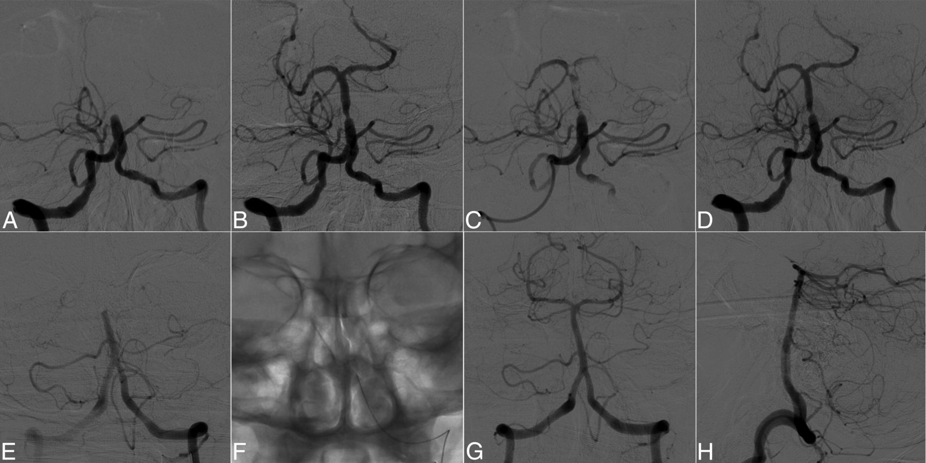
- Fig 1.
Sequential angiographic images of a patient with an atherosclerotic basilar occlusion (A–D) and a patient with an embolic basilar occlusion (E–H). An occlusion in the basilar artery in a 70-year-old woman (A). Note significant focal stenosis at the occlusion site after the first pass of thrombectomy (B). Follow-up angiography after 10 minutes demonstrates that the vessel is occluding again (C). Significant fixed focal stenosis in the final angiogram after repeat thrombectomy (D). An occlusion in the basilar artery of a 63-year-old man (E). A single forced arterial suction thrombectomy (F), and complete reperfusion of the basilar artery without residual stenosis as shown on the Towne and lateral views, respectively (G and H).
- Fig 2.
Steps to angiographically diagnosing intracranial atherosclerotic disease in an occlusion.
- Fig 3.
Flowchart of the present study.
- Fig 4.
Modified Rankin Scale score at 3 months for each group. A favorable outcome is significantly less frequent in the intracranial atherosclerotic disease group compared with the embolism group, despite a similar mortality (mRS, 0–3: 26.3% versus 56.2%; P = .038).


Tables
IAD Embolism P Value No. 19 32 Age (yr) (mean) 66.89 ± 10.90 67.84 ± 10.79 .763 Female sex (No.) (%) 6 (31.6) 11 (34.4) .838 Hypertension 17 (89.5%) 21 (65.6%) .096a Diabetes 8 (42.1%) 6 (18.8%) .071 Hyperlipidemia 11 (57.9%) 12 (37.5%) .157 Atrial fibrillation 2 (10.5%) 13 (40.6%) .023 Baseline NIHSS (median) (IQR) 14 (6–23) 22 (14.5–26.5) .097b Intravenous rtPA 4 (21.1%) 20 (62.5%) .004 Calcification on brain CT .018 None 3 (16.7%) 18 (58.1%) Calcification in situ 5 (27.8%) 4 (12.9%) Calcification proximal to the occlusion 10 (55.6%) 9 (29.0%) Occlusion location on CTA <.001 Distal BA 2 (15.4%) 21 (87.5%) Middle BA 5 (38.5%) 2 (8.3%) Proximal BA 4 (30.8%) 0 (0%) Intracranial VA 2 (15.4%) 0 (0%) Other and mixed 0 (0%) 1 (4.2%) Presence of PcomA 16 (84.2%) 27 (84.4%) 1.000 IAD Embolism P Value Primary endovascular treatment methods .711a FAST (No.) (%) 15 (78.9%) 27 (84.4%) Stent retriever 4 (21.1%) 5 (15.6%) Frequency of rescue treatment 13 (68.4%) 6 (18.8%) <.001 Switching strategy 3 (16.7%) 2 (6.2%) .348a Intra-arterial tirofiban 6 (31.6%) 0 (0%) .002a Stenting 6 (31.6%) 4 (12.5%)b .146a Angioplasty 6 (31.6%) 4 (12.5%)b .146a Reocclusion during the procedure 11 (57.9%) 5 (15.6%) .002 Onset-to-groin puncture time (mean) (min) 310.47 ± 136.57 263.13 ± 146.28 .258 Door-to-groin puncture time (mean) (min) 131.78 ± 46.94 137.13 ± 44.36 .694 Procedure time (mean) (min) 96.42 ± 46.03 61.16 ± 31.84 .002 Onset-to-recanalization time (mean) (min) 415.74 ± 128.51 330.66 ± 154.33 .049 Successful recanalization 17 (89.5%) 28 (87.5%) 1.000a Intracerebral hemorrhage .867 Hemorrhagic infarction type 1 1 (5.3%) 3 (9.4%) Hemorrhagic infarction type 2 2 (10.5%) 3 (9.4%) Parenchymal hematoma type 1 – – Parenchymal hematoma type 2 – – Subarachnoid hemorrhage 0 3 (9.4%) .285a NIHSS at 7 days (median) (IQR) 21 (9–26) 8 (5–21.5) .060c mRS 0–2 at 3 mo 2 (10.5%) 12 (37.5%) .037 mRS 0–3 at 3 mo 5 (26.3%) 18 (56.2%) .038 Mortality at 3 mo 4 (21.1%) 7 (21.9%) 1.000a - Table 3:
Multiple logistic regression analyses for IAD as a poor prognostic factor at 3 months
Variables For Predicting mRS 4–6 For Predicting mRS 3–6 OR (95% CI) P Value OR (95% CI) P Value Age 1.036 (0.972–1.105) .275 1.023 (0.947–1.105) .568 Baseline NIHSS 1.060 (0.976–1.151) .164 1.049 (0.956–1.151) .316 Intravenous rtPA, not infused 1.818 (0.456–7.247) .397 6.065 (0.998–36.860) .050 IAD 5.469 (1.085–27.580) .040 8.738 (0.960–79.570) .054 Onset-to-reperfusion time 0.999 (0.994–1.003) .593 0.995 (0.988–1.002) .179 Failed revascularization (TICI 0–2a) 8.531 (0.724–100.580) .089 5.711 (0.300–108.677) .246 Absence of the collateral PcomA 1.330 (0.210–8.444) .762 2.316E+9 (0.000 to −1.797e+308) .999 Note:—PcomA indicates posterior communicating artery.


Jump to section
Related Articles
Cited By...
- Angioplasty and/or stenting following successful mechanical thrombectomy for intracranial atherosclerosis-related emergent large vessel occlusive stroke (ASSET): protocol of a multicentre randomised trial
- The Rapid Occluded MCA Vessel Etiology (ROME) Score - Identifying the Etiology of Large Vessel Occlusions of the Middle Cerebral Artery
- Optimal Endovascular Therapy Technique for Isolated Intracranial Atherothrombotic Stroke-Related Large-Vessel Occlusion in the Acute-to-Subacute Stage
- Endovascular therapy for acute intracranial large vessel occlusion due to atherothrombosis: Multicenter historical registry
- European Stroke Organisation (ESO) and European Society for Minimally Invasive Neurological Therapy (ESMINT) guideline on acute management of basilar artery occlusion
- Balloon Angioplasty as the First-Choice Treatment for Intracranial Atherosclerosis-Related Emergent Large Vessel Occlusion Involving the Microcatheter "First-Pass Effect"
- Association of Plaque Morphology With Stroke Mechanism in Patients With Symptomatic Posterior Circulation ICAD
- Intracranial non-occlusive intraluminal thrombus may indicate underlying etiology of large vessel occlusion in patients undergoing endovascular therapy
- Clot Meniscus Sign: An Angiographic Clue for Choosing between Stent Retriever and Contact Aspiration in Acute Basilar Artery Occlusion
- Choice of ANaesthesia for EndoVAScular treatment of acute ischaemic stroke at posterior circulation (CANVAS II): protocol for an exploratory randomised controlled study
- Significance of angiographic clot meniscus sign in mechanical thrombectomy of basilar artery stroke
- Endovascular treatment of tandem occlusions in vertebrobasilar stroke: technical aspects and outcome compared with isolated basilar artery occlusion
- Stroke mechanisms and outcomes of isolated symptomatic basilar artery stenosis
- Mechanical thrombectomy for basilar artery occlusion: efficacy, outcomes, and futile recanalization in comparison with the anterior circulation
- First attempt recanalization with ADAPT: rate, predictors, and outcome
- Single-center experience with the Tigertriever device for the recanalization of large vessel occlusions in acute ischemic stroke
- Mechanical thrombectomy and rescue therapy for intracranial large artery occlusion with underlying atherosclerosis
- First Pass Effect: A New Measure for Stroke Thrombectomy Devices
- Acute Basilar Artery Occlusion: Differences in Characteristics and Outcomes after Endovascular Therapy between Patients with and without Underlying Severe Atherosclerotic Stenosis