
Article Figures & Data
Figures
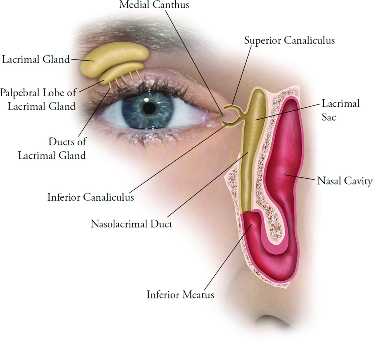
- Fig 1.
Normal anatomy of the lacrimal drainage system apparatus, which includes the canaliculi, lacrimal sac, and nasolacrimal duct.
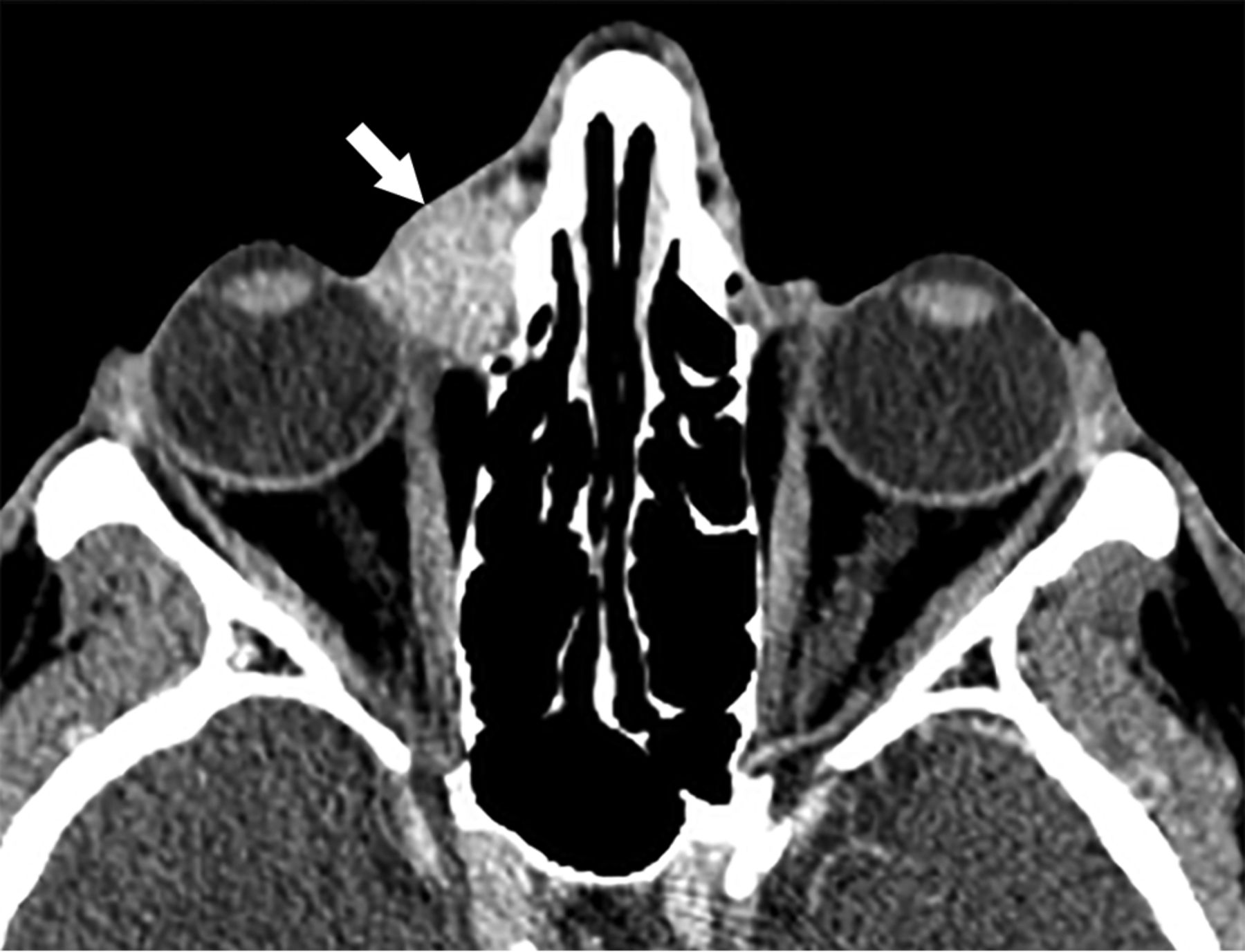
- Fig 2.
A 46-year-old woman with moderately differentiated invasive SCCA of the right lacrimal sac and nasolacrimal duct. Post-contrast-enhanced CT shows tumor extension from the lacrimal sac into the medial canthus region (arrow), which is a common site of tumor spread.
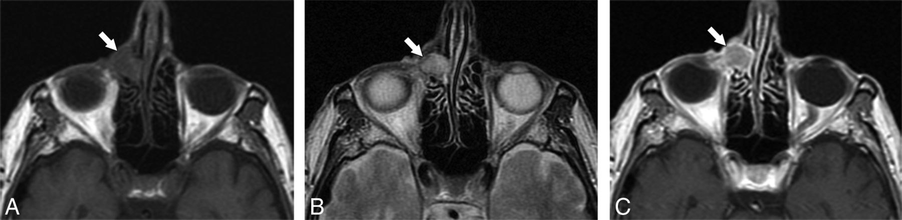
- Fig 3.
An 85-year-old man with metastatic melanoma to the lacrimal sac and nasolacrimal duct with extension into the medial canthus region. Axial MR imaging demonstrates an isointense mass in the right medial canthus on T1WI (A), isointensity on T2WI (B), and enhancement on postcontrast T1WI (C).
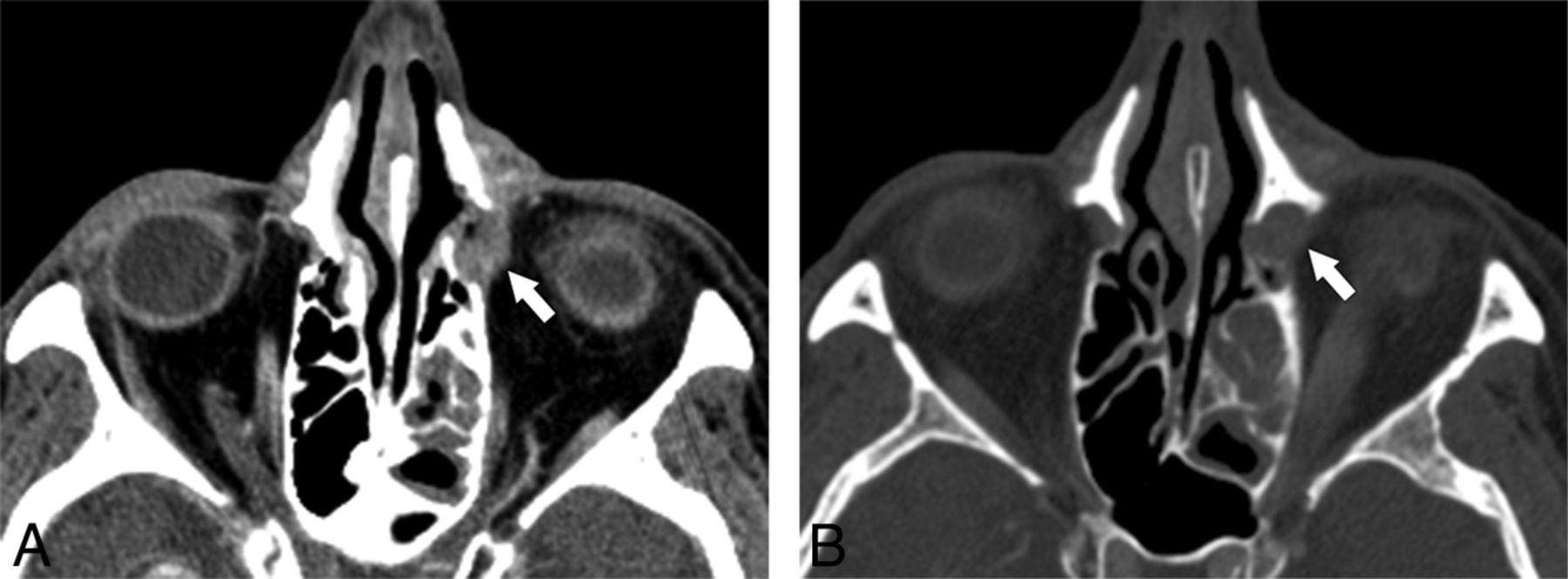
- Fig 4.
A 50-year-old man with adenocarcinoma of the left lacrimal sac and nasolacrimal duct. A, Postcontrast thin-section coronal CT reformatted images demonstrate tumor in the lacrimal sac and duct (white arrow) with direct extension into the inferior intraconal orbital space (black arrow). B, Due to the extent of tumor spread, the patient underwent left orbital exenteration, maxillectomy, and free flap reconstruction (white arrowheads) with adjuvant radiation therapy and has been disease-free for 3 years.
- Fig 5.
A 73-year-old woman with well-differentiated SCCA of the lacrimal sac and nasolacrimal duct. A, Post-contrast-enhanced CT demonstrates an enhancing tumor within the left lacrimal sac (arrow). B, At a slightly more inferior level (bone window), note the mild expansion of the lacrimal bony canal by tumor (arrow).


Tables
Imaging features and regional spread of malignant lacrimal sac and nasolacrimal duct tumors
Features No. of Patients Tumor location along lacrimal system on CT and MRI (n = 18) Lacrimal sac only 3 Nasolacrimal duct only 0 Involving lacrimal sac and nasolacrimal duct 15 Tumor involvement of the nasolacrimal duct bony canal on CT (n = 16) No duct dilation 2 Smoothly expanded duct 11 Erosive/lytic changes to duct 2 Iatrogenic changes to duct from prior dacryocystorhinostomy 1 Orbit involvement by tumor (n = 18) Medial cantus/extraconal space of orbit 16 Intraconal space of orbit 2 Sinonasal involvement by tumor in primary malignant tumors of the lacrimal sac or duct (n = 5)a Ethmoid sinus 4 Maxillary sinus 3 Nasal cavity 5 Other findings (n = 18) Nodal metastasis 1 Distant metastasis 0 Perineural tumor spread along the infraorbital nerve 0 Intracranial extension 0 Dacryocystocele formation 0 ↵a Please note that some patients had >1 subsite of sinonasal tumor extension.


Jump to section
Related Articles
Cited By...
- No citing articles found.