Advertisement

Main menu

  • Home
  • Content
    • Current Issue
    • Accepted Manuscripts
    • Article Preview
    • Past Issue Archive
    • AJNR Case Collection
    • Case of the Week Archive
    • Classic Case Archive
    • Case of the Month Archive
  • Special Collections
    • Spinal CSF Leak Articles (Jan 2020-June 2024)
    • 2024 AJNR Journal Awards
    • Most Impactful AJNR Articles
  • Multimedia
    • AJNR Podcast
    • AJNR Scantastics
    • Video Articles
  • For Authors
    • Submit a Manuscript
    • Author Policies
    • Fast publishing of Accepted Manuscripts
    • Graphical Abstract Preparation
    • Manuscript Submission Guidelines
    • Imaging Protocol Submission
    • Submit a Case for the Case Collection
  • About Us
    • About AJNR
    • Editorial Board
  • More
    • Become a Reviewer/Academy of Reviewers
    • Subscribers
    • Permissions
    • Alerts
    • Feedback
    • Advertisers
    • ASNR Home
  • Other Publications
    • ajnr

User menu

  • Alerts
  • Log in

Search

  • Advanced search
American Journal of Neuroradiology

American Journal of Neuroradiology

ASHNR American Society of Functional Neuroradiology ASHNR American Society of Pediatric Neuroradiology ASSR
  • Alerts
  • Log in

Advanced Search

  • Home
  • Content
    • Current Issue
    • Accepted Manuscripts
    • Article Preview
    • Past Issue Archive
    • AJNR Case Collection
    • Case of the Week Archive
    • Classic Case Archive
    • Case of the Month Archive
  • Special Collections
    • Spinal CSF Leak Articles (Jan 2020-June 2024)
    • 2024 AJNR Journal Awards
    • Most Impactful AJNR Articles
  • Multimedia
    • AJNR Podcast
    • AJNR Scantastics
    • Video Articles
  • For Authors
    • Submit a Manuscript
    • Author Policies
    • Fast publishing of Accepted Manuscripts
    • Graphical Abstract Preparation
    • Manuscript Submission Guidelines
    • Imaging Protocol Submission
    • Submit a Case for the Case Collection
  • About Us
    • About AJNR
    • Editorial Board
  • More
    • Become a Reviewer/Academy of Reviewers
    • Subscribers
    • Permissions
    • Alerts
    • Feedback
    • Advertisers
    • ASNR Home
  • Follow AJNR on Twitter
  • Visit AJNR on Facebook
  • Follow AJNR on Instagram
  • Join AJNR on LinkedIn
  • RSS Feeds

Welcome to the new AJNR, Updated Hall of Fame, and more. Read the full announcements.


AJNR is seeking candidates for the position of Associate Section Editor, AJNR Case Collection. Read the full announcement.


Letter

Embryologic Anatomic Variations: Challenges in Intra-Arterial Chemotherapy for Intraocular Retinoblastoma

C. Paruthi, V. Gupta and N. Khandelwal
American Journal of Neuroradiology November 2016, 37 (11) E71-E72; DOI: https://doi.org/10.3174/ajnr.A4905
C. Paruthi
aDepartment of Radio-Diagnosis Postgraduate Institute of Medical Education and Research Chandigarh, India
  • Find this author on Google Scholar
  • Find this author on PubMed
  • Search for this author on this site
  • ORCID record for C. Paruthi
V. Gupta
aDepartment of Radio-Diagnosis Postgraduate Institute of Medical Education and Research Chandigarh, India
  • Find this author on Google Scholar
  • Find this author on PubMed
  • Search for this author on this site
  • ORCID record for V. Gupta
N. Khandelwal
aDepartment of Radio-Diagnosis Postgraduate Institute of Medical Education and Research Chandigarh, India
  • Find this author on Google Scholar
  • Find this author on PubMed
  • Search for this author on this site
  • ORCID record for N. Khandelwal
  • Article
  • Figures & Data
  • Info & Metrics
  • Responses
  • References
  • PDF
Loading

We read the article on intraocular chemotherapy for retinoblastoma by Bertelli et al,1 e-published in February 2016, with considerable interest. It gives excellent insight into the adaptable approaches of the procedure described. The authors have mainly described 3 patterns of drug delivery in their patients: a fixed pattern through the ophthalmic artery, a fixed pattern through branches of the external carotid artery (ECA), and a variable pattern via either of these. They have also mentioned the “Japanese technique.”2 The authors have also highlighted difficulties in direct ophthalmic artery catheterization due to its acute takeoff from the internal carotid artery, where primary shaping of the catheter can be attempted. In cases in which the choroid blush via the ophthalmic artery was not seen, catheterization of the ECA branches was also performed.

In our experience, sometimes even shaping the microcatheter does not allow ophthalmic artery catheterization. In such cases, if a choroid blush is seen only via the ophthalmic artery and not through the middle meningeal artery, the Japanese technique might be helpful. However, sometimes even this may be challenging due to presence of embryologic vascular variations. The case in point was an 11-month-old female child with grade D retinoblastoma in the left eye, refractory to systemic chemotherapy. A left internal carotid artery angiogram revealed a type 1 persistent trigeminal artery (PTA) arising from the vertical portion of the cavernous segment, with an approximate 4-mm caliber (Fig 1A). The left ophthalmic artery had an acute angled takeoff from the ICA (Fig 1A), and attempts to cannulate it with straight and angled microcatheters were unsuccessful. Tumor blush and choroid blush were, however, seen only via the ophthalmic artery on the left ICA injection. Left ECA and selective left middle meningeal artery runs did not show any choroid blush or reformation of the ophthalmic artery. Therefore, a 4 × 7 mm hypercompliant balloon was inflated in the left ICA, distal to the ophthalmic artery origin. A microcatheter run with its tip in the ICA at the level of left ophthalmic artery origin revealed no opacification of the ophthalmic artery due to preferential retrograde flow across the PTA into the posterior circulation (Fig 1B). Ophthalmic artery flow was re-established on restoration of forward flow after balloon deflation. The only option left was using 2 balloons, one at the former position and the other at the PTA origin, followed by injection of the drug in the segment between these 2 balloons. However, due to the excessive complication risk anticipated due to the presence of 2 balloons in a single artery of an infant,3 this was not attempted. The patient was thus referred for surgical management.

Fig 1.
  • Download figure
  • Open in new tab
  • Download powerpoint
Fig 1.

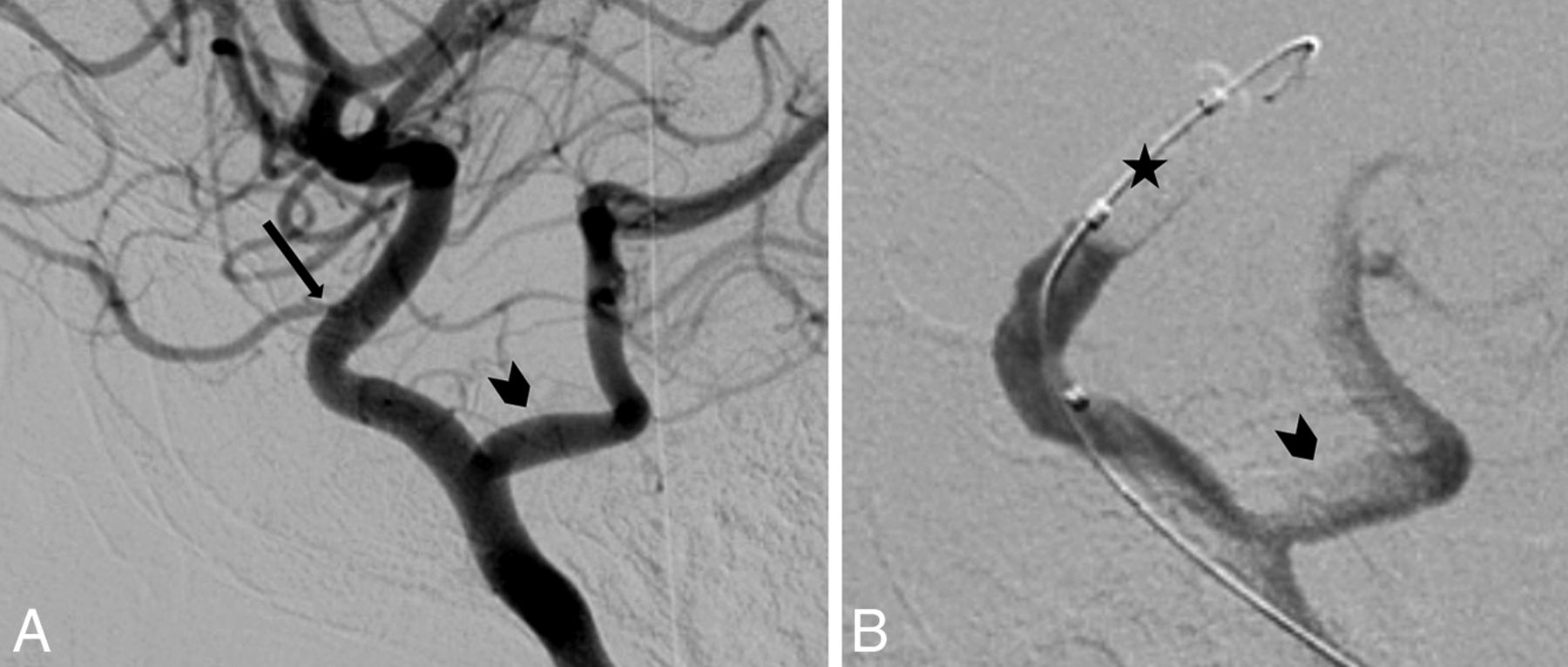
A, Left internal carotid artery run showing ophthalmic artery takeoff at an acute angle (arrow) and a type 1 persistent trigeminal artery (arrowhead). B, Left internal carotid artery microcatheter run after inflation of a hypercompliant balloon within its lumen distal to the origin of ophthalmic artery (star), showing no opacification of the ophthalmic artery and preferential flow across the persistent trigeminal artery (arrowhead).

Although the authors have highlighted different approaches to successfully treating intraocular retinoblastomas via the endovascular route, knowledge of embryologic variations in the involved vascular territory and their hemodynamic effects is also essential to prevent failure.

References

  1. 1.↵
    1. Bertelli E,
    2. Leonini S,
    3. Galimberti D, et al
    . Hemodynamic and anatomic variations require an adaptable approach during intra-arterial chemotherapy for intraocular retinoblastoma: alternative routes, strategies, and follow-up. AJNR Am J Neuroradiol 2016;37:1289–95 doi:10.3174/ajnr.A4741 pmid:26915566
    Abstract/FREE Full Text
  2. 2.↵
    1. Yamane T,
    2. Kaneko A,
    3. Mohri M
    . The technique of ophthalmic arterial infusion therapy for patients with intraocular retinoblastoma. Int J Clin Oncol 2004;9:69–73 doi:10.1007/s10147-004-0392-6 pmid:15108036
    CrossRefPubMed
  3. 3.↵
    1. Suzuki S,
    2. Yamane T,
    3. Mohri M
    . Selective ophthalmic arterial injection therapy for intraocular retinoblastoma: the long-term prognosis. Ophthalmology 2011;118:2081–87 doi:10.1016/j.ophtha.2011.03.013 pmid:21715012
    CrossRefPubMed
  • © 2016 by American Journal of Neuroradiology
PreviousNext
Back to top

In this issue

American Journal of Neuroradiology: 37 (11)
American Journal of Neuroradiology
Vol. 37, Issue 11
1 Nov 2016
  • Table of Contents
  • Index by author
  • Complete Issue (PDF)
Advertisement
Print
Download PDF
Email Article

Thank you for your interest in spreading the word on American Journal of Neuroradiology.

NOTE: We only request your email address so that the person you are recommending the page to knows that you wanted them to see it, and that it is not junk mail. We do not capture any email address.

Enter multiple addresses on separate lines or separate them with commas.
Embryologic Anatomic Variations: Challenges in Intra-Arterial Chemotherapy for Intraocular Retinoblastoma
(Your Name) has sent you a message from American Journal of Neuroradiology
(Your Name) thought you would like to see the American Journal of Neuroradiology web site.
CAPTCHA
This question is for testing whether or not you are a human visitor and to prevent automated spam submissions.
Cite this article
C. Paruthi, V. Gupta, N. Khandelwal
Embryologic Anatomic Variations: Challenges in Intra-Arterial Chemotherapy for Intraocular Retinoblastoma
American Journal of Neuroradiology Nov 2016, 37 (11) E71-E72; DOI: 10.3174/ajnr.A4905

Citation Manager Formats

  • BibTeX
  • Bookends
  • EasyBib
  • EndNote (tagged)
  • EndNote 8 (xml)
  • Medlars
  • Mendeley
  • Papers
  • RefWorks Tagged
  • Ref Manager
  • RIS
  • Zotero
0 Responses
Respond to this article
Share
Bookmark this article
Embryologic Anatomic Variations: Challenges in Intra-Arterial Chemotherapy for Intraocular Retinoblastoma
C. Paruthi, V. Gupta, N. Khandelwal
American Journal of Neuroradiology Nov 2016, 37 (11) E71-E72; DOI: 10.3174/ajnr.A4905
del.icio.us logo Twitter logo Facebook logo Mendeley logo
  • Tweet Widget
  • Facebook Like
  • Google Plus One
Purchase

Jump to section

  • Article
    • References
  • Figures & Data
  • Info & Metrics
  • Responses
  • References
  • PDF

Related Articles

  • Reply:
  • PubMed
  • Google Scholar

Cited By...

  • No citing articles found.
  • Crossref
  • Google Scholar

This article has not yet been cited by articles in journals that are participating in Crossref Cited-by Linking.

Similar Articles

Advertisement

Indexed Content

  • Current Issue
  • Accepted Manuscripts
  • Article Preview
  • Past Issues
  • Editorials
  • Editors Choice
  • Fellow Journal Club
  • Letters to the Editor

Cases

  • Case Collection
  • Archive - Case of the Week
  • Archive - Case of the Month
  • Archive - Classic Case

Special Collections

  • Special Collections

Resources

  • News and Updates
  • Turn around Times
  • Submit a Manuscript
  • Author Policies
  • Manuscript Submission Guidelines
  • Evidence-Based Medicine Level Guide
  • Publishing Checklists
  • Graphical Abstract Preparation
  • Imaging Protocol Submission
  • Submit a Case
  • Become a Reviewer/Academy of Reviewers
  • Get Peer Review Credit from Publons

Multimedia

  • AJNR Podcast
  • AJNR SCANtastic
  • Video Articles

About Us

  • About AJNR
  • Editorial Board
  • Not an AJNR Subscriber? Join Now
  • Alerts
  • Feedback
  • Advertise with us
  • Librarian Resources
  • Permissions
  • Terms and Conditions

American Society of Neuroradiology

  • Not an ASNR Member? Join Now

© 2025 by the American Society of Neuroradiology All rights, including for text and data mining, AI training, and similar technologies, are reserved.
Print ISSN: 0195-6108 Online ISSN: 1936-959X

Powered by HighWire