
Article Figures & Data
Figures
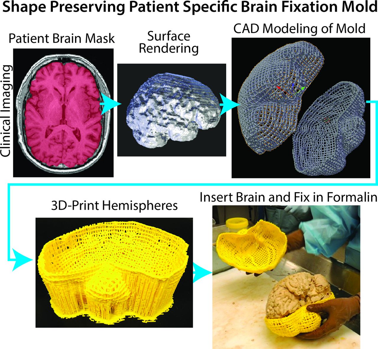
- Fig 1.
Demonstration of the creation of a patient-specific brain mold for minimizing tissue distortion during fixation. The patient's MR imaging is used to generate a brain mask, which is then used as a guide for generating the mold in 3D modeling software. Molds are then 3D-printed in plastic.
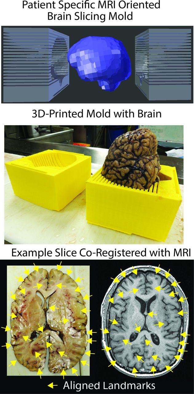
- Fig 2.
Demonstration of the use of a custom 3D-printed slicing jig for sectioning the brain in the same axial orientation as the imaging. Shown on the lower right are examples of gyri and sulci that align well with the imaging (yellow arrows).
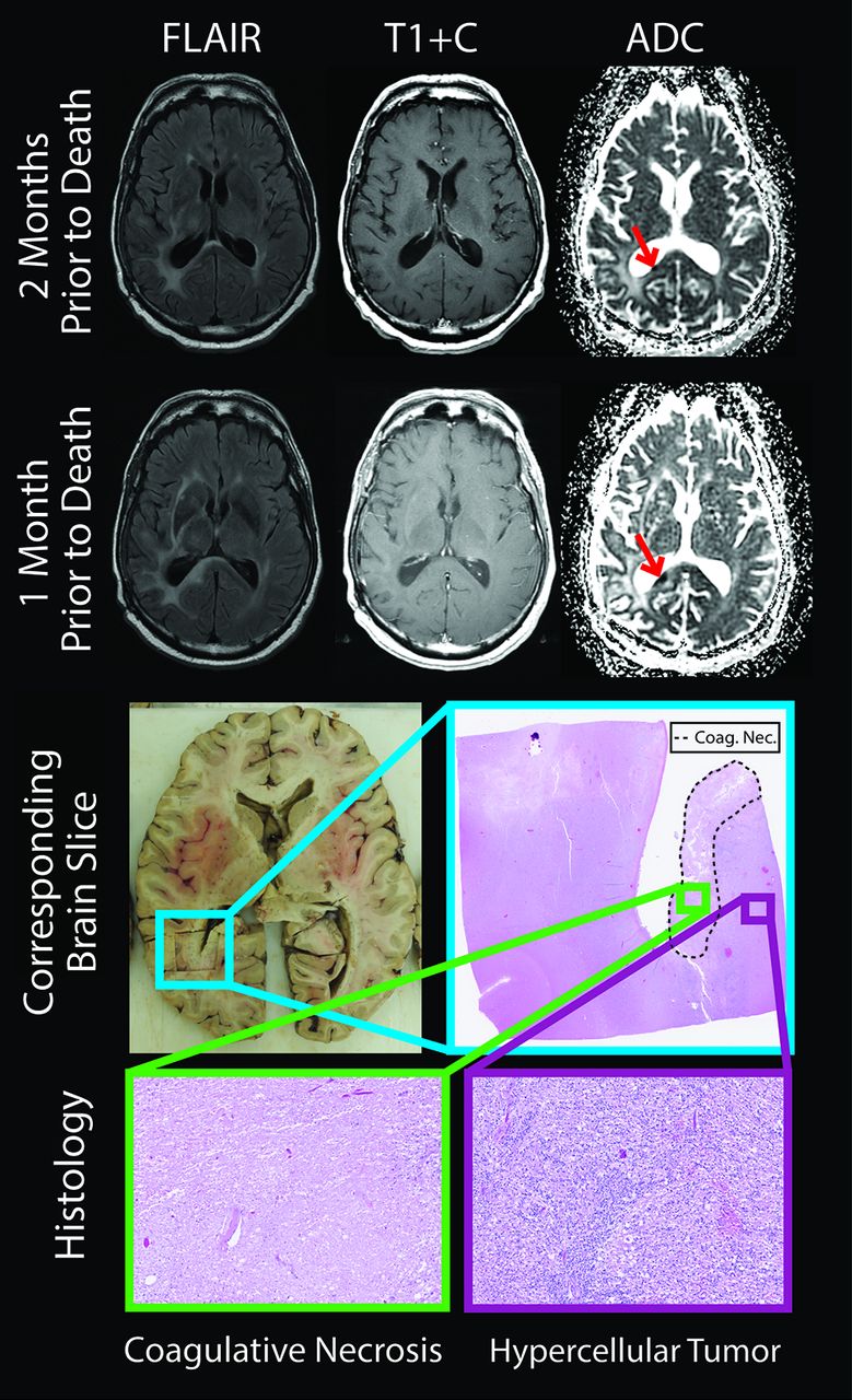
- Fig 3.
The brain section and corresponding imaging from a representative patient. The diffusion-restricted lesion (red arrows) was growing between the 2 imaging sessions, shown 2 months and 1 month before death. Histology revealed coagulative necrosis surrounded by viable hypercellular tumor (lower section). T1+C indicates T1 + gadolinium contrast.
- Fig 4.
Examples of stable and progressive diffusion-restricted lesions occurring following the onset of bevacizumab treatment.
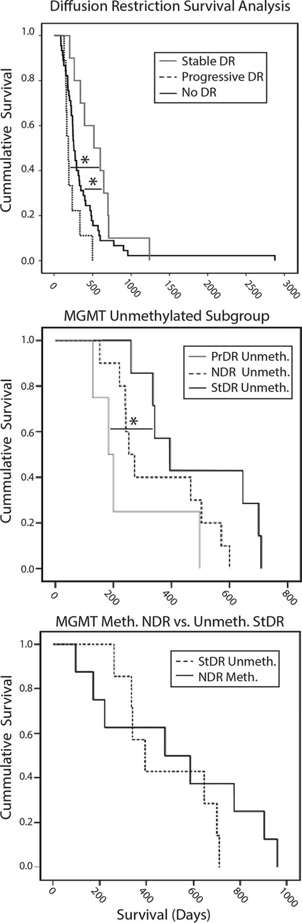
- Fig 5.
Survival analyses comparing groups. Upper Section, Overall survival is significantly greater in patients with stable diffusion restriction compared with those with no diffusion restriction (P < .05). OS is significantly lower in patients with progressive diffusion restriction compared with those with stable diffusion restriction (P < .05). Middle Section, Unmethylated tumors show survival trends similar to those in the overall population, in which patients with stable diffusion restriction survived longer than those with no diffusion restriction and progressive diffusion restriction (P < .05). Lower Section, Patients with unmethylated stable diffusion restriction show survival similar to those with no diffusion-restriction methylation. DR indicates diffusion restriction; PrDR, progressive diffusion restriction; NDR, no diffusion restriction; StDR, stable diffusion restriction; Meth, methylation; Unmeth, no methylation.


Tables
Patient No. Last MRI to Death (day) Age at Death (yr) Tumor Type Surgery XRT TMZ Location of Focal Region of Diffusion Restriction Bevacizumab (day) Before Death Before Focal Region Appears Between Focal Region and Death 1 10 40 Grade III mixed glioma + + + Corpus callosum 44 34 10 2 3 68 GBM + + + Corona radiata 435 306 129 3 23 53 GBM + + + Corona radiata 85 34 51 4 37 65 GBM + + + Centrum semiovale 343 264 79 5 29 58 GBM + + + Corpus callosum 827 728 99 6 62 42 GBM + + + Centrum semiovale 700 534 166 Note:—XRT indicates radiation therapy; TMZ, temozolomide; +, yes.
NDR (n = 45),a (Meth [n = 8]/Unmeth [n = 10]) StDR (n = 10),b (Unmeth [n = 7]) PrDR (n = 9),c (Unmeth [n = 4]) Age at death (yr) (mean) (SD) 55 (13) (58/63) 55 (11) (55) 52 (8) (58) Sex Male 23 (4/4) 6 (4) 8 (4) Female 22 (4/6) 4 (3) 1 (0) Days between bevacizumab initiation and death (median) (lower/upper CI) 256 (213–298) (524–352) 516 (197–835) (484) 183 (125–241) (252) Recurrences/progression (median) (range) 2 (1–5) (2.5/2.2) 2 (1–5) (2.57) 2 (1–3) (1.5) Initial pathology Grade II 5 (1/1) 1 (1) 0 (0) Grade III 2 (0/1) 2 (2) 1 (0) GBM 38 (7/8) 7 (4) 8 (4) Therapeutic regimen Surgery + XRT/TMZ + adjuvant TMZ 45 (8/10) 10 (7) 9 (4) Reoperation 22 (2/5) 6 (4) 2 (0) Bevacizumab 45 (8/10) 10 (7) 9 (4) Stopped?a 10 (1/3) 3 (1) 1 (0) Irinotecan 6 (0) 2 (0) 3 (0) Isotretinoin 15 (2/3) 2 (2) 1 (0) CCNU/BCNU 8 (3/1) 2 (2) 3 (2) Interferon 2 (0) 0 (0) 0 (0) Optuned TTF 0 (0) 1 (1) 0 (0) PLDR 12 (5/4) 5 (4) 3 (2) Note:—PLDR indicates pulsed low-dose rate radiation; NDR, no diffusion restriction; StDR, stable diffusion restriction; PrDR, progressive diffusion restriction; TTF, tumor treating fields; Meth, methylation; Unmeth, no methylation; CCNU/BCNU, carmustine/lomustine; XRT, radiation therapy; TMZ, temozolomide.
↵a Bevacizumab was stopped for further surgery (for resection, n = 5; for infection, n = 1; for shunting, n = 1; for hemorrhage, n = 1; buttock abscess, n = 1; and hip fracture, n = 1).
↵b Bevacizumab was stopped for neutropenia/thrombocytopenia (n = 1), fatigue (n = 1), and hemorrhage (n = 1).
↵c Bevacizumab was stopped for further surgery (resection, n = 1).
↵d Novocure, Portsmouth, New Hampshire.


Jump to section
Related Articles
Cited By...
- Non-invasive tumor probability maps developed using autopsy tissue identify novel areas of tumor beyond the imaging-defined margin
- Non-invasive tumor probability maps developed using autopsy tissue identify novel areas of tumor beyond the imaging-defined margin
- Radio-Pathomic Maps of Cell Density Identify Brain Tumor Invasion beyond Traditional MRI-Defined Margins
- Radio-pathomic maps of cell density identify glioma invasion beyond traditional MR imaging defined margins
- Response Assessment in Neuro-Oncology Criteria for Gliomas: Practical Approach Using Conventional and Advanced Techniques
- Diagnostic Accuracy of Centrally Restricted Diffusion in the Differentiation of Treatment-Related Necrosis from Tumor Recurrence in High-Grade Gliomas