
Article Figures & Data
Figures
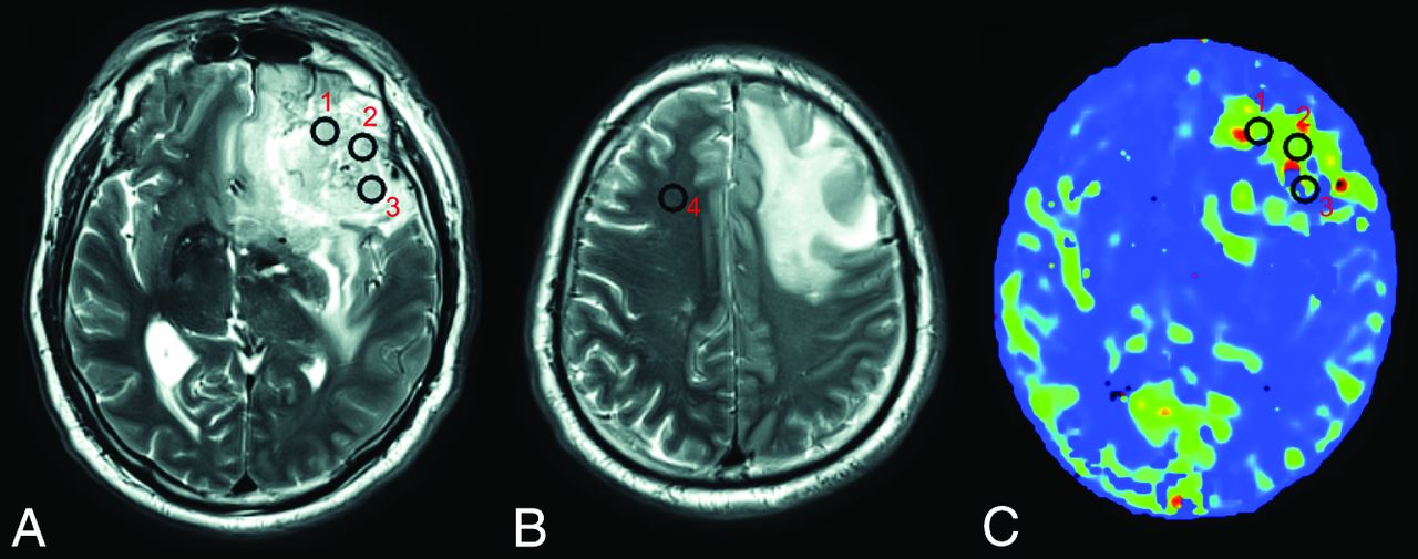
- Fig 1.
VOI positioning. Three VOIs are placed on the solid part of the tumor (VOI 1, 2, and 3) (A), and 1 is placed on the contralateral normal-appearing white matter of the frontal lobe (VOI 4) (B) on T2-weighted images by each of the 2 neuroradiologists. The VOIs are projected onto the maps derived from ASL (eg, the CBF-mTI map) (C).
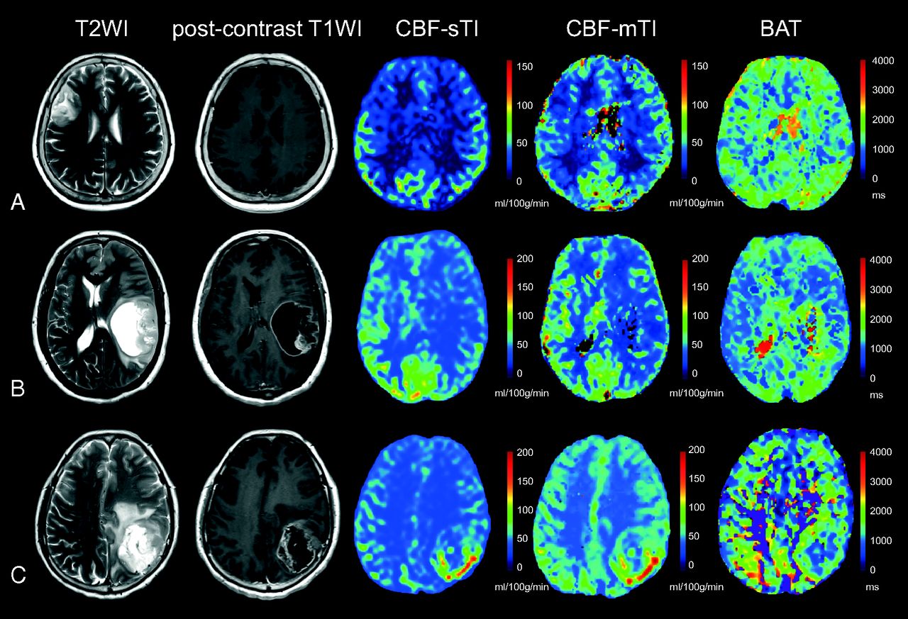
- Fig 2.
Examples of the 3 different astrocytomas. A, A 60-year-old female patient with a diffuse astrocytoma (WHO II) in the right frontal-parietal region. The lesion demonstrates high signal on the T2-weighted image, a relatively low CBF value on both the CBF-sTI and CBF-mTI maps, and moderate signal intensity on the BAT map. No obvious enhancement is visible on the postcontrast T1-weighted image. B, Images from a 50-year-old woman with an anaplastic astrocytoma (WHO III) in the left temporal lobe demonstrate high signal intensity in the T2-weighted image and moderate enhancement in the postcontrast T1-weighted image for the solid part of the tumor. Both the CBF-sTI and CBF-mTI maps demonstrate low signal intensity, but the BAT value for the tumor areas is longer than that for the corresponding contralateral normal cerebral region. C, A 56-year-old female patient with a glioblastoma (WHO IV) in the left parietal lobe. The solid part of the tumor appears as an area of high signal intensity in the T2-weighted image and as obvious enhancement in the postcontrast T1-weighted image. Hyperperfusion is visible in both the CBF-sTI and CBF-mTI maps, and the BAT value is shortened.
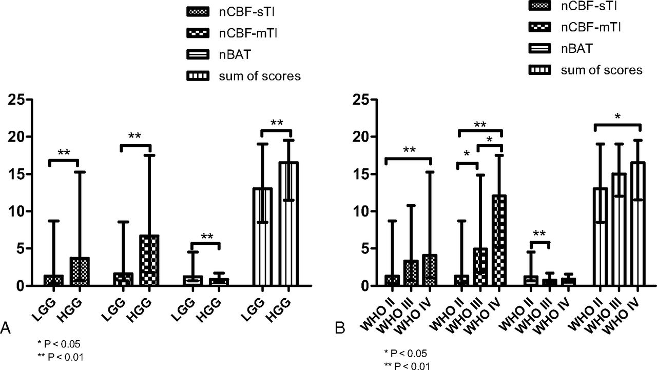
- Fig 3.
The values of the different parameters are plotted as bar graphs by median and range. Significant differences between groups are indicated by asterisks. Error bars indicate the range. A, The bar graph shows the 4 indices of nCBF-mTI, nBAT, and nCBF-sTI and the sum of scores for the LGG and HGG groups. All indices indicate a significant difference between the LGG and HGG groups. B, The values in the bar graph show the parameters of each tumor grade. The nCBF-mTI shows significant differences between each pair of grades. The nBAT shows a significant difference between the WHO II and III groups. The nCBF-sTI and sum of scores show significant differences between only the WHO II and IV groups.
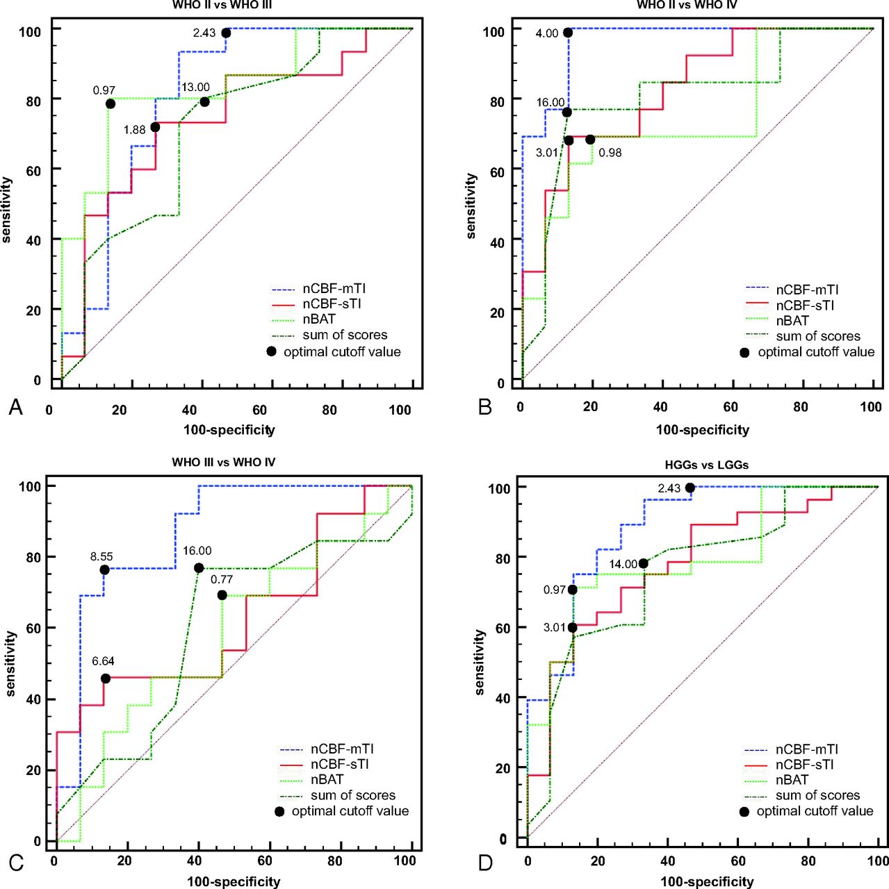
- Fig 4.
Results of the receiver operating characteristic curve analyses. A, The receiver operating characteristic (ROC) curve for the nCBF-mTI, nCBF-sTI, nBAT, and sum of scores for MR imaging features in differentiating the WHO II and III groups. The nBAT has the best performance with an AUC of 0.836, followed by the nCBF-mTI with an AUC of 0.813, the nCBF-sTI with an AUC of 0.742, and the sum of scores with an AUC of 0.716. B, The ROC curves for the nCBF-mTI, nCBF-sTI, nBAT, and the sum of scores in differentiating the WHO II and IV groups. The nCBF-mTI has the best performance with an AUC of 0.964, followed by the nCBF-sTI, sum of scores, and nBAT, with AUCs of 0.826, 0.805, and 0.744, respectively. C, The ROC curves for the nCBF-mTI, nCBF-sTI, nBAT, and the sum of scores in differentiating the WHO III and IV groups. The nCBF-mTI has the best performance with an AUC of 0.872, followed by the nCBF-sTI, sum of scores, and nBAT, with AUCs of 0.631, 0.603, and 0.585, respectively. D, The ROC curves for the nCBF-mTI, nCBF-sTI, nBAT, and the sum of scores in differentiating the LGG and HGG groups. The nCBF-mTI has the best performance with an AUC of 0.883, followed by the nBAT, nCBF-sTI, and the sum of scores with AUCs of 0.793, 0.781, and 0.757, respectively.
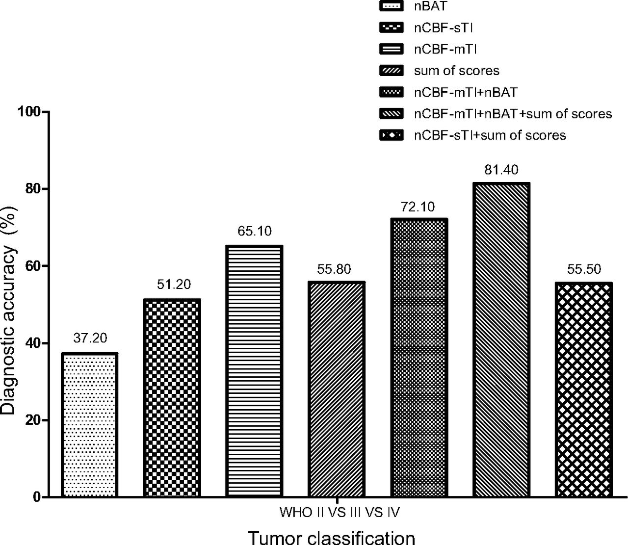
- Fig 5.
The independent and combined diagnostic accuracies (as percentages) of nCBF-mTI, nBAT, nCBF-sTI, and the sum of scores for discriminating among the WHO II, III, and IV astrocytoma grades, as calculated. All of the diagnostic accuracy values are indicated at the top of the columns.


Tables
Histologic Diagnosis Diffuse Astrocytoma (WHO II) Anaplastic Astrocytoma (WHO III) Glioblastoma (WHO IV) No. of patients 15 15 13 Male/female ratio 7:8 6:9 8:5 Median age (yr) (range) 54 (17–70) 52 (15–73) 57 (28–67) Tumor Feature Scores Edema None or mild (1); smaller than tumor volume (2); larger than tumor volume (3) Mass effect Subarachnoid space effacement (1); ventricular system compression (2); midline shift (3) Contrast enhancement None (1); mild or nodular (2); marked and heterogeneous (3) Borders Well-defined (1); poorly defined (2) Heterogeneity Homogeneous (1); heterogeneous on T2WI (2); heterogeneous on T1WI and T2WI (3) Necrosis/cyst None (1); involving less than half the volume of the tumoral mass (2); involving more than half the volume of the tumoral mass (3) Hemorrhage None (1); present (2) Flow void None (1); present (2) ↵a The numbers in parentheses are the scores assigned for the grades of tumor features.
- Table 3:
Comparison of the sum of scores for the MR imaging features and ASL parameters (average values obtained by the 2 readers) among the 3 pathologic gradesa
WHO Grade Sum of Scores (Median) (Range) nCBF-sTI (Median) (Range) nCBF-mTI (Median) (Range) nBAT (Median) (Range) II (LGGs) 13 (8.5–19) 1.28 (0.36–8.7) 1.62 (0.68–8.56) 1.21 (0.68–4.52) III 15 (12–19) 3.33 (0.72–10.78) 4.93 (1.78–14.85) 0.77 (0.53–1.70) IV 16.5 (11.5–19.5) 4.07 (1.07–15.23) 13.40 (5.18–22.36) 0.90 (0.53–1.55) III and IV (HGGs) 16.5 (11.5–19.5) 3.7 (0.72–14.51) 6.7 (1.78–22.36) 0.86 (0.53–1.70) ↵a Values are presented as median and range.
- Table 4:
P values of comparisons for the sum of scores for the MR imaging features and ASL parameters among the 3 pathologic grades
WHO Grade Sum of Scores nCBF-sTI nCBF-mTI nBAT II vs III .139 .098 .021a .005a II vs IV .015a .006a <.001a .078 III vs IV 1 .905 .023a 1 LGGs vs HGGs .006a .003a <.001a .002a ↵a Significant at 95% (for intergroup comparisons among the WHO II, III, and IV grades. P values have been multiplied by 3 for Bonferroni correction).


Jump to section
Related Articles
Cited By...
- No citing articles found.