
Article Figures & Data
Figures
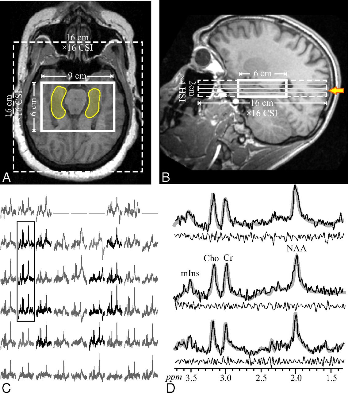
- Fig 1.
Upper: Axial (A) and sagittal (B) T1-weighted MR imaging from a 23-year-old male patient (17 in Table 1) superimposed on the 9 × 6 × 2 cm3 (left-right × anteroposterior × inferior-superior) VOI, 16 × 16 cm2 axial CSI FOV (solid and dashed lines), and the hippocampal outline (transparent yellow on A). The yellow arrow in B indicates the level of A, C, and D. Lower left: C, Real part of the 9 × 6 axial (left-right × anteroposterior) 1H spectra matrix from the VOI section shown in A and marked with the solid yellow arrow on B. Spectra within the hippocampus in A are black, while the remaining ones (not included in the analyses) are gray. All are on a common frequency (parts per million) and intensity scale. The 3 spectra in the black frame over the right hippocampus are expanded on the right (D) for greater detail. Note that the hippocampi do not include voxels at the edges of the VOI (that may have relative VOI chemical shift displacement); note also the good SNR and excellent spectral resolution (8.1 ± 3.0 Hz linewidth) from the high spatial resolution (0.5 cm3) voxels. Right: D, The 3 spectra from the solid frame on C (black line) overlaid on the spectral fit (thick gray lines) and the residual (experimental − fit) underneath (thin black line). Note the spectral resolution and fidelity of the fit, reflected by the residual.
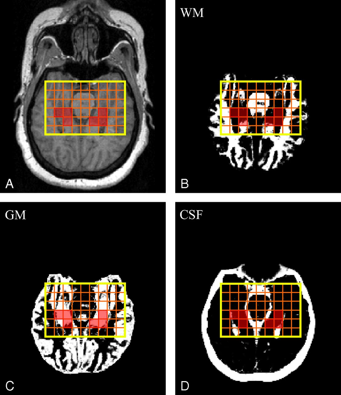
- Fig 2.
Upper: A, Axial MPRAGE image from a 51-year-old female patient (16 in Table 1) superimposed on the VOI (in yellow). Orange lines show the 9 × 6 voxel CSI grid; voxels that passed the selection criteria to calculate the NAA concentration are highlighted in transparent red. B–D, SPM12-generated WM (B), GM (C), and CSF (D) masks also superimposed on the VOI CSI grid and selected voxels. Note the n ≥ 2 voxels that “passed” the selection criteria described in the “Materials and Methods” section.
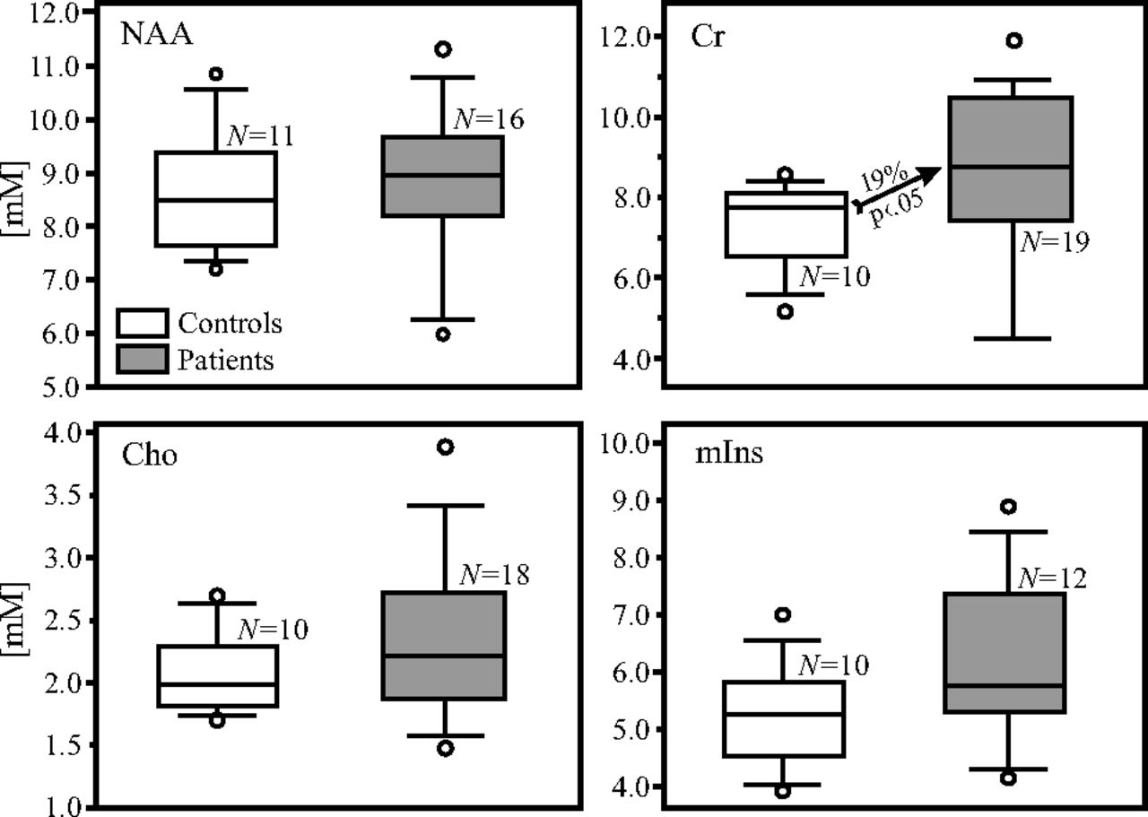
- Fig 3.
Boxplots showing the first, second (median), and third quartiles; 5th and 95th percentiles (whiskers); and outliers (dots) of the distribution of the bilateral hippocampal NAA, Cr, Cho, and mIns concentrations (millimolar) in the patient (shaded) and control (white) boxes. Numbers of controls and patients included in the analyses for each metabolite, N, are listed. Note that the NAA, Cho, and mIns concentrations do not differ significantly between patients and controls (Table 2), whereas the Cr concentration is 19% higher in the bilateral hippocampi of patients than in controls (arrow).


Tables
Subject Status Age (yr)/Sex Disease Duration (yr) Psychotropic Medication 1 C 45/M NA NA 2 C 31/M NA NA 3 C 29/F NA NA 4 C 36/M NA NA 5 C 43/M NA NA 6 C 24/F NA NA 7 C 55/F NA NA 8 C 26/F NA NA 9 C 29/F NA NA 10 C 22/M NA NA 11 C 31/F NA NA 12 P 41/M 22 Fluphenazine 13 P 44/F 27 Quetiapine 14 P 43/M 18 Haloperidol, quetiapine 15 P 52/M 32 Citalopram 16 P 51/F 15 Gabapentin, lithium, ziprasidone 17 P 23/M 3 Risperidone 18 P 47/M 31 Fluoxetine, risperidone, valproic acid, trazodone 19 P 44/M 26 Clozapine, valproic acid 20 P 26/M 8 Ziprasidone 21 P 29/F 8 Bupropion, aripiprazole, fluphenazine 22 P 42/F 23 Ziprasidone, bupropion, eszopiclone 23 P 22/M 4 Clozapine 24 P 48/M 23 Quetiapine 25 P 34/M 5 Risperidone 26 P 30/F 10 Risperidone 27 P 49/F 31 Aripiprazole 28 P 51/F 35 NA 29 P 43/F 20 Aripiprazole, escitalopram, fluphenazine 30 P 52/M 30 Aripiprazole, valproic acid, hydroxyzine, paroxetine, trazodone Note:—NA indicates not applicable; C, controls; P, patients.
- Table 2:
Means, number of subjects from whom the data was derived (in parentheses), and P values (from unequal variance t tests) of the absolute NAA, Cr, Cho, and mIns hippocampal GM concentrations and the volumes of the bilateral hippocampi in controls and patientsa
Metabolite Controls Patients P Value NAA (mM) 8.7 ± 1.2 (n = 11) 8.8 ± 1.6 (n = 16) .876 Cr (mM) 7.4 ± 1.2 (n = 10)b 8.7 ± 2.2 (n = 19)b .035b Cho (mM) 2.1 ± 0.3 (n = 10) 2.3 ± 0.7 (n = 18) .189 mIns (mM) 5.2 ± 0.9 (n = 10) 6.1 ± 1.5 (n = 12) .161 Volume (cm3) 8.4 ± 0.5 (n = 11)b 7.5 ± 0.9 (n = 19)b .003b


