Advertisement

Main menu

  • Home
  • Content
    • Current Issue
    • Accepted Manuscripts
    • Article Preview
    • Past Issue Archive
    • Video Articles
    • AJNR Case Collection
    • Case of the Week Archive
    • Case of the Month Archive
    • Classic Case Archive
  • Special Collections
    • AJNR Awards
    • Low-Field MRI
    • Alzheimer Disease
    • ASNR Foundation Special Collection
    • Photon-Counting CT
    • View All
  • Multimedia
    • AJNR Podcasts
    • AJNR SCANtastic
    • Trainee Corner
    • MRI Safety Corner
    • Imaging Protocols
  • For Authors
    • Submit a Manuscript
    • Submit a Video Article
    • Submit an eLetter to the Editor/Response
    • Manuscript Submission Guidelines
    • Statistical Tips
    • Fast Publishing of Accepted Manuscripts
    • Graphical Abstract Preparation
    • Imaging Protocol Submission
    • Author Policies
  • About Us
    • About AJNR
    • Editorial Board
    • Editorial Board Alumni
  • More
    • Become a Reviewer/Academy of Reviewers
    • Subscribers
    • Permissions
    • Alerts
    • Feedback
    • Advertisers
    • ASNR Home

User menu

  • Alerts
  • Log in

Search

  • Advanced search
American Journal of Neuroradiology

American Journal of Neuroradiology

ASHNR American Society of Functional Neuroradiology ASHNR American Society of Pediatric Neuroradiology ASSR
  • Alerts
  • Log in

Advanced Search

  • Home
  • Content
    • Current Issue
    • Accepted Manuscripts
    • Article Preview
    • Past Issue Archive
    • Video Articles
    • AJNR Case Collection
    • Case of the Week Archive
    • Case of the Month Archive
    • Classic Case Archive
  • Special Collections
    • AJNR Awards
    • Low-Field MRI
    • Alzheimer Disease
    • ASNR Foundation Special Collection
    • Photon-Counting CT
    • View All
  • Multimedia
    • AJNR Podcasts
    • AJNR SCANtastic
    • Trainee Corner
    • MRI Safety Corner
    • Imaging Protocols
  • For Authors
    • Submit a Manuscript
    • Submit a Video Article
    • Submit an eLetter to the Editor/Response
    • Manuscript Submission Guidelines
    • Statistical Tips
    • Fast Publishing of Accepted Manuscripts
    • Graphical Abstract Preparation
    • Imaging Protocol Submission
    • Author Policies
  • About Us
    • About AJNR
    • Editorial Board
    • Editorial Board Alumni
  • More
    • Become a Reviewer/Academy of Reviewers
    • Subscribers
    • Permissions
    • Alerts
    • Feedback
    • Advertisers
    • ASNR Home
  • Follow AJNR on Twitter
  • Visit AJNR on Facebook
  • Follow AJNR on Instagram
  • Join AJNR on LinkedIn
  • RSS Feeds

AJNR Awards, New Junior Editors, and more. Read the latest AJNR updates

Research ArticleAdult Brain

T1 Signal-Intensity Increase in the Dentate Nucleus after Multiple Exposures to Gadodiamide: Intraindividual Comparison between 2 Commonly Used Sequences

J. Ramalho, M. Ramalho, M. AlObaidy, R.H. Nunes, M. Castillo and R.C. Semelka
American Journal of Neuroradiology August 2016, 37 (8) 1427-1431; DOI: https://doi.org/10.3174/ajnr.A4757
J. Ramalho
aFrom the Department of Radiology (J.R., M.R., M.A., R.H.N., M.C., R.C.S.), University of North Carolina at Chapel Hill, Chapel Hill, North Carolina
bDepartment of Neuroradiology (J.R.), Centro Hospitalar de Lisboa Central, Lisbon, Portugal
  • Find this author on Google Scholar
  • Find this author on PubMed
  • Search for this author on this site
  • ORCID record for J. Ramalho
M. Ramalho
aFrom the Department of Radiology (J.R., M.R., M.A., R.H.N., M.C., R.C.S.), University of North Carolina at Chapel Hill, Chapel Hill, North Carolina
cDepartment of Radiology (M.R.), Hospital Garcia de Orta, Almada, Portugal
  • Find this author on Google Scholar
  • Find this author on PubMed
  • Search for this author on this site
  • ORCID record for M. Ramalho
M. AlObaidy
aFrom the Department of Radiology (J.R., M.R., M.A., R.H.N., M.C., R.C.S.), University of North Carolina at Chapel Hill, Chapel Hill, North Carolina
dDepartment of Radiology (M.A.), King Faisal Specialist Hospital and Research Center, Riyadh, Saudi Arabia
  • Find this author on Google Scholar
  • Find this author on PubMed
  • Search for this author on this site
  • ORCID record for M. AlObaidy
R.H. Nunes
aFrom the Department of Radiology (J.R., M.R., M.A., R.H.N., M.C., R.C.S.), University of North Carolina at Chapel Hill, Chapel Hill, North Carolina
eDivision of Neuroradiology (R.H.N.), Santa Casa de Misericórdia de São Paulo, São Paulo, Brazil.
  • Find this author on Google Scholar
  • Find this author on PubMed
  • Search for this author on this site
  • ORCID record for R.H. Nunes
M. Castillo
aFrom the Department of Radiology (J.R., M.R., M.A., R.H.N., M.C., R.C.S.), University of North Carolina at Chapel Hill, Chapel Hill, North Carolina
  • Find this author on Google Scholar
  • Find this author on PubMed
  • Search for this author on this site
  • ORCID record for M. Castillo
R.C. Semelka
aFrom the Department of Radiology (J.R., M.R., M.A., R.H.N., M.C., R.C.S.), University of North Carolina at Chapel Hill, Chapel Hill, North Carolina
  • Find this author on Google Scholar
  • Find this author on PubMed
  • Search for this author on this site
  • ORCID record for R.C. Semelka
  • Article
  • Figures & Data
  • Info & Metrics
  • Responses
  • References
  • PDF
Loading

Article Figures & Data

Figures

  • Tables
  • Fig 1.
    • Download figure
    • Open in new tab
    • Download powerpoint
    Fig 1.

    Axial MR images in a 40-year-old male patient with a right frontal low-grade astrocytoma. Unenhanced axial T1-weighted spin-echo (A and C) and 3D MPRAGE MR images (B and D) of the first (A and B) and fifth (2 years later, C and D) gadolinium-enhanced MR imaging examinations at the level of the dentate nuclei of the cerebellum. The images show progressively increased T1 signal of the dentate nuclei (white arrows, C and D). Note that the qualitative analysis was slightly different between the 2 sequences.

  • Fig 2.
    • Download figure
    • Open in new tab
    • Download powerpoint
    Fig 2.

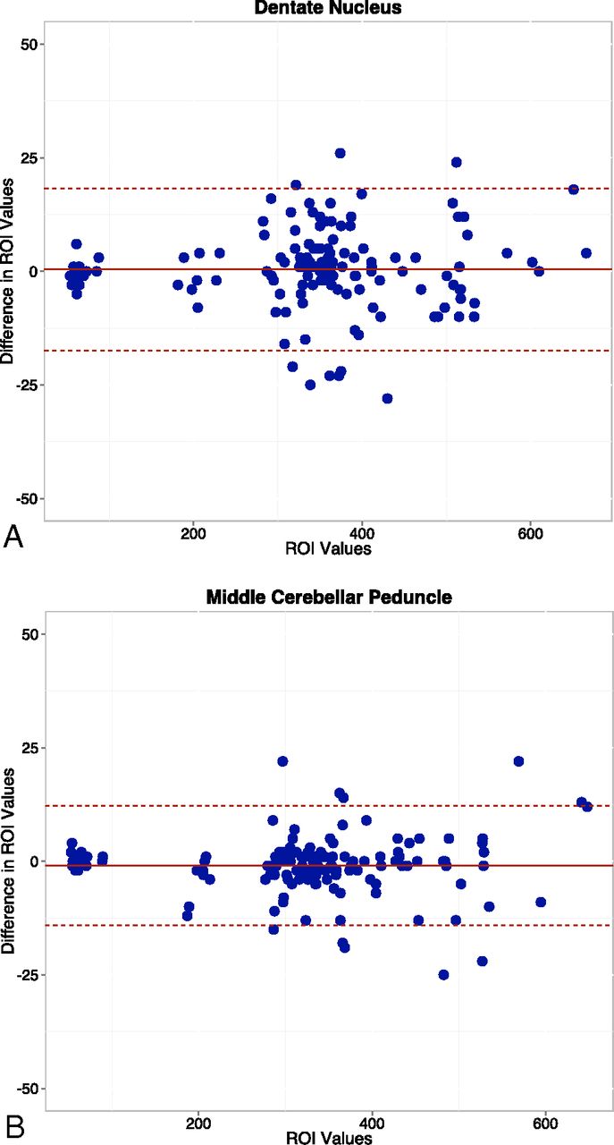
    Bland-Altman plots show the differences in average ROI measurements between the 2 readers for the dentate nucleus (A) and middle cerebellar peduncle (B).

  • Fig 3.
    • Download figure
    • Open in new tab
    • Download powerpoint
    Fig 3.

    Intraindividual linear graphic representation of the DN/MCP ratios, with error bars, for spin-echo and 3D MPRAGE sequences. Note the higher ratios with SE on both baseline and final examinations.

  • Fig 4.
    • Download figure
    • Open in new tab
    • Download powerpoint
    Fig 4.

    Intraindividual linear graphic representation demonstrating the correlation between qualitative and quantitative (DN/MCP ratio) evaluations of the increased T1 signal intensity within the dentate nucleus for SE and 3D MPRAGE sequences. Note the stronger correlation with MPRAGE (0.9) compared with SE (0.63). Values on the y-axis are represented on an ordinal scale with random vertical offset (jitter) to minimize overlapping.

Tables

  • Figures
  • Patients' demographic and clinical characteristics

    Demographics
    Patients (number)18
    Sex12 Women
    Age (mean ± SD) (range) (yrs)52.56 ± 15.21 (17–76)
    eMRIs performed (mean ± SD) (range)4.78 ± 2.51 (2–10)
    Interval (MRIbaseline − MRIx) (mean ± SD) (range)933 ± 610.78 (96–1905 days)
    Diagnosis (number)
        Meningioma12
        Glioblastoma2
        Low-grade glioma1
        Oligodendroglioma1
        Chordoma1
        Spinal hemangioblastoma1
    • Note:—eMRI indicates enhanced MR imaging; yrs, years.

PreviousNext
Back to top

In this issue

American Journal of Neuroradiology: 37 (8)
American Journal of Neuroradiology
Vol. 37, Issue 8
1 Aug 2016
  • Table of Contents
  • Index by author
  • Complete Issue (PDF)
Advertisement
Print
Download PDF
Email Article

Thank you for your interest in spreading the word on American Journal of Neuroradiology.

NOTE: We only request your email address so that the person you are recommending the page to knows that you wanted them to see it, and that it is not junk mail. We do not capture any email address.

Enter multiple addresses on separate lines or separate them with commas.
T1 Signal-Intensity Increase in the Dentate Nucleus after Multiple Exposures to Gadodiamide: Intraindividual Comparison between 2 Commonly Used Sequences
(Your Name) has sent you a message from American Journal of Neuroradiology
(Your Name) thought you would like to see the American Journal of Neuroradiology web site.
CAPTCHA
This question is for testing whether or not you are a human visitor and to prevent automated spam submissions.
Cite this article
J. Ramalho, M. Ramalho, M. AlObaidy, R.H. Nunes, M. Castillo, R.C. Semelka
T1 Signal-Intensity Increase in the Dentate Nucleus after Multiple Exposures to Gadodiamide: Intraindividual Comparison between 2 Commonly Used Sequences
American Journal of Neuroradiology Aug 2016, 37 (8) 1427-1431; DOI: 10.3174/ajnr.A4757

Citation Manager Formats

  • BibTeX
  • Bookends
  • EasyBib
  • EndNote (tagged)
  • EndNote 8 (xml)
  • Medlars
  • Mendeley
  • Papers
  • RefWorks Tagged
  • Ref Manager
  • RIS
  • Zotero
0 Responses
Respond to this article
Share
Bookmark this article
T1 Signal-Intensity Increase in the Dentate Nucleus after Multiple Exposures to Gadodiamide: Intraindividual Comparison between 2 Commonly Used Sequences
J. Ramalho, M. Ramalho, M. AlObaidy, R.H. Nunes, M. Castillo, R.C. Semelka
American Journal of Neuroradiology Aug 2016, 37 (8) 1427-1431; DOI: 10.3174/ajnr.A4757
del.icio.us logo Twitter logo Facebook logo Mendeley logo
  • Tweet Widget
  • Facebook Like
  • Google Plus One
Purchase

Jump to section

  • Article
    • Abstract
    • ABBREVIATIONS:
    • Materials and Methods
    • Results
    • Discussion
    • Conclusions
    • References
  • Figures & Data
  • Info & Metrics
  • Responses
  • References
  • PDF

Related Articles

  • No related articles found.
  • PubMed
  • Google Scholar

Cited By...

  • Age-Dependent Signal Intensity Changes in the Structurally Normal Pediatric Brain on Unenhanced T1-Weighted MR Imaging
  • Retention of Gadolinium-Based Contrast Agents in Multiple Sclerosis: Retrospective Analysis of an 18-Year Longitudinal Study
  • Crossref
  • Google Scholar

This article has not yet been cited by articles in journals that are participating in Crossref Cited-by Linking.

More in this TOC Section

  • Diagnostic Neuroradiology of Monoclonal Antibodies
  • Segmentation of Brain Metastases with BLAST
  • Cerebral ADC Changes in Fabry Disease
Show more Adult Brain

Similar Articles

Advertisement

Indexed Content

  • Current Issue
  • Accepted Manuscripts
  • Article Preview
  • Past Issues
  • Editorials
  • Editor's Choice
  • Fellows' Journal Club
  • Letters to the Editor
  • Video Articles

Cases

  • Case Collection
  • Archive - Case of the Week
  • Archive - Case of the Month
  • Archive - Classic Case

More from AJNR

  • Trainee Corner
  • Imaging Protocols
  • MRI Safety Corner
  • Book Reviews

Multimedia

  • AJNR Podcasts
  • AJNR Scantastics

Resources

  • Turnaround Time
  • Submit a Manuscript
  • Submit a Video Article
  • Submit an eLetter to the Editor/Response
  • Manuscript Submission Guidelines
  • Statistical Tips
  • Fast Publishing of Accepted Manuscripts
  • Graphical Abstract Preparation
  • Imaging Protocol Submission
  • Evidence-Based Medicine Level Guide
  • Publishing Checklists
  • Author Policies
  • Become a Reviewer/Academy of Reviewers
  • News and Updates

About Us

  • About AJNR
  • Editorial Board
  • Editorial Board Alumni
  • Alerts
  • Permissions
  • Not an AJNR Subscriber? Join Now
  • Advertise with Us
  • Librarian Resources
  • Feedback
  • Terms and Conditions
  • AJNR Editorial Board Alumni

American Society of Neuroradiology

  • Not an ASNR Member? Join Now

© 2025 by the American Society of Neuroradiology All rights, including for text and data mining, AI training, and similar technologies, are reserved.
Print ISSN: 0195-6108 Online ISSN: 1936-959X

Powered by HighWire