Advertisement

Main menu

  • Home
  • Content
    • Current Issue
    • Accepted Manuscripts
    • Article Preview
    • Past Issue Archive
    • Video Articles
    • AJNR Case Collection
    • Case of the Week Archive
    • Case of the Month Archive
    • Classic Case Archive
  • Special Collections
    • AJNR Awards
    • Low-Field MRI
    • Alzheimer Disease
    • ASNR Foundation Special Collection
    • Photon-Counting CT
    • View All
  • Multimedia
    • AJNR Podcasts
    • AJNR SCANtastic
    • Trainee Corner
    • MRI Safety Corner
    • Imaging Protocols
  • For Authors
    • Submit a Manuscript
    • Submit a Video Article
    • Submit an eLetter to the Editor/Response
    • Manuscript Submission Guidelines
    • Statistical Tips
    • Fast Publishing of Accepted Manuscripts
    • Graphical Abstract Preparation
    • Imaging Protocol Submission
    • Author Policies
  • About Us
    • About AJNR
    • Editorial Board
    • Editorial Board Alumni
  • More
    • Become a Reviewer/Academy of Reviewers
    • Subscribers
    • Permissions
    • Alerts
    • Feedback
    • Advertisers
    • ASNR Home

User menu

  • Alerts
  • Log in

Search

  • Advanced search
American Journal of Neuroradiology

American Journal of Neuroradiology

ASHNR American Society of Functional Neuroradiology ASHNR American Society of Pediatric Neuroradiology ASSR
  • Alerts
  • Log in

Advanced Search

  • Home
  • Content
    • Current Issue
    • Accepted Manuscripts
    • Article Preview
    • Past Issue Archive
    • Video Articles
    • AJNR Case Collection
    • Case of the Week Archive
    • Case of the Month Archive
    • Classic Case Archive
  • Special Collections
    • AJNR Awards
    • Low-Field MRI
    • Alzheimer Disease
    • ASNR Foundation Special Collection
    • Photon-Counting CT
    • View All
  • Multimedia
    • AJNR Podcasts
    • AJNR SCANtastic
    • Trainee Corner
    • MRI Safety Corner
    • Imaging Protocols
  • For Authors
    • Submit a Manuscript
    • Submit a Video Article
    • Submit an eLetter to the Editor/Response
    • Manuscript Submission Guidelines
    • Statistical Tips
    • Fast Publishing of Accepted Manuscripts
    • Graphical Abstract Preparation
    • Imaging Protocol Submission
    • Author Policies
  • About Us
    • About AJNR
    • Editorial Board
    • Editorial Board Alumni
  • More
    • Become a Reviewer/Academy of Reviewers
    • Subscribers
    • Permissions
    • Alerts
    • Feedback
    • Advertisers
    • ASNR Home
  • Follow AJNR on Twitter
  • Visit AJNR on Facebook
  • Follow AJNR on Instagram
  • Join AJNR on LinkedIn
  • RSS Feeds

AJNR Awards, New Junior Editors, and more. Read the latest AJNR updates

Research ArticleSpine Imaging and Spine Image-Guided Interventions

SAPHO Syndrome: Imaging Findings of Vertebral Involvement

A.M. McGauvran, A.L. Kotsenas, F.E. Diehn, J.T. Wald, C.M. Carr and J.M. Morris
American Journal of Neuroradiology August 2016, 37 (8) 1567-1572; DOI: https://doi.org/10.3174/ajnr.A4736
A.M. McGauvran
aFrom the Department of Radiology, Mayo Clinic, Rochester, Minnesota.
  • Find this author on Google Scholar
  • Find this author on PubMed
  • Search for this author on this site
  • ORCID record for A.M. McGauvran
A.L. Kotsenas
aFrom the Department of Radiology, Mayo Clinic, Rochester, Minnesota.
  • Find this author on Google Scholar
  • Find this author on PubMed
  • Search for this author on this site
  • ORCID record for A.L. Kotsenas
F.E. Diehn
aFrom the Department of Radiology, Mayo Clinic, Rochester, Minnesota.
  • Find this author on Google Scholar
  • Find this author on PubMed
  • Search for this author on this site
  • ORCID record for F.E. Diehn
J.T. Wald
aFrom the Department of Radiology, Mayo Clinic, Rochester, Minnesota.
  • Find this author on Google Scholar
  • Find this author on PubMed
  • Search for this author on this site
  • ORCID record for J.T. Wald
C.M. Carr
aFrom the Department of Radiology, Mayo Clinic, Rochester, Minnesota.
  • Find this author on Google Scholar
  • Find this author on PubMed
  • Search for this author on this site
  • ORCID record for C.M. Carr
J.M. Morris
aFrom the Department of Radiology, Mayo Clinic, Rochester, Minnesota.
  • Find this author on Google Scholar
  • Find this author on PubMed
  • Search for this author on this site
  • ORCID record for J.M. Morris
  • Article
  • Figures & Data
  • Info & Metrics
  • Responses
  • References
  • PDF
Loading

Article Figures & Data

Figures

  • Tables
  • Fig 1.
    • Download figure
    • Open in new tab
    • Download powerpoint
    Fig 1.

    Flow chart demonstrating how patients were narrowed from a large subset to 18 patients with SAPHO syndrome and spinal involvement. *SAPHO criteria were based on Benhamou et al.14
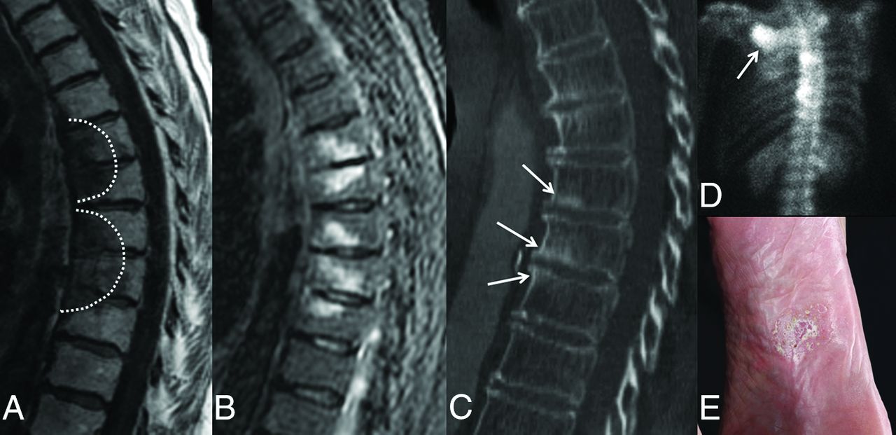
  • Fig 2.
    • Download figure
    • Open in new tab
    • Download powerpoint
    Fig 2.

    MR imaging, CT, and bone scan of the thoracic spine in a 74-year-old woman with back pain. Sagittal T1-weighted (A) and STIR (B) images demonstrate hypo- and hyperintensity, respectively, in a curvilinear or semicircular pattern (dashed line) in contiguous vertebral body segments. Note the absence of abnormal signal within the intervertebral disc spaces. C, Sagittal CT image shows associated sclerosis (arrows) corresponding to levels of abnormal increased signal on MR imaging. D, Bone scan, posteroanterior view, demonstrates focal areas of increased radiotracer uptake within thoracic vertebral bodies and the right sternoclavicular joint (arrow). E, Previously undiagnosed plantar pustulosis was evident on physical examination.

  • Fig 3.
    • Download figure
    • Open in new tab
    • Download powerpoint
    Fig 3.

    MR images in a 32-year-old woman with back pain. Sagittal T1-weighted fat-saturated images of the thoracic spine following administration of gadolinium demonstrate multilevel enhancement (arrows) of the spinous processes (B) and bilateral facet joints (A and C).

  • Fig 4.
    • Download figure
    • Open in new tab
    • Download powerpoint
    Fig 4.

    MR images in a 69-year-old woman with cervical and thoracic back pain. A, Sagittal T2-weighted fat-suppressed image of the cervical spine demonstrates paravertebral soft-tissue thickening (arrows), extending from the C3–C4 interspace to T3. Note the abnormally high T2-weighted signal within the C5–C6 disc (arrowhead). B, Sagittal T1-weighted gadolinium-enhanced fat-suppressed image demonstrates corresponding enhancement of the paravertebral soft tissue (arrows) with disc space enhancement (arrowhead). C, Axial T1-weighted gadolinium-enhanced fat-suppressed image demonstrates enhancement of the thickened paravertebral soft tissues (arrows). D, Axial T2-weighted fat-suppressed image further demonstrates the extent of the paravertebral soft-tissue thickening (arrows).

Tables

  • Figures
  • Vertebral body findings per imaging modality

    Imaging ModalityVertebral Body FindingNo. of Patients
    MRI (n = 16)
        T1-weighted images (n = 16)T1 hypointensity15
        T1 hypointensity → T1 hyperintensity1
        T2-weighted images (n = 15)T2 hyperintensity12
        Mixed T2 hypo- and hyperintensity3
        Gadolinium-enhanced images (n = 9)Enhancement corresponding to areas of T1 hypointensity/T2 hyperintensity9
    CT (n = 14)Sclerosis corresponding to MR signal abnormalities14
    Technetium Tc99m methylene diphosphonate 3-phase bone scan (n = 10)Increased radiotracer uptake10
    Whole-body FDG-PET/CT (n = 2)No abnormal FDG uptake2
PreviousNext
Back to top

In this issue

American Journal of Neuroradiology: 37 (8)
American Journal of Neuroradiology
Vol. 37, Issue 8
1 Aug 2016
  • Table of Contents
  • Index by author
  • Complete Issue (PDF)
Advertisement
Print
Download PDF
Email Article

Thank you for your interest in spreading the word on American Journal of Neuroradiology.

NOTE: We only request your email address so that the person you are recommending the page to knows that you wanted them to see it, and that it is not junk mail. We do not capture any email address.

Enter multiple addresses on separate lines or separate them with commas.
SAPHO Syndrome: Imaging Findings of Vertebral Involvement
(Your Name) has sent you a message from American Journal of Neuroradiology
(Your Name) thought you would like to see the American Journal of Neuroradiology web site.
CAPTCHA
This question is for testing whether or not you are a human visitor and to prevent automated spam submissions.
Cite this article
A.M. McGauvran, A.L. Kotsenas, F.E. Diehn, J.T. Wald, C.M. Carr, J.M. Morris
SAPHO Syndrome: Imaging Findings of Vertebral Involvement
American Journal of Neuroradiology Aug 2016, 37 (8) 1567-1572; DOI: 10.3174/ajnr.A4736

Citation Manager Formats

  • BibTeX
  • Bookends
  • EasyBib
  • EndNote (tagged)
  • EndNote 8 (xml)
  • Medlars
  • Mendeley
  • Papers
  • RefWorks Tagged
  • Ref Manager
  • RIS
  • Zotero
0 Responses
Respond to this article
Share
Bookmark this article
SAPHO Syndrome: Imaging Findings of Vertebral Involvement
A.M. McGauvran, A.L. Kotsenas, F.E. Diehn, J.T. Wald, C.M. Carr, J.M. Morris
American Journal of Neuroradiology Aug 2016, 37 (8) 1567-1572; DOI: 10.3174/ajnr.A4736
del.icio.us logo Twitter logo Facebook logo Mendeley logo
  • Tweet Widget
  • Facebook Like
  • Google Plus One
Purchase

Jump to section

  • Article
    • Abstract
    • ABBREVIATION:
    • Materials and Methods
    • Results
    • Discussion
    • Conclusions
    • Acknowledgments
    • Footnotes
    • References
  • Figures & Data
  • Info & Metrics
  • Responses
  • References
  • PDF

Related Articles

  • No related articles found.
  • PubMed
  • Google Scholar

Cited By...

  • Whole-spine Computed Tomography Findings in SAPHO Syndrome
  • Comment on "SAPHO Syndrome: Imaging Findings of Vertebral Involvement"
  • Reply:
  • Crossref
  • Google Scholar

This article has not yet been cited by articles in journals that are participating in Crossref Cited-by Linking.

More in this TOC Section

  • Management Outcomes For VO Spine Biopsy
  • Characteristics of SIH Type I Culprit Lesions
  • Advanced Imaging of Type 2 Spinal CSF Leaks
Show more Spine Imaging and Spine Image-Guided Interventions

Similar Articles

Advertisement

Indexed Content

  • Current Issue
  • Accepted Manuscripts
  • Article Preview
  • Past Issues
  • Editorials
  • Editor's Choice
  • Fellows' Journal Club
  • Letters to the Editor
  • Video Articles

Cases

  • Case Collection
  • Archive - Case of the Week
  • Archive - Case of the Month
  • Archive - Classic Case

More from AJNR

  • Trainee Corner
  • Imaging Protocols
  • MRI Safety Corner
  • Book Reviews

Multimedia

  • AJNR Podcasts
  • AJNR Scantastics

Resources

  • Turnaround Time
  • Submit a Manuscript
  • Submit a Video Article
  • Submit an eLetter to the Editor/Response
  • Manuscript Submission Guidelines
  • Statistical Tips
  • Fast Publishing of Accepted Manuscripts
  • Graphical Abstract Preparation
  • Imaging Protocol Submission
  • Evidence-Based Medicine Level Guide
  • Publishing Checklists
  • Author Policies
  • Become a Reviewer/Academy of Reviewers
  • News and Updates

About Us

  • About AJNR
  • Editorial Board
  • Editorial Board Alumni
  • Alerts
  • Permissions
  • Not an AJNR Subscriber? Join Now
  • Advertise with Us
  • Librarian Resources
  • Feedback
  • Terms and Conditions
  • AJNR Editorial Board Alumni

American Society of Neuroradiology

  • Not an ASNR Member? Join Now

© 2025 by the American Society of Neuroradiology All rights, including for text and data mining, AI training, and similar technologies, are reserved.
Print ISSN: 0195-6108 Online ISSN: 1936-959X

Powered by HighWire