Advertisement

Main menu

  • Home
  • Content
    • Current Issue
    • Accepted Manuscripts
    • Article Preview
    • Past Issue Archive
    • AJNR Case Collection
    • Case of the Week Archive
    • Classic Case Archive
    • Case of the Month Archive
  • Special Collections
    • Spinal CSF Leak Articles (Jan 2020-June 2024)
    • 2024 AJNR Journal Awards
    • Most Impactful AJNR Articles
  • Multimedia
    • AJNR Podcast
    • AJNR Scantastics
    • Video Articles
  • For Authors
    • Submit a Manuscript
    • Author Policies
    • Fast publishing of Accepted Manuscripts
    • Graphical Abstract Preparation
    • Manuscript Submission Guidelines
    • Imaging Protocol Submission
    • Submit a Case for the Case Collection
  • About Us
    • About AJNR
    • Editorial Board
  • More
    • Become a Reviewer/Academy of Reviewers
    • Subscribers
    • Permissions
    • Alerts
    • Feedback
    • Advertisers
    • ASNR Home
  • Other Publications
    • ajnr

User menu

  • Alerts
  • Log in

Search

  • Advanced search
American Journal of Neuroradiology

American Journal of Neuroradiology

ASHNR American Society of Functional Neuroradiology ASHNR American Society of Pediatric Neuroradiology ASSR
  • Alerts
  • Log in

Advanced Search

  • Home
  • Content
    • Current Issue
    • Accepted Manuscripts
    • Article Preview
    • Past Issue Archive
    • AJNR Case Collection
    • Case of the Week Archive
    • Classic Case Archive
    • Case of the Month Archive
  • Special Collections
    • Spinal CSF Leak Articles (Jan 2020-June 2024)
    • 2024 AJNR Journal Awards
    • Most Impactful AJNR Articles
  • Multimedia
    • AJNR Podcast
    • AJNR Scantastics
    • Video Articles
  • For Authors
    • Submit a Manuscript
    • Author Policies
    • Fast publishing of Accepted Manuscripts
    • Graphical Abstract Preparation
    • Manuscript Submission Guidelines
    • Imaging Protocol Submission
    • Submit a Case for the Case Collection
  • About Us
    • About AJNR
    • Editorial Board
  • More
    • Become a Reviewer/Academy of Reviewers
    • Subscribers
    • Permissions
    • Alerts
    • Feedback
    • Advertisers
    • ASNR Home
  • Follow AJNR on Twitter
  • Visit AJNR on Facebook
  • Follow AJNR on Instagram
  • Join AJNR on LinkedIn
  • RSS Feeds

Welcome to the new AJNR, Updated Hall of Fame, and more. Read the full announcements.


AJNR is seeking candidates for the position of Associate Section Editor, AJNR Case Collection. Read the full announcement.


  • Getting new auth cookie, if you see this message a lot, tell someone!
  • Getting new auth cookie, if you see this message a lot, tell someone!
Research ArticleADULT BRAIN
Open Access

Temporal and Spatial Variances in Arterial Spin-Labeling Are Inversely Related to Large-Artery Blood Velocity

A.D. Robertson, G. Matta, V.S. Basile, S.E. Black, C.K. Macgowan, J.A. Detre and B.J. MacIntosh
American Journal of Neuroradiology August 2017, 38 (8) 1555-1561; DOI: https://doi.org/10.3174/ajnr.A5257
A.D. Robertson
aFrom the Heart and Stroke Foundation Canadian Partnership for Stroke Recovery (A.D.R., G.M., S.E.B., B.J.M.)
bHurvitz Brain Sciences (A.D.R., S.E.B., B.J.M.), Sunnybrook Research Institute, University of Toronto, Toronto, Ontario, Canada
  • Find this author on Google Scholar
  • Find this author on PubMed
  • Search for this author on this site
  • ORCID record for A.D. Robertson
G. Matta
aFrom the Heart and Stroke Foundation Canadian Partnership for Stroke Recovery (A.D.R., G.M., S.E.B., B.J.M.)
  • Find this author on Google Scholar
  • Find this author on PubMed
  • Search for this author on this site
  • ORCID record for G. Matta
V.S. Basile
cDivision of Neurology, Department of Medicine (V.S.B., S.E.B.), University of Toronto, Toronto, Ontario, Canada
dMackenzie Health (V.S.B.), Richmond Hill, Ontario, Canada
  • Find this author on Google Scholar
  • Find this author on PubMed
  • Search for this author on this site
  • ORCID record for V.S. Basile
S.E. Black
aFrom the Heart and Stroke Foundation Canadian Partnership for Stroke Recovery (A.D.R., G.M., S.E.B., B.J.M.)
bHurvitz Brain Sciences (A.D.R., S.E.B., B.J.M.), Sunnybrook Research Institute, University of Toronto, Toronto, Ontario, Canada
cDivision of Neurology, Department of Medicine (V.S.B., S.E.B.), University of Toronto, Toronto, Ontario, Canada
eL.C. Campbell Cognitive Neurology Research Unit (S.E.B.), Sunnybrook Health Sciences Centre, Toronto, Ontario, Canada
  • Find this author on Google Scholar
  • Find this author on PubMed
  • Search for this author on this site
  • ORCID record for S.E. Black
C.K. Macgowan
fHospital for Sick Children (C.K.M.), Toronto, Ontario, Canada
gDepartment of Medical Biophysics (C.K.M., B.J.M.), University of Toronto, Toronto, Ontario, Canada
  • Find this author on Google Scholar
  • Find this author on PubMed
  • Search for this author on this site
  • ORCID record for C.K. Macgowan
J.A. Detre
hDepartment of Neurology (J.A.D.), University of Pennsylvania, Philadelphia, Pennsylvania.
  • Find this author on Google Scholar
  • Find this author on PubMed
  • Search for this author on this site
  • ORCID record for J.A. Detre
B.J. MacIntosh
aFrom the Heart and Stroke Foundation Canadian Partnership for Stroke Recovery (A.D.R., G.M., S.E.B., B.J.M.)
bHurvitz Brain Sciences (A.D.R., S.E.B., B.J.M.), Sunnybrook Research Institute, University of Toronto, Toronto, Ontario, Canada
gDepartment of Medical Biophysics (C.K.M., B.J.M.), University of Toronto, Toronto, Ontario, Canada
  • Find this author on Google Scholar
  • Find this author on PubMed
  • Search for this author on this site
  • ORCID record for B.J. MacIntosh
  • Article
  • Figures & Data
  • Info & Metrics
  • Responses
  • References
  • PDF
Loading

Article Figures & Data

Figures

  • Tables
  • Fig 1.
    • Download figure
    • Open in new tab
    • Download powerpoint
    Fig 1.

    Vascular characteristics of the bilateral (left and right) internal carotid (LICA and RICA) and vertebral arteries (LVA and RVA) for each group. Panel 3 shows the relative contribution to total CBF by each vessel.
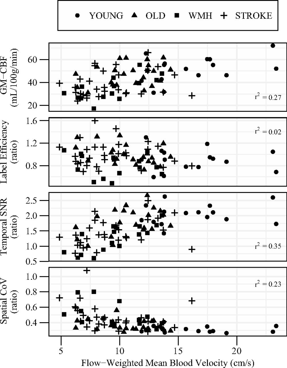
  • Fig 2.
    • Download figure
    • Open in new tab
    • Download powerpoint
    Fig 2.

    The association of mean blood velocity with GM-CBF (panel 1), ASL-to-PC ratio for whole-brain CBF (panel 2), ASL temporal SNR (panel 3), and CBF spatial CoV (panel 4). Velocity is shown as a weighted mean of all 4 extracranial vessels relative to their contribution to total CBF. GM-CBF is uncorrected for labeling efficiency. Age, sex, and group-adjusted linear models are reported in Table 3.
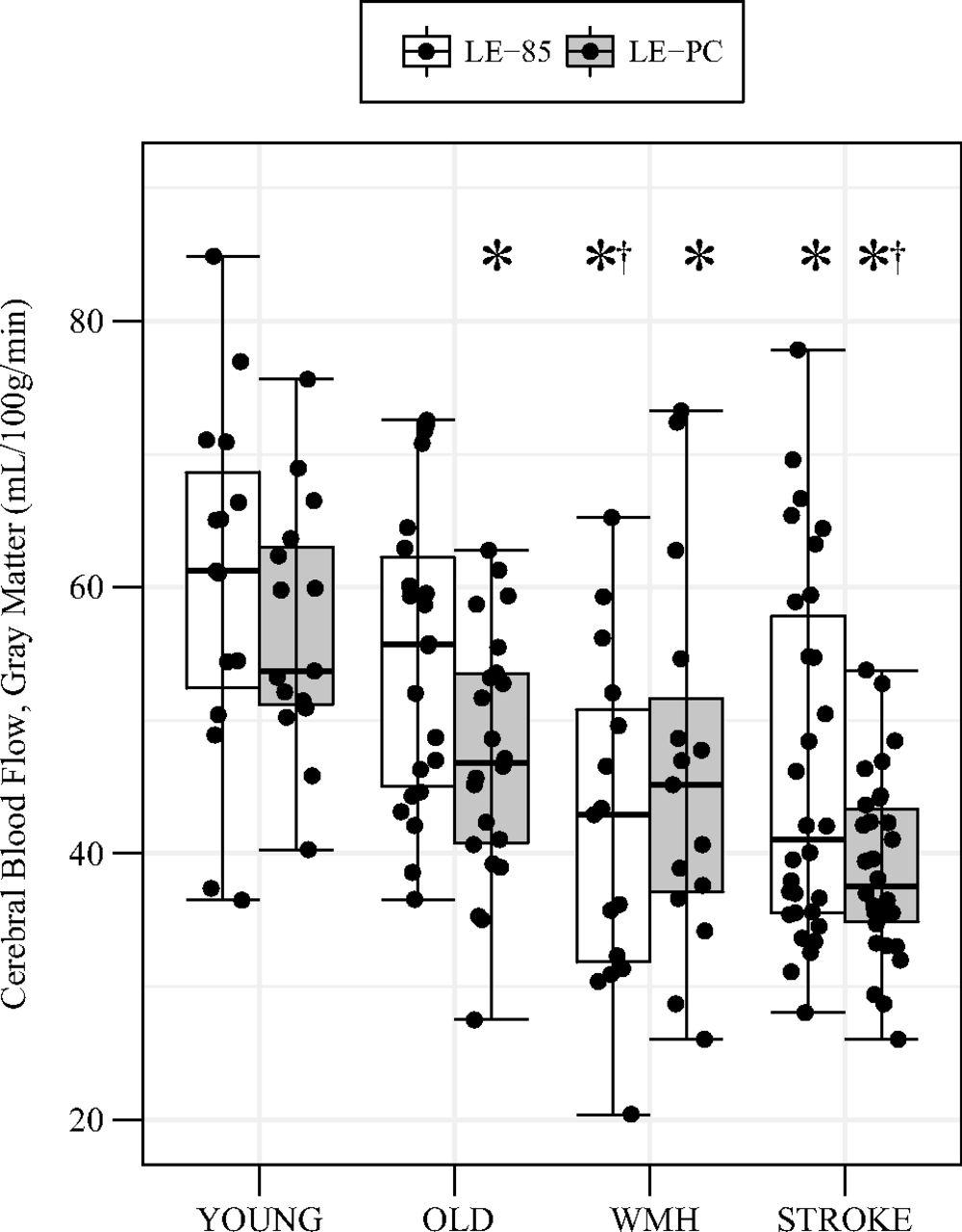
  • Fig 3.
    • Download figure
    • Open in new tab
    • Download powerpoint
    Fig 3.

    GM-CBF between groups calculated with 2 labeling efficiency estimates. White bars incorporate a constant labeling efficiency (ie, 0.85), and gray bars incorporate individual labeling efficiency based on the ASL-to-PC ratio for whole-brain CBF. Post hoc group comparisons are indicated at PBonferroni < .05 for differences from the young group (asterisk) and differences from old group (dagger) within the same calibration technique.

Tables

  • Figures
    • View popup
    Table 1:

    Participant characteristicsa

    YoungOldWMHStrokeP Value
    No.15221530
    Age (range) (yr)25.7 ± 4.6 (20–36)69.1 ± 7.0b (55–81)72.0 ± 8.2b (51–83)67.9 ± 10.4b (51–88)<.001
    Sex, female (%)7 (47)17 (77)8 (53)9 (30).009
    Hemodynamics
        ICA mean velocity (cm/s)18.2 ± 4.511.9 ± 2.9b10.3 ± 3.3b9.7 ± 3.1b<.001
        ICA area (cm2)0.21 ± 0.040.24 ± 0.060.26 ± 0.06026 ± 0.09.102
        VA mean velocity (cm/s)10.0 ± 2.56.7 ± 1.6b6.5 ± 1.7b5.6 ± 2.1b<.001
        VA area (cm2)0.13 ± 0.030.13 ± 0.030.14 ± 0.040.12 ± 0.03.190
        PC-CBF (mL/100 g/min)49.0 ± 8.541.4 ± 8.140.6 ± 12.933.8 ± 6.3b,c<.001
        ASL-CBF (mL/100 g/min)44.1 ± 10.240.8 ± 8.831.4 ± 9.6b,c34.3 ± 10.5b<.001
    • Note:—VA indicates vertebral artery.

    • ↵a Data are mean ± SD or count (proportion). Area and velocity are reported as the mean of bilateral vessels. ASL-CBF is uncorrected for labeling efficiency.

    • ↵b Group differences compared with the young group at PBonferroni < .05.

    • ↵c Group differences compared with the old group at PBonferroni < .05.

    • View popup
    Table 2:

    Comparison of MRI methods for whole-brain CBF estimationa

    No.ASL:PC (ratio)ASL-PC Difference (mL/100 g/min)
    Young150.91 ± 0.20−4.9 ± 9.8
    Old221.00 ± 0.18b−0.6 ± 7.0b
    WMH150.80 ± 0.20−9.2 ± 9.4
    Stroke301.01 ± 0.23b0.5 ± 7.9b
    All820.95 ± 0.22−2.6 ± 9.0
    • ↵a Data are mean ± SD.

    • ↵b Group differences compared with WMH at PBonferroni < .05.

    • View popup
    Table 3:

    Linear regression parameters for the association of extracranial mean blood velocity and cross-sectional area with ASL characteristicsa

    ModelIndependent Variablesβ95% CIT-StatisticP Value
    GM-CBFArea (cm2)46.5(8.2–84.7)2.03.046
        Adjusted R2 = 0.43Velocity (cm/s)1.5(0.8–2.3)3.42.001
    ASL-CBF:PC-CBFArea (cm2)−0.79(−1.58–0.01)−1.65.103
        Adjusted R2 = 0.21Velocity (cm/s)−0.02(−0.04–0.01)−2.78.007
    GM-temporal SNRArea (cm2)1.78(0.24–3.32)1.93.058
        Adjusted R2 = 0.47Velocity (cm/s)0.07(0.04–0.10)3.99<.001
    GM-spatial CoVArea (cm2)−0.11(−0.61–0.39)−0.36.720
        Adjusted R2 = 0.31Velocity (cm/s)−0.01(−0.02–0.00)−2.01.048
    • ↵a All models were adjusted for age, sex, and group. GM-CBF is uncorrected for labeling efficiency.

PreviousNext
Back to top

In this issue

American Journal of Neuroradiology: 38 (8)
American Journal of Neuroradiology
Vol. 38, Issue 8
1 Aug 2017
  • Table of Contents
  • Index by author
  • Complete Issue (PDF)
Advertisement
Print
Download PDF
Email Article

Thank you for your interest in spreading the word on American Journal of Neuroradiology.

NOTE: We only request your email address so that the person you are recommending the page to knows that you wanted them to see it, and that it is not junk mail. We do not capture any email address.

Enter multiple addresses on separate lines or separate them with commas.
Temporal and Spatial Variances in Arterial Spin-Labeling Are Inversely Related to Large-Artery Blood Velocity
(Your Name) has sent you a message from American Journal of Neuroradiology
(Your Name) thought you would like to see the American Journal of Neuroradiology web site.
CAPTCHA
This question is for testing whether or not you are a human visitor and to prevent automated spam submissions.
Cite this article
A.D. Robertson, G. Matta, V.S. Basile, S.E. Black, C.K. Macgowan, J.A. Detre, B.J. MacIntosh
Temporal and Spatial Variances in Arterial Spin-Labeling Are Inversely Related to Large-Artery Blood Velocity
American Journal of Neuroradiology Aug 2017, 38 (8) 1555-1561; DOI: 10.3174/ajnr.A5257

Citation Manager Formats

  • BibTeX
  • Bookends
  • EasyBib
  • EndNote (tagged)
  • EndNote 8 (xml)
  • Medlars
  • Mendeley
  • Papers
  • RefWorks Tagged
  • Ref Manager
  • RIS
  • Zotero
0 Responses
Respond to this article
Share
Bookmark this article
Temporal and Spatial Variances in Arterial Spin-Labeling Are Inversely Related to Large-Artery Blood Velocity
A.D. Robertson, G. Matta, V.S. Basile, S.E. Black, C.K. Macgowan, J.A. Detre, B.J. MacIntosh
American Journal of Neuroradiology Aug 2017, 38 (8) 1555-1561; DOI: 10.3174/ajnr.A5257
del.icio.us logo Twitter logo Facebook logo Mendeley logo
  • Tweet Widget
  • Facebook Like
  • Google Plus One
Purchase

Jump to section

  • Article
    • Abstract
    • ABBREVIATIONS:
    • Materials and Methods
    • Results
    • Discussion
    • Conclusions
    • Acknowledgments
    • Footnotes
    • REFERENCES
  • Figures & Data
  • Info & Metrics
  • Responses
  • References
  • PDF

Related Articles

  • No related articles found.
  • PubMed
  • Google Scholar

Cited By...

  • Mid-life association between cardiovascular risk factors and cerebral blood flow in a multi-ethnic population
  • Effects of Acquisition Parameter Modifications and Field Strength on the Reproducibility of Brain Perfusion Measurements Using Arterial Spin-Labeling
  • The effects of age on resting-state BOLD signal variability is explained by cardiovascular and cerebrovascular factors
  • ExploreASL: an image processing pipeline for multi-center ASL perfusion MRI studies
  • Crossref (18)
  • Google Scholar

This article has been cited by the following articles in journals that are participating in Crossref Cited-by Linking.

  • ExploreASL: An image processing pipeline for multi-center ASL perfusion MRI studies
    Henk J.M.M. Mutsaerts, Jan Petr, Paul Groot, Pieter Vandemaele, Silvia Ingala, Andrew D. Robertson, Lena Václavů, Inge Groote, Hugo Kuijf, Fernando Zelaya, Owen O’Daly, Saima Hilal, Alle Meije Wink, Ilse Kant, Matthan W.A. Caan, Catherine Morgan, Jeroen de Bresser, Elisabeth Lysvik, Anouk Schrantee, Astrid Bjørnebekk, Patricia Clement, Zahra Shirzadi, Joost P.A. Kuijer, Viktor Wottschel, Udunna C. Anazodo, Dasja Pajkrt, Edo Richard, Reinoud P.H. Bokkers, Liesbeth Reneman, Mario Masellis, Matthias Günther, Bradley J. MacIntosh, Eric Achten, Michael A. Chappell, Matthias J.P. van Osch, Xavier Golay, David L. Thomas, Enrico De Vita, Atle Bjørnerud, Aart Nederveen, Jeroen Hendrikse, Iris Asllani, Frederik Barkhof
    NeuroImage 2020 219
  • The effects of age on resting‐state BOLD signal variability is explained by cardiovascular and cerebrovascular factors
    Kamen A. Tsvetanov, Richard N. A. Henson, P. Simon Jones, Henk Mutsaerts, Delia Fuhrmann, Lorraine K. Tyler, James B. Rowe
    Psychophysiology 2021 58 7
  • Exercise Training Increases Parietal Lobe Cerebral Blood Flow in Chronic Stroke: An Observational Study
    Andrew D. Robertson, Susan Marzolini, Laura E. Middleton, Vincenzo S. Basile, Paul I. Oh, Bradley J. MacIntosh
    Frontiers in Aging Neuroscience 2017 9
  • The Meta VCI Map consortium for meta‐analyses on strategic lesion locations for vascular cognitive impairment using lesion‐symptom mapping: Design and multicenter pilot study
    Nick A. Weaver, Lei Zhao, J. Matthijs Biesbroek, Hugo J. Kuijf, Hugo P. Aben, Hee‐Joon Bae, Miguel Á.A. Caballero, Francesca M. Chappell, Christopher P.L.H. Chen, Martin Dichgans, Marco Duering, Marios K. Georgakis, Ruben S. van der Giessen, Bibek Gyanwali, Olivia K.L. Hamilton, Saima Hilal, Elise M. vom Hofe, Paul L.M. de Kort, Peter J. Koudstaal, Bonnie Y.K. Lam, Jae‐Sung Lim, Stephen D.J. Makin, Vincent C.T. Mok, Lin Shi, Maria C. Valdés Hernández, Narayanaswamy Venketasubramanian, Joanna M. Wardlaw, Frank A. Wollenweber, Adrian Wong, Xu Xin, C. DeCarli, E.A. Fletcher, P. Maillard, J. Barnes, C.H. Sudre, J.M. Schott, M.A. Ikram, J.M. Papma, R.M.E. Steketee, M.W. Vernooij, R. Bordet, R. Lopes, C.‐W. Huang, R. Frayne, C.R. McCreary, E.E. Smith, W. Backes, S. Köhler, R.J. van Oostenbrugge, J. Staals, F. Verhey, C.Y. Cheng, R.N. Kalaria, D. Werring, J.L. Hsu, K.‐L. Huang, J. van der Grond, J.W. Jukema, R.C. van der Mast, T.C.W. Nijboer, K.‐H. Yu, R. Schmidt, L. Pirpamer, B.J. MacIntosh, A.D. Robertson, F.‐E. de Leeuw, A.M. Tuladhar, N. Chaturvedi, T. Tillin, H. Brodaty, P. Sachdev, F. Barkhof, W.M. van der Flier, L.J. Kappelle, Geert Jan Biessels
    Alzheimer's & Dementia: Diagnosis, Assessment & Disease Monitoring 2019 11 1
  • Spatial coefficient of variation in pseudo-continuous arterial spin labeling cerebral blood flow images as a hemodynamic measure for cerebrovascular steno-occlusive disease: A comparative 15O positron emission tomography study
    Masanobu Ibaraki, Kazuhiro Nakamura, Hideto Toyoshima, Kazuhiro Takahashi, Keisuke Matsubara, Atsushi Umetsu, Josef Pfeuffer, Hideto Kuribayashi, Toshibumi Kinoshita
    Journal of Cerebral Blood Flow & Metabolism 2019 39 1
  • Classifying cognitive impairment based on the spatial heterogeneity of cerebral blood flow images
    Zahra Shirzadi, Bojana Stefanovic, Henri J.M.M. Mutsaerts, Mario Masellis, Bradley J. MacIntosh
    Journal of Magnetic Resonance Imaging 2019 50 3
  • Spatial coefficient of variation of arterial spin labeling MRI as a cerebrovascular correlate of carotid occlusive disease
    Henri J. M. M. Mutsaerts, Jan Petr, Reinoud P. H. Bokkers, Ronald M. Lazar, Randolph S. Marshall, Iris Asllani, Ona Wu
    PLOS ONE 2020 15 2
  • Cerebral blood flow predicts multiple demand network activity and fluid intelligence across the adult lifespan
    Shuyi Wu, Lorraine K. Tyler, Richard N.A. Henson, James B. Rowe, Cam-CAN, Kamen A. Tsvetanov
    Neurobiology of Aging 2023 121
  • Effects of Acquisition Parameter Modifications and Field Strength on the Reproducibility of Brain Perfusion Measurements Using Arterial Spin-Labeling
    K.P.A. Baas, J. Petr, J.P.A. Kuijer, A.J. Nederveen, H.J.M.M. Mutsaerts, K.C.C. van de Ven
    American Journal of Neuroradiology 2021 42 1
  • Specificities of arterial spin labeling (ASL) abnormalities in acute seizure
    Mathieu Schertz, Joseph Benzakoun, Nadya Pyatigorskaya, Samia Belkacem, Melika Sahli-Amor, Vincent Navarro, Clément Cholet, Delphine Leclercq, Didier Dormont, Bruno Law-ye
    Journal of Neuroradiology 2020 47 1

More in this TOC Section

  • Diagnostic Neuroradiology of Monoclonal Antibodies
  • ML for Glioma Molecular Subtype Prediction
  • Segmentation of Brain Metastases with BLAST
Show more Adult Brain

Similar Articles

Advertisement

Indexed Content

  • Current Issue
  • Accepted Manuscripts
  • Article Preview
  • Past Issues
  • Editorials
  • Editors Choice
  • Fellow Journal Club
  • Letters to the Editor

Cases

  • Case Collection
  • Archive - Case of the Week
  • Archive - Case of the Month
  • Archive - Classic Case

Special Collections

  • Special Collections

Resources

  • News and Updates
  • Turn around Times
  • Submit a Manuscript
  • Author Policies
  • Manuscript Submission Guidelines
  • Evidence-Based Medicine Level Guide
  • Publishing Checklists
  • Graphical Abstract Preparation
  • Imaging Protocol Submission
  • Submit a Case
  • Become a Reviewer/Academy of Reviewers
  • Get Peer Review Credit from Publons

Multimedia

  • AJNR Podcast
  • AJNR SCANtastic
  • Video Articles

About Us

  • About AJNR
  • Editorial Board
  • Not an AJNR Subscriber? Join Now
  • Alerts
  • Feedback
  • Advertise with us
  • Librarian Resources
  • Permissions
  • Terms and Conditions

American Society of Neuroradiology

  • Not an ASNR Member? Join Now

© 2025 by the American Society of Neuroradiology All rights, including for text and data mining, AI training, and similar technologies, are reserved.
Print ISSN: 0195-6108 Online ISSN: 1936-959X

Powered by HighWire