
Article Figures & Data
Figures
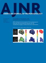
- Fig 1.
A, Measured proton-density values as a function of R1 relaxation rate values of a slice of a brain of a patient with glioma grade IV before administration of GBCA (at 3T). The solid line traverses the average position of gray matter and white matter, indicating the predetermined, linear relationship between R1 and PD for the native brain parenchyma. The dotted line indicates a threshold of 0.2 seconds−1 from the solid line. B, PD and R1 of the same slice after GBCA administration in which the present glioma exhibits enhancement. Some R1 values are substantially increased above the dotted threshold line. The estimated R1 enhancement corresponds to the measured R1 value minus the corresponding R1 value on the predetermined solid line.
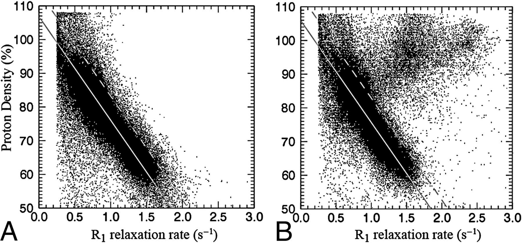
- Fig 2.
Images of the same slice as in Fig 1: synthetic T1-weighted imaging using native data (A), synthetic T1-weighted imaging using post-GBCA data (B), the native R1 map (C), the post-GBCA R1 map (D), the difference map of the coregistered native map (E), and the post-GBCA R1 synthetic-difference map based on the post-GBCA acquisition only (F).
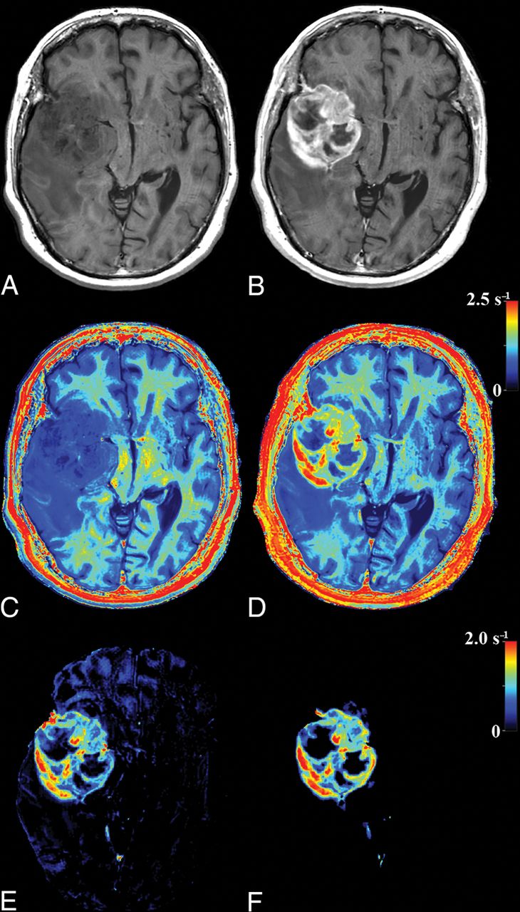
- Fig 3.
2D histogram of the R1 enhancement found using the subtraction of native and post-GBCA R1 maps as a function of the synthetic R1 enhancement, based on the post-GBCA acquisition only, of all included patients. The black and white intensity in the plot is proportional to the number of times an x, y coordinate occurred. The diagonal line indicates equivalence.
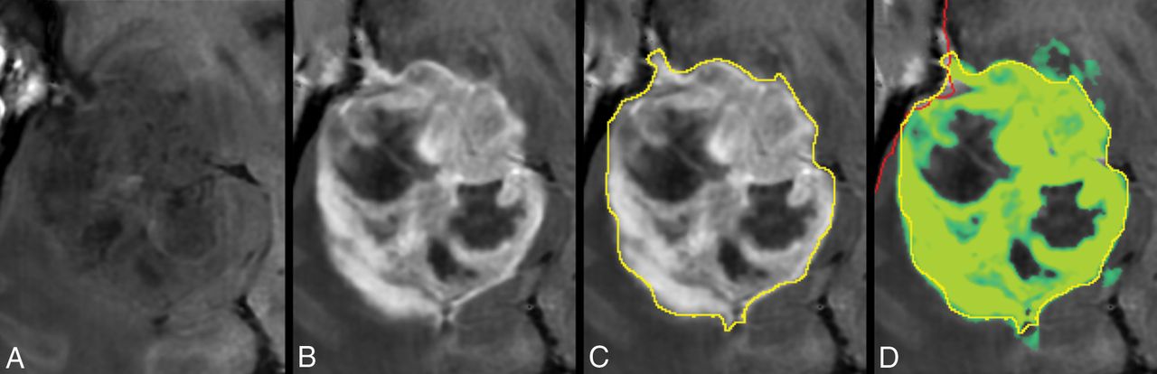
- Fig 4.
Zoomed part around the tumor displayed in Fig 2. Synthetic T1-weighted imaging using native data (A), synthetic T1-weighted imaging using post-GBCA data (B), the ROI line as drawn by a neuroradiologist to encapsulate the border of the enhancing tumor (C). D, Synthetic R1 enhancement map shown as a green overlay on the synthetic T1-weighted image in which full color corresponds to dR1 = 1.0 seconds−1. The minimum enhancement was set at dR1 = 0.2 seconds−1. Some low-intensity enhancement is visible outside the yellow ROI. The red line indicates the edge of the intracranial volume.
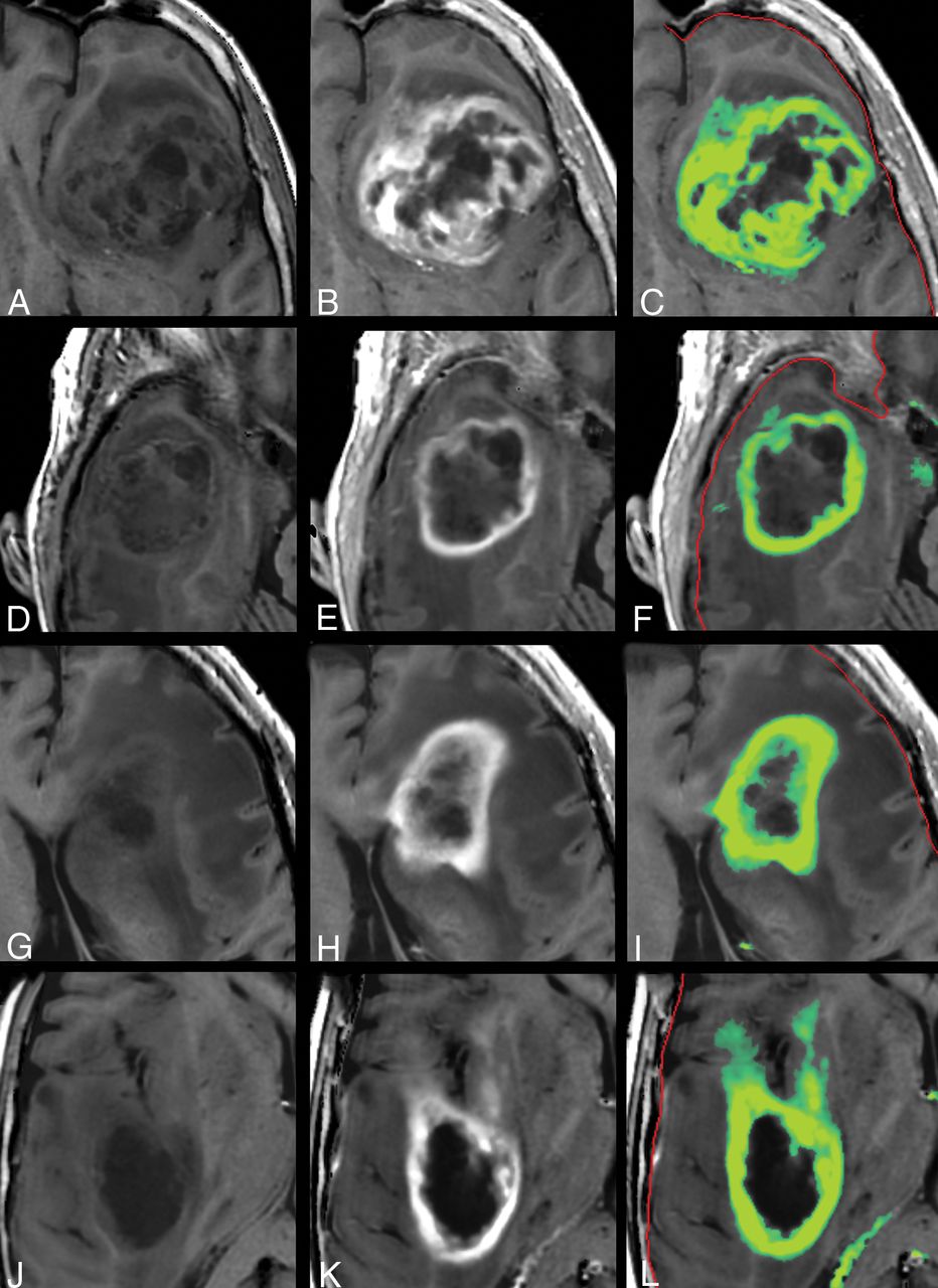
- Fig 5.
Other examples of the synthetic R1 enhancement map and low-intensity enhancement at the edges of high-intensity enhancement in gliomas. Left: native synthetic T1-weighted image. Center: post-GBCA synthetic T1-weighted image. Right: synthetic R1 enhancement map as a green overlay. The color indicates a range of dR1 of 0.2–1.0 seconds−1. The red line indicates the edge of the intracranial volume.


Tables
Patient Sex Age (yr) WHO 2007 Male 68 Glioblastoma IV Female 57 Glioblastoma IV Male 63 Anaplastic oligodendroglioma III Female 58 Glioblastoma Male 69 Glioblastoma Male 71 Glioblastoma Male 65 Gliosarcoma Female 65 Anaplastic oligodendroglioma Female 45 Glioblastoma Male 65 Glioblastoma Male 79 Glioblastoma Male 45 Glioblastoma Male 72 Glioblastoma Male 74 Glioblastoma Note:—WHO indicates World Health Organization.
- Table 2:
Observed R1 enhancement of the pixels of the ROI line drawn by a neuroradiologist to encapsulate the enhancing tumor, as a percentage of all values above dR1 = 0.2 seconds−1, the mean dR1, and the mean dR1 of all values of >0.2 seconds−1a
dR1 >0.2 s−1 (%) Mean dR1 (s−1) Mean dR1 (>0.2 s−1 only) (s−1) Synthetic Subtraction Synthetic Subtraction Synthetic Subtraction On ROI line 35.8 ± 14.3 50.3 ± 10.2 0.19 ± 0.09 0.27 ± 0.08 0.48 ± 0.12 0.46 ± 0.11 + 1 mm 8.0 ± 5.8 17.4 ± 9.0 0.03 ± 0.02 0.08 ± 0.05 0.35 ± 0.08 0.37 ± 0.12 + 2 mm 2.3 ± 1.5 8.6 ± 4.9 0.01 ± 0.01 0.04 ± 0.04 0.32 ± 0.15 0.37 ± 0.17 a Results are listed for the R1 difference generated by synthesizing the R1 difference map and subtraction of the pre- and post-GBCA R1 maps. Two more ROI lines were created at 1- and 2-mm outward to analyze the results if a larger margin around the enhancing tumor had been drawn.


