
Article Figures & Data
Figures
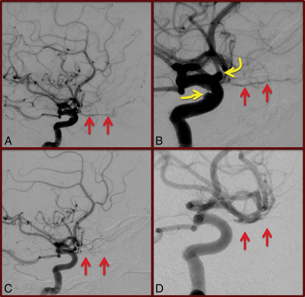
- Fig 1.
Global and magnified lateral views (A and B) of pre-PED right internal carotid artery angiography demonstrating gross patency of the right ophthalmic artery (straight arrows). The target paraophthalmic and cavernous aneurysms (curved arrows, B) are also visualized. Conventional angiography performed 6 months following PED deployment (C and D) demonstrates occlusion of the target aneurysms; however, the ophthalmic artery is also occluded (straight arrows). The patient was asymptomatic, and the ophthalmic artery was found to fill retrogradely on external carotid artery injections (not shown).
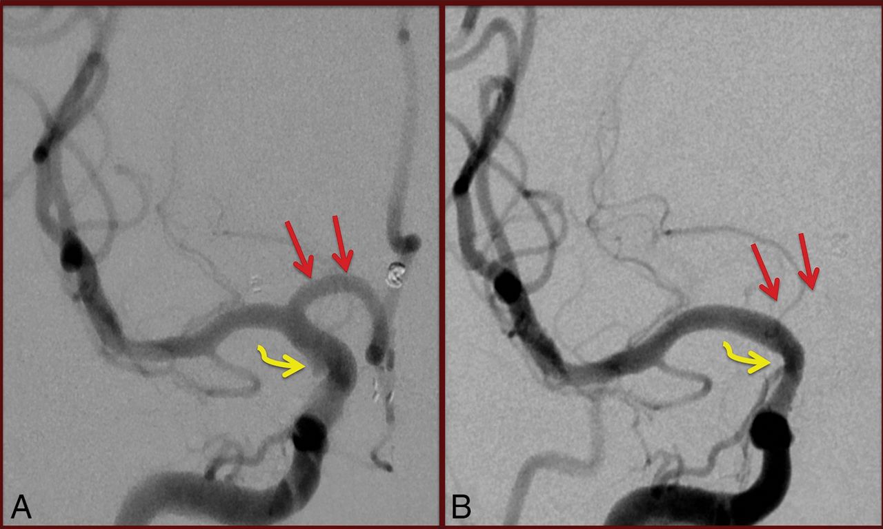
- Fig 2.
Anteroposterior magnified view (A) of pre-PED right internal carotid artery angiography demonstrating gross patency of the A1 segment of the right anterior cerebral artery (straight arrows). The target, irregular right anterior choroidal artery aneurysm is only partially visualized (curved arrow). On conventional angiography performed 6 months following PED deployment (B), the A1 segment of the right anterior cerebral artery no longer fills on right internal carotid artery injection. The target aneurysm is closed (curved arrow). The patient was asymptomatic, and the bilateral anterior cerebral arteries are noted to be filling via the left A1 segment on left internal carotid artery injections (not shown).
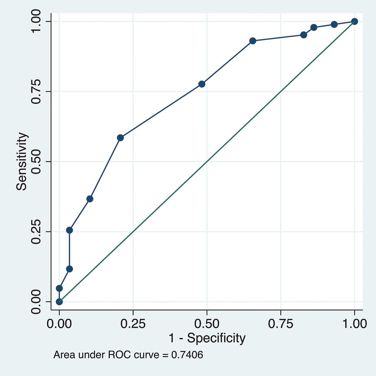
- Fig 3.
Receiver operating characteristic (ROC) curve demonstrating the diagnostic accuracy for parent vessel diameter to predict branch vessel occlusion in the entire cohort. The reference line is depicted as the solid line along the middle of the plot.


Tables
Demographics No. of patients 137 No. of PED treatments 148 Average patient age (yr) 54.7 % Male per treatment 6.6% No. of aneurysms treated 178 Average aneurysm size (mm) 5.2 % Aneurysms treated acutely/subacutely after rupture 3.3% % Aneurysms with branch vessel incorporation 35% Mean No. of PEDs per treatment 1.07 % Aneurysms adjunctively coiled 9.2% % Treatments using J-tip microcatheter 17% % Treatments using balloon angioplasty 6.1% % Treatments requiring abciximab 4.8% Mean length of DSA follow-up (mo) 18.4 Type No. Patent Stenotic Occluded Ophthalmic artery 125 72% 22% 5.8% PcomA 39 56.7% 10% 33.3% Anterior choroidal artery 31 96.8% 0% 3.2% A1 arterial segment 11 28% 36% 36% PICA 3 33.4% 66.6% 0% AcomA 3 33.4% 0% 66.6% Othera 5 80% 0% 20% Note:—PcomA indicates posterior communicating artery; AcomA, anterior communicating artery.
↵a Detailed in Results section.
Patent Branch Vessel Occluded Branch Vessel Univariate P Value Multivariate P Value OR (95% CI) Mean patient age (yr) 54.4 ± 12.2 59.3 ± 10.2 .04 .179 1.03 (0.99–1.07) Male 26 (13.8%) 4 (13.8%) .996 Acutely ruptured target lesion 7/188 (3.7%) 1/29 (3%) .942 Mean max aneurysm sac size 5.6 ± 3.8 4.2 ± 2.3 .067 .117 0.87 (0.73–1.04) Mean aneurysm neck size 3.4 ± 1.7 3.0 ± 1.2 .139 Mean procedural time (min) 206 ± 66 214 ± 60 .513 Mean fluoroscopy time (min) 64 ± 36 62 ± 27 .764 >1 PED deployed 17/188 (9.0%) 1/29 (3.4%) .309 % PED classic 107/125 (86%) 18/29 (62.1%) .601 Balloon angioplasty 13/188 (7%) 1/29 (7%) .479 Mean PED diameter (mm) 4.0 ± 0.5 3.5 ± 0.5 <0.001 .001 0.16 (0.06–0.45) Mean Δmax size of parent vessel and PED 0.22 ± 0.19 0.26 ± 0.26 .405 Mean Δmin size of parent vessel and PED 0.77 ± 0.41 0.67 ± 0.40 .230 Branch vessel incorporation into aneurysm sac 52/188 (28%) 9/29 (31%) .707 Mean periprocedural P2Y12 127 ± 68 122 ± 80 .379 Intraprocedural administration of abciximab 17/188 (9.0%) 3/29 (10.3%) .821 Presence of endothelial hyperplasia 43/185 (23%) 10/29 (34%) .192 Mean length of DSA follow-up (mo) 18.3 ± 9.9 20.1 ± 8.8 .351 A1 ACA 7/11 (63.6%) 4/11 (36.3%) .021 .813 1.29 (0.16–10.56) Anterior choroidal artery 30/31 (96.8%) 1/31 (3.2%) .073 .092 0.10 (0.01–1.46) PcomA 26/39 (66.6%) 13/39 (33.3%) <.001 .205 3.26 (0.52–20.28) Ophthalmic artery 118/125 (94%) 8/125 (6%) <.001 0.449 0.49 (0.08–3.07) Note:—ACA indicates anterior cerebral artery; max, maximum; min, minimum; PcomA, posterior communicating artery.
↵a Percentages reflect analysis per branch vessel.
Patent Branch Vessel Occluded Branch Vessel Univariate P Value Multivariate P Value OR (95% CI) Mean patient age (yr) 53.8 60.1 .119 Male 15 (12.7%) 0 (0%) .283 Mean max aneurysm sac 5.9 ± 3.8 5.8 ± 3.3 .977 Mean aneurysm neck size 3.6 ± 1.6 3.6 ± 0.5 .998 Acutely ruptured target lesion 4/118 (3.7%) 0/7 (0%) .597 Prior coiling 29/118 (24.6%) 1/7 (14.3%) .438 Mean procedural time (min) 200 ± 63 212 ± 60 .628 Mean fluoroscopy time (min) 63 ± 37 53 ± 28 .429 >1 PED deployed 10/118 (8.5%) 0/7 (0%) .391 % PED classic 70/75 (93.3%) 5/75 (6.7%) .859 Balloon angioplasty 9/118 (7.6%) 0/7 (0%) .418 Mean PED diameter (mm) 4.1 ± 0.5 3.4 ± 0.6 <0.001 <.001 0.10 (0.04–0.21) Mean Δmax size of parent vessel and PED 0.23 ± 0.18 0.24 ± 0.21 .887 Mean Δmin size of parent vessel and PED 0.79 ± 0.41 0.79 ± 0.48 .979 Branch vessel incorporation into aneurysm sac 31/118 (26.3%) 2/7 (29%) .937 Mean periprocedural P2Y12 136 ± 68 150 ± 63 .597 Intraprocedural administration of abciximab 5/118 (4.2%) 2/7 (29%) .013 .194 4.09 (0.49–34.19) Presence of endothelial hyperplasia 26/115 (22.6%) 3/7 (43%) .337 Mean length of DSA follow-up (mo) 17.9 ± 9.9 21.4 ± 13.8 .682 ↵a Percentages reflect analysis per branch vessel.
Patent Branch Vessel Occluded/Stenotic Branch Vessel Univariate P Value Multivariate P Value OR (95% CI) Mean patient age (yr) 54.6 ± 12.6 56.0 ± 10.9 .426 Male 19/148 (12.8%) 11/69 (15.9%) .537 Mean max aneurysm sac size 6.0 ± 4.0 5.6 ± 3.7 .096 .415 0.94 (0.82–1.09) Mean aneurysm neck size 3.6 ± 1.7 3.4 ± 1.2 .054 .534 0.91 (0.67–1.22) Acutely ruptured target lesion 4/148 (2.7%) 4/69 (5.8%) .260 Mean procedural time (min) 205 ± 65 210 ± 66 .658 Mean fluoroscopy time (min) 65 ± 37 60 ± 30 .365 >1 PED deployed 14/148 (9.5%) 4/69 (5.8%) .362 % PED classic 60/148 (40.5%) 32/69 (46.3%) .418 Balloon angioplasty 12/148 (8.1%) 2/69 (2.9%) .146 Mean PED diameter (mm) 4.0 ± 0.5 3.8 ± 0.5 .013 .029 0.46 (0.23–0.92) Mean Δmax size of parent vessel and PED 0.21 ± 0.17 0.27 ± 0.25 .057 .142 3.60 (0.65–19.89) Mean Δmin size of parent vessel and PED 0.78 ± 0.4 0.70 ± 0.42 .226 Branch vessel incorporation into aneurysm sac 42/148 (28.3%) 16/69 (27.5%) .898 Mean periprocedural P2Y12 127 ± 68 125 ± 74 .855 Intraprocedural administration of abciximab 9/148 (6.1%) 11/69 (15.9%) .019 .030 4.26 (1.15–15.78) Presence of endothelial hyperplasia 31/146 (21.2%) 22/68 (32.4%) .079 .504 1.31 (0.59–2.94) Mean length of DSA follow-up (mo) 18.8 ± 19.4 18.0 ± 8.2 .584 A1 ACA 3/11 (27.2%) 8/11 (72.7%) 0.003 .114 3.12 (0.76–12.81) Anterior choroidal artery 30/31 (96.8%) 1/31 (3.2%) <.001 .003 0.04 (0.004–0.32) PcomA 22/39 (56.4%) 17/39 (43.6%) .081 .248 1.57 (0.73–3.40) Ophthalmic artery 90/125 (71.2%) 36/125 (28.8%) .23 Note:—PcomA indicates posterior communicating artery; AcomA, anterior communicationg.
↵a Percentages reflect analysis per branch vessel.


Jump to section
Related Articles
Cited By...
- Correlation of Flow Diverter Malapposition at the Aneurysm Neck with Incomplete Aneurysm Occlusion in Patients with Small Intracranial Aneurysms: A Single-Center Experience
- The utility of platelet inhibition testing in patients undergoing Pipeline embolization of intracranial aneurysms
- Pipeline embolization device diameter is an important factor determining the efficacy of flow diversion treatment of small intracranial saccular aneurysms