
Article Figures & Data
Figures
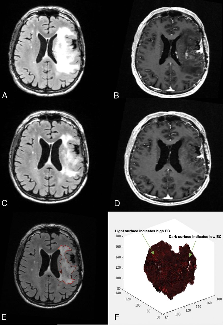
- Fig 1.
A 58-year-old man's MR imaging post-subtotal resection with low edge contrast. The patient had a poor survival estimation, with a PFS of 1.8 months and OS of 7.3 months. A, FLAIR prebevacizumab. B, T1 postcontrast prebevacizumab. C, FLAIR postbevacizumab. D, T1 postcontrast postbevacizumab. E, Overlay of the EC contour over the postbevacizumab FLAIR image. F, 3D presentation of the surface of the hyperintense region. Darker areas on the surface indicate lower EC/more indistinct border, whereas the lighter areas toward red show higher EC/more distinct border.
- Fig 2.
A 60-year-old woman's MR imaging post-subtotal resection with high edge contrast. The patient had a high survival estimation with PFS of 11.5 months and OS of 13.6 months. A, FLAIR prebevacizumab. B, T1 postcontrast prebevacizumab. C, FLAIR postbevacizumab. D, T1 postcontrast postbevacizumab. E, Overlay of the EC contour over the postbevacizumab FLAIR image. F, 3D presentation of the surface of the hyperintense region. Darker areas on the surface indicate lower EC and more indistinct border, whereas the lighter areas toward red show higher EC and more distinct border.
- Fig 3.
Stratification of patients based on MSRS analysis for splits in post-EC100%. Kaplan-Meier curves for high and low change groups for PFS (A) and OS (B).


Tables
Parameter Prebevacizumab Mean (SD) × 103 Postbevacizumab Mean (SD) × 103 P Value (T Value) EC100% 2.52 (0.57) 2.43 (0.57) .256 (1.15) EC75% 1.91 (0.53) 1.77 (0.47) .281 (1.08) EC50% 1.51 (0.48) 1.38 (0.40) .277 (1.11) EC25% 1.06 (0.38) 0.98 (0.30) .184 (1.35) FLAIRVOLa 121.88 (70.68) 77.317 (38.55) .002 (4.28) CEVOLa 24.90 (15.26) 9.16 (9.17) <.001 (6.34) ↵a P < .05.
- Table 2:
Summary of the multivariate CPH models with age and surgical extent (subtotal, gross total resection) as covariates
Edge Time PFS P Value HR 95% CI OS P Value HR 95% CI EC100% Postbevacizumab .009a 0.37 0.18–0.78 .046b 0.42 0.18–0.98 EC75% Postbevacizumab .013b 0.33 0.13–0.79 .041b 0.34 0.12–0.96 EC50% Postbevacizumab .015b 0.29 0.10–0.78 .026b 0.25 0.07–0.85 EC25% Postbevacizumab .018b 0.21 0.05–0.77 .022b 0.16 0.03–0.77 FLAIRVOL Postbevacizumab .872 1.000 1.000–1.000 .204 1.000 1.000–1.000 CEVOL Postbevacizumab .758 1.000 1.000–1.000 .258 1.000 1.000–1.000 ΔEC100% Pre- and postbevacizumab .018b 2.81 1.92–6.65 .033b 2.98 1.09–8.16 ΔEC75% Pre- and postbevacizumab .039b 2.75 1.05–7.19 .050 3.06 0.99–9.39 ΔEC50% Pre- and postbevacizumab .058 2.91 0.96–8.08 .054 3.54 0.97–12.88 ΔEC25% Pre- and postbevacizumab .067 3.74 0.090–15.42 .056 4.76 0.95–23.64 ΔFLAIRVOL Pre- and postbevacizumab .360 1.000 1.000–1.000 .487 1.000 1.000–1.000 ΔCEVOL Pre- and postbevacizumab .951 1.000 1.000–1.000 .926 1.000 1.000–1.000


