
Article Figures & Data
Figures
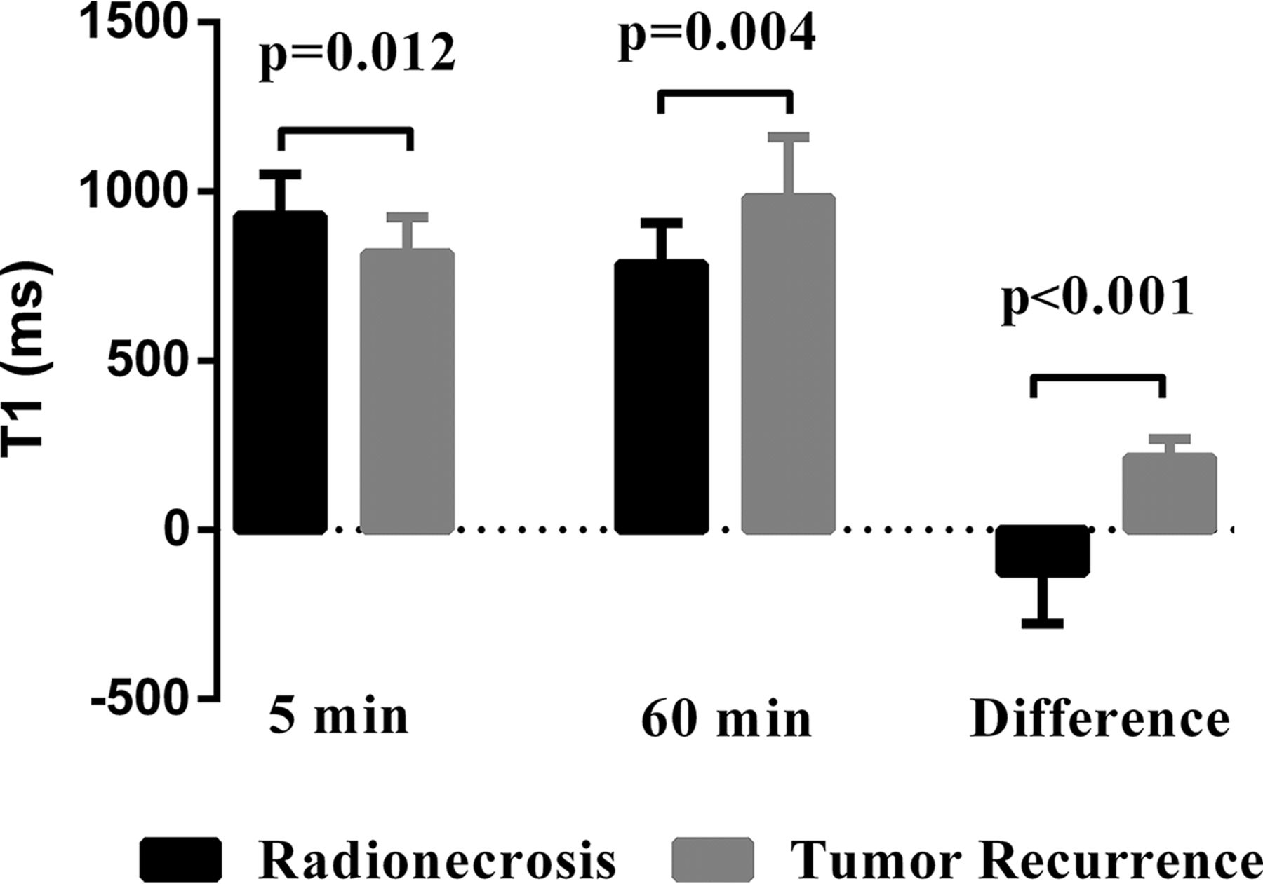
- Fig 1.
Comparison of T15min, T160min, and T1differ between the radionecrosis and tumor recurrence groups. There were significant differences in each of the 3 parameters (P = .012, P = .004, and P < .001, respectively) between the 2 groups.
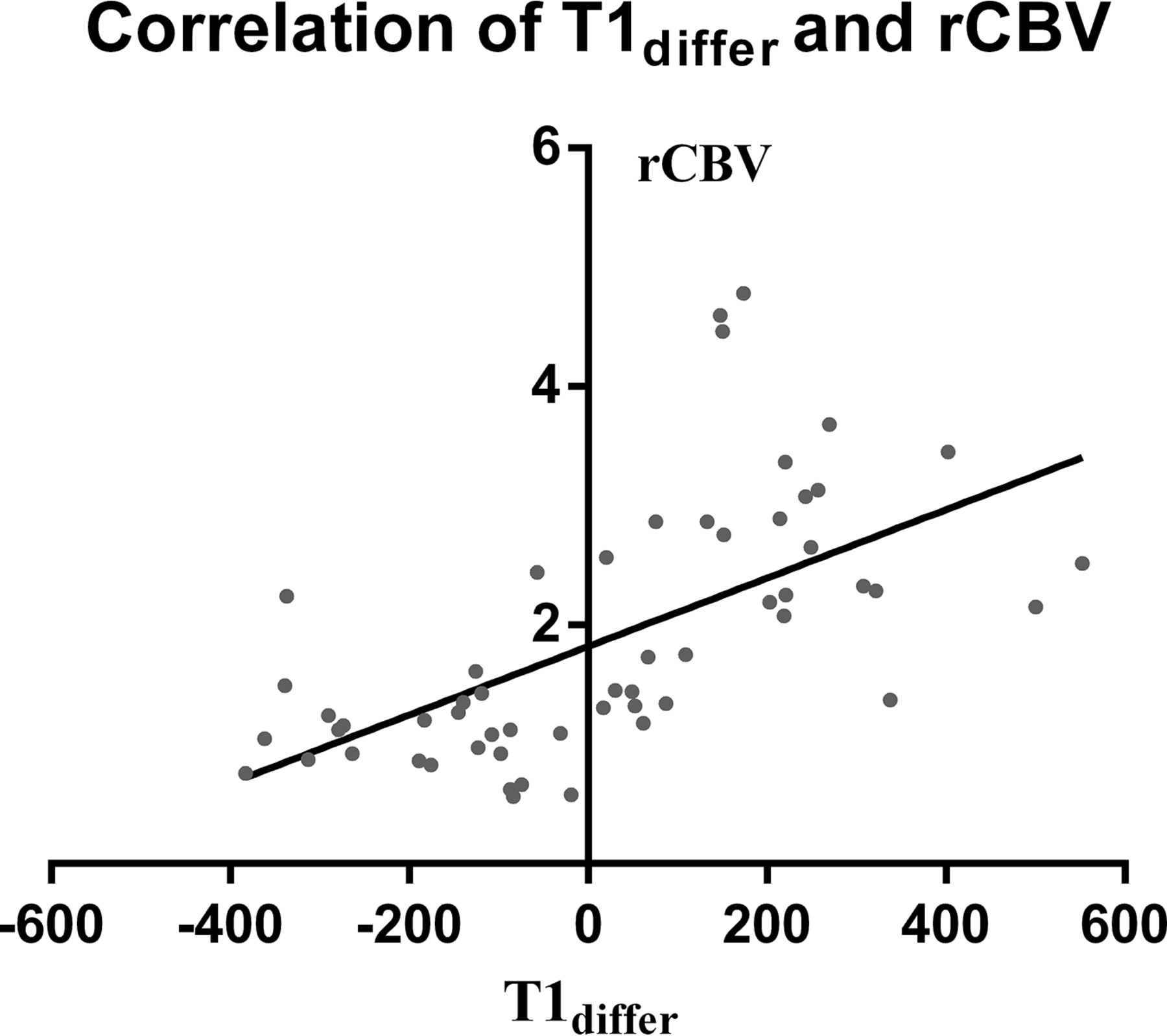
- Fig 2.
Correlation between T1differ and rCBV. T1differ significantly correlated with rCBV (r = 0.70; 95% CI, 0.53–0.82; P < .001).
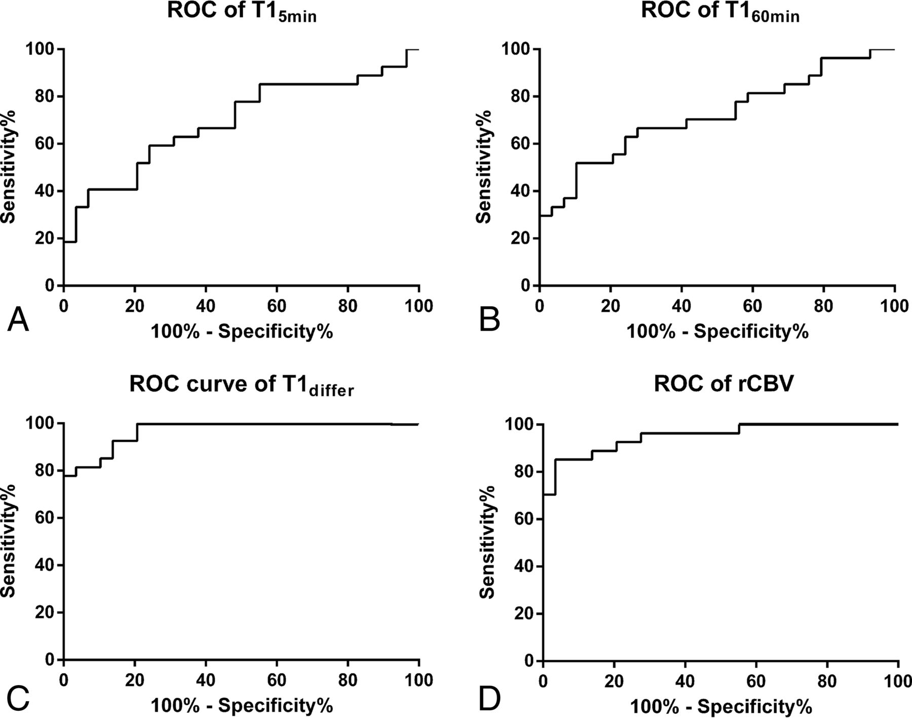
- Fig 3.
Receiver operating characteristic curves of T15min (A), T160min (B), T1differ (C), and rCBV (D) for radionecrosis after stereotactic radiosurgery reveals that T1differ has a similar diagnostic performance compared with rCBV (AUC = 0.97; 95% CI, 0.93–1.00 versus AUC = 0.95; 95% CI, 0.90–1.00).
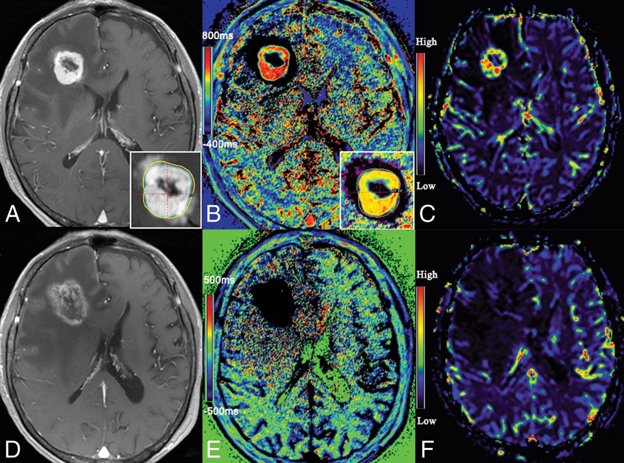
- Fig 4.
A 48-year-old male patient with cerebral metastasis from the lungs was treated with GKR (A). Follow-up MR imaging shows that enhancement returned at 5 months after GKR (B), while T1 mapping 5 minutes (C) and 60 minutes (D) after contrast administration reveals T15min and T160min values of 539 ms and 1064 ms, respectively, in the area of enhancement. The T1differ map revealed a positive area in the lesion (E). Histopathologic finings reveal lung cancer cells in the lesion (F).
- Fig 5.
A 62-year-old female patient with brain metastasis from the breast was treated by GKR (A). Follow-up MR imaging shows a 210% increment in maximal diameter at 6 months after GKR (B). The results of T1 mapping at 5 minutes (C) and 60 minutes (D) after contrast administration reveal T15min and T160min values of 1035 and 771 ms, respectively, in the area of enhancement. The T1differ map reveals negative areas in the lesion (E). Histopathologic findings confirmed them as radionecrosis (F).
- Fig 6.
MR image of a 54-year-old female patient with cerebral metastasis from the digestive tract. Contrast-enhanced T1-weighted image shows enhancement in the previously irradiated lesion 6 months later, after the first GKR (A). The T1differ map demonstrates it as a mixed lesion and detects the parts of tumor recurrence to guide the treatment plan (B). The recurrent part is confirmed by a cerebral blood volume map (C). Follow-up MR imaging (6 months after re-irradiation) shows a 90% increment in maximal diameter (D). The T1differ map demonstrates it as radionecrosis (E). The radionecrosis is confirmed by a CBV map (F).


Tables
Variables Radionecrosis Recurrence Summary Age (IQR) (yr) 58 (52.5–66) 59 (53–67) 58.5 (53–65.8) Sex (M/F) 13:16 13:14 26:30 Primary tumor history Lung (No.) (%) 18 (62.0) 16 (59.3) 34 (60.7) Digestive tract (No.) (%) 2 (6.9) 4 (14.8) 6 (10.6) Breast (No.) (%) 4 (13.8) 4 (14.8) 8 (14.3) Kidney (No.) (%) 2 (6.9) 2 (7.4) 4 (7.1) Skin (No.) (%) 3 (10.3) 1 (3.7) 4 (7.1) Location Occipital (No.) (%) 7 (24.1) 4 (14.8) 11 (19.6) Parietal (No.) (%) 5 (17.2) 7 (25.9) 12 (21.4) Frontal (No.) (%) 7 (24.1) 5 (18.5) 12 (21.4) Temporal (No.) (%) 3 (10.3) 2 (7.4) 5 (18.5) Cerebellum (No.) (%) 4 (13.8) 6 (22.2) 10 (37.0) Brain stem (No.) (%) 1 (3.4) 1 (3.7) 2 (7.4) Basal ganglia (No.) (%) 2 (6.9) 2 (7.4) 4 (14.8) MD (median) (IQR) (cm) 2.2 (1.6–2.5) 2.5 (1.7–3.4) 2.3 (1.6–2.8) Dose (median) (IQR) (Gy) 18 (18–21) 18 (18–21) 18 (18–21) KPS (median) (IQR) 70 (70–90) 70 (60–90) 70 (70–90) Note:—KPS indicates Karnofsky Performance Status Scale; MD, maximum diameters; IQR, interquartile range.
↵a Data are presented as descriptive statistics (median or count). Numbers in parentheses represent the range of data.
Parameters Radionecrosis Tumor Recurrence P Value T15min (median) (IQR) (ms) 914 (824–1055) 817 (567–924) .012 T160min (median) (IQR) (ms) 798 (682–920) 981 (774–1160) .004 T1differ (median) (IQR) (ms) −126 (−276 to −79) 214 (109–269) <.001 rCBV (median) (IQR) 1.12 (0.86–1.35) 2.57 (2.15–3.13) <.001


Jump to section
Related Articles
Cited By...
- No citing articles found.