
Article Figures & Data
Figures
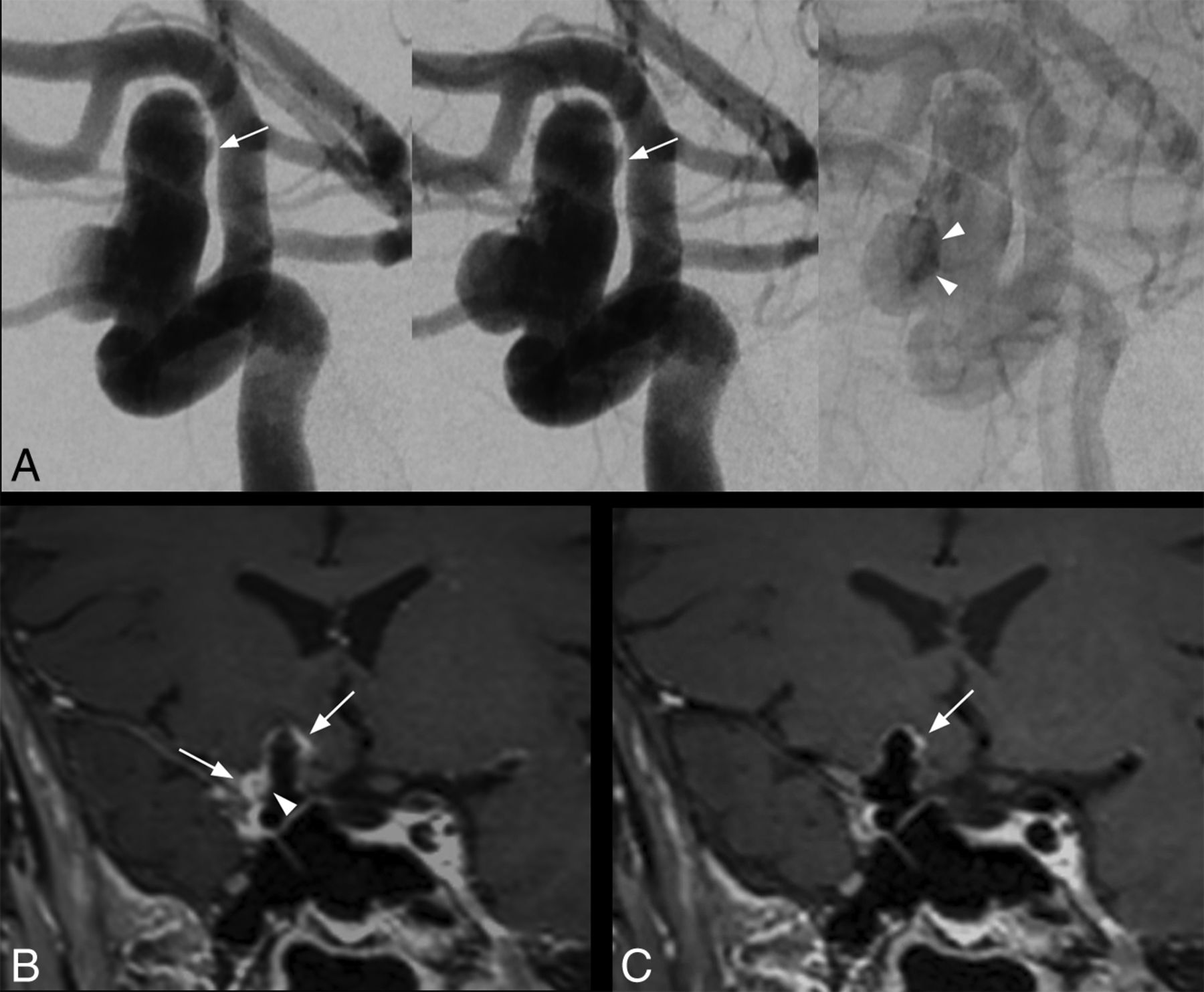
- Fig 1.
Saccular intracranial aneurysm in a 63-year-old patient. DSA angiogram (A, from left to right: early arterial phase, arterial phase, and venous phase) shows a 14-mm right internal carotid artery aneurysm with intrasaccular slow flowing/stagnant blood (arrowheads) and irregular shape (arrow). With conventional SPACE imaging (B), extensive AWE is visible, more prominent at the apex and at the lateral portion of the aneurysm (B, arrows). Note the enhancement within the aneurysm lumen visible only using conventional SPACE imaging (B, arrowhead), matching the stagnant blood on DSA (A, arrowheads). With MSDE SPACE imaging, AWE is only visible at the apex (C, arrow).
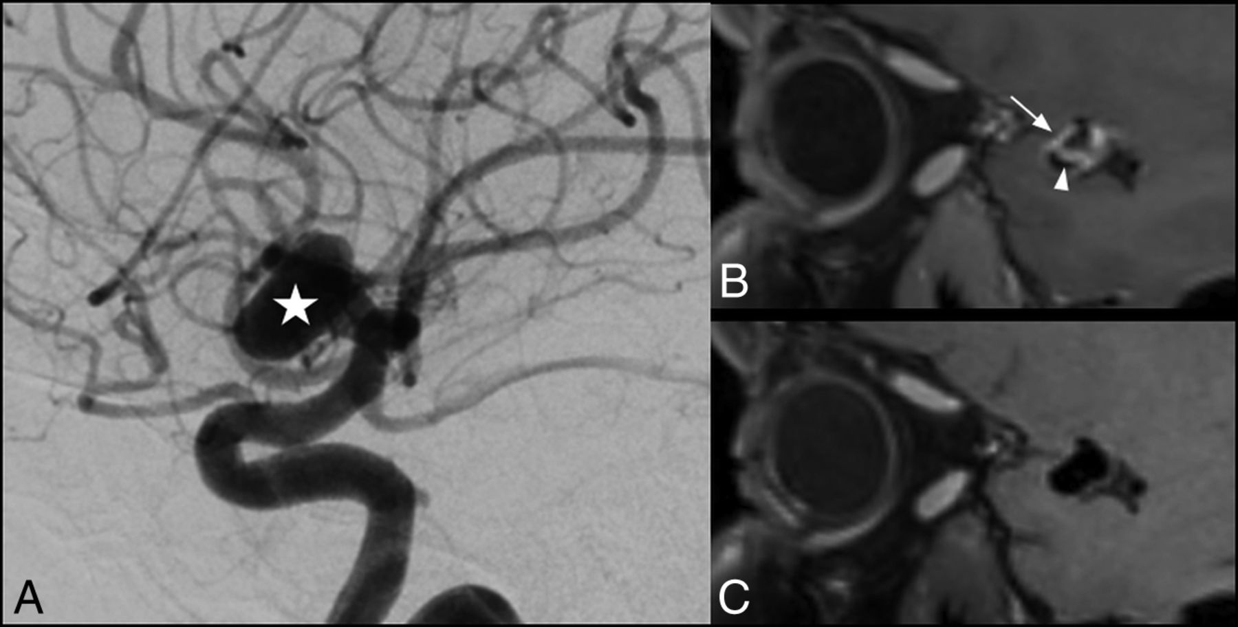
- Fig 2.
Saccular intracranial aneurysm in a 42-year-old patient. DSA angiogram shows an 11-mm aneurysm (A, star) arising from the M1 segment of the right middle cerebral artery. AWE is visible on the conventional SPACE image (B, arrow), while there is no enhancement visible on the corresponding MSDE SPACE image (C). Enhancement within the aneurysm lumen was also visible using a conventional SPACE image (B, arrowhead).


Jump to section
Related Articles
Cited By...
- DANTE-CAIPI Accelerated Contrast-Enhanced 3D T1: Deep Learning-Based Image Quality Improvement for Vessel Wall MRI
- Improved Blood Suppression of Motion-Sensitized Driven Equilibrium in High-Resolution Whole-Brain Vessel Wall Imaging: Comparison of Contrast-Enhanced 3D T1-Weighted FSE with Motion-Sensitized Driven Equilibrium and Delay Alternating with Nutation for Tailored Excitation
- Quantitative analysis of unruptured intracranial aneurysm wall thickness and enhancement using 7T high resolution, black blood magnetic resonance imaging
- Image-Quality Assessment of 3D Intracranial Vessel Wall MRI Using DANTE or DANTE-CAIPI for Blood Suppression and Imaging Acceleration
- Vessel wall imaging in intracranial aneurysms
- Reply:
- Comment on "Blood Flow Mimicking Aneurysmal Wall Enhancement: A Diagnostic Pitfall of Vessel Wall MRI Using the Postcontrast 3D Turbo Spin-Echo MR Imaging Sequence"