Advertisement

Main menu

  • Home
  • Content
    • Current Issue
    • Accepted Manuscripts
    • Article Preview
    • Past Issue Archive
    • AJNR Case Collection
    • Case of the Week Archive
    • Classic Case Archive
    • Case of the Month Archive
  • Special Collections
    • Spinal CSF Leak Articles (Jan 2020-June 2024)
    • 2024 AJNR Journal Awards
    • Most Impactful AJNR Articles
  • Multimedia
    • AJNR Podcast
    • AJNR Scantastics
    • Video Articles
  • For Authors
    • Submit a Manuscript
    • Author Policies
    • Fast publishing of Accepted Manuscripts
    • Graphical Abstract Preparation
    • Manuscript Submission Guidelines
    • Imaging Protocol Submission
    • Submit a Case for the Case Collection
  • About Us
    • About AJNR
    • Editorial Board
  • More
    • Become a Reviewer/Academy of Reviewers
    • Subscribers
    • Permissions
    • Alerts
    • Feedback
    • Advertisers
    • ASNR Home
  • Other Publications
    • ajnr

User menu

  • Alerts
  • Log in

Search

  • Advanced search
American Journal of Neuroradiology

American Journal of Neuroradiology

ASHNR American Society of Functional Neuroradiology ASHNR American Society of Pediatric Neuroradiology ASSR
  • Alerts
  • Log in

Advanced Search

  • Home
  • Content
    • Current Issue
    • Accepted Manuscripts
    • Article Preview
    • Past Issue Archive
    • AJNR Case Collection
    • Case of the Week Archive
    • Classic Case Archive
    • Case of the Month Archive
  • Special Collections
    • Spinal CSF Leak Articles (Jan 2020-June 2024)
    • 2024 AJNR Journal Awards
    • Most Impactful AJNR Articles
  • Multimedia
    • AJNR Podcast
    • AJNR Scantastics
    • Video Articles
  • For Authors
    • Submit a Manuscript
    • Author Policies
    • Fast publishing of Accepted Manuscripts
    • Graphical Abstract Preparation
    • Manuscript Submission Guidelines
    • Imaging Protocol Submission
    • Submit a Case for the Case Collection
  • About Us
    • About AJNR
    • Editorial Board
  • More
    • Become a Reviewer/Academy of Reviewers
    • Subscribers
    • Permissions
    • Alerts
    • Feedback
    • Advertisers
    • ASNR Home
  • Follow AJNR on Twitter
  • Visit AJNR on Facebook
  • Follow AJNR on Instagram
  • Join AJNR on LinkedIn
  • RSS Feeds

Welcome to the new AJNR, Updated Hall of Fame, and more. Read the full announcements.


AJNR is seeking candidates for the position of Associate Section Editor, AJNR Case Collection. Read the full announcement.


Research ArticleAdult Brain
Open Access

Imaging Differences between Neuromyelitis Optica Spectrum Disorders and Multiple Sclerosis: A Multi-Institutional Study in Japan

H. Tatekawa, S. Sakamoto, M. Hori, Y. Kaichi, A. Kunimatsu, K. Akazawa, T. Miyasaka, H. Oba, T. Okubo, K. Hasuo, K. Yamada, T. Taoka, S. Doishita, T. Shimono and Y. Miki
American Journal of Neuroradiology July 2018, 39 (7) 1239-1247; DOI: https://doi.org/10.3174/ajnr.A5663
H. Tatekawa
aFrom the Department of Diagnostic and Interventional Radiology (H.T., S.S., S.D., T.S., Y.M.), Osaka City University Graduate School of Medicine, Osaka, Japan
  • Find this author on Google Scholar
  • Find this author on PubMed
  • Search for this author on this site
  • ORCID record for H. Tatekawa
S. Sakamoto
aFrom the Department of Diagnostic and Interventional Radiology (H.T., S.S., S.D., T.S., Y.M.), Osaka City University Graduate School of Medicine, Osaka, Japan
  • Find this author on Google Scholar
  • Find this author on PubMed
  • Search for this author on this site
  • ORCID record for S. Sakamoto
M. Hori
bDepartment of Radiology (M.H.), Graduate School of Medicine, Juntendo University, Tokyo, Japan
  • Find this author on Google Scholar
  • Find this author on PubMed
  • Search for this author on this site
  • ORCID record for M. Hori
Y. Kaichi
cDepartment of Diagnostic Radiology (Y.K.), Graduate School and Institute of Biomedical and Health Sciences, Hiroshima University, Hiroshima, Japan
  • Find this author on Google Scholar
  • Find this author on PubMed
  • Search for this author on this site
  • ORCID record for Y. Kaichi
A. Kunimatsu
dDepartment of Radiology (A.K.), Graduate School of Medicine, University of Tokyo, Tokyo, Japan
  • Find this author on Google Scholar
  • Find this author on PubMed
  • Search for this author on this site
  • ORCID record for A. Kunimatsu
K. Akazawa
eDepartment of Radiology (K.A., K.Y.), Graduate School of Medical Science, Kyoto Prefectural University of Medicine, Kyoto, Japan
  • Find this author on Google Scholar
  • Find this author on PubMed
  • Search for this author on this site
  • ORCID record for K. Akazawa
T. Miyasaka
fDepartment of Radiology (T.M.), Nara Medical University, Nara, Japan
  • Find this author on Google Scholar
  • Find this author on PubMed
  • Search for this author on this site
  • ORCID record for T. Miyasaka
H. Oba
gDepartment of Radiology (H.O.), Teikyo University School of Medicine, Tokyo, Japan
  • Find this author on Google Scholar
  • Find this author on PubMed
  • Search for this author on this site
  • ORCID record for H. Oba
T. Okubo
hDepartment of Radiology (T.O.), Teikyo University Chiba Medical Center, Chiba, Japan
  • Find this author on Google Scholar
  • Find this author on PubMed
  • Search for this author on this site
  • ORCID record for T. Okubo
K. Hasuo
iDepartment of Diagnostic Radiology (K.H.), National Center for Global Health and Medicine, Tokyo, Japan
  • Find this author on Google Scholar
  • Find this author on PubMed
  • Search for this author on this site
  • ORCID record for K. Hasuo
K. Yamada
eDepartment of Radiology (K.A., K.Y.), Graduate School of Medical Science, Kyoto Prefectural University of Medicine, Kyoto, Japan
  • Find this author on Google Scholar
  • Find this author on PubMed
  • Search for this author on this site
  • ORCID record for K. Yamada
T. Taoka
jDepartment of Radiology (T.T.), Nagoya University Graduate School of Medicine, Nagoya, Japan.
  • Find this author on Google Scholar
  • Find this author on PubMed
  • Search for this author on this site
  • ORCID record for T. Taoka
S. Doishita
aFrom the Department of Diagnostic and Interventional Radiology (H.T., S.S., S.D., T.S., Y.M.), Osaka City University Graduate School of Medicine, Osaka, Japan
  • Find this author on Google Scholar
  • Find this author on PubMed
  • Search for this author on this site
  • ORCID record for S. Doishita
T. Shimono
aFrom the Department of Diagnostic and Interventional Radiology (H.T., S.S., S.D., T.S., Y.M.), Osaka City University Graduate School of Medicine, Osaka, Japan
  • Find this author on Google Scholar
  • Find this author on PubMed
  • Search for this author on this site
  • ORCID record for T. Shimono
Y. Miki
aFrom the Department of Diagnostic and Interventional Radiology (H.T., S.S., S.D., T.S., Y.M.), Osaka City University Graduate School of Medicine, Osaka, Japan
  • Find this author on Google Scholar
  • Find this author on PubMed
  • Search for this author on this site
  • ORCID record for Y. Miki
  • Article
  • Figures & Data
  • Supplemental
  • Info & Metrics
  • Responses
  • References
  • PDF
Loading

Article Figures & Data

Figures

  • Tables
  • Fig 1.
    • Download figure
    • Open in new tab
    • Download powerpoint
    Fig 1.

    Patient selection flowchart. Consecutive patients, 89 with NMOSD and 89 with MS, are examined. According to the eligibility criteria, 79 patients with NMOSD and 87 with MS are assessed for brain analyses, 57 patients with NMOSD and 55 with MS are assessed for spinal cord analyses, and 42 patients with NMOSD and 14 with MS are assessed for optic nerve analyses.
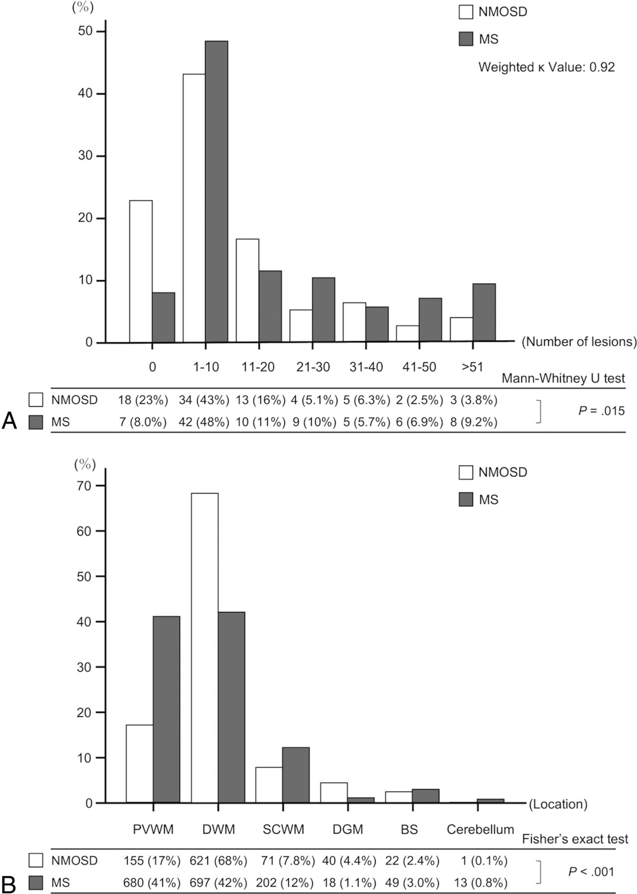
  • Fig 2.
    • Download figure
    • Open in new tab
    • Download powerpoint
    Fig 2.

    Bar graphs show the proportion of patients classified by the number of brain lesions in bins of 10 lesions (A), and the distribution of brain lesions categorized by the location (PVWM, DWM, SCWM, DGM, BS, cerebellum) (B) for rater 1. A total of 911 brain lesions in 79 patients with NMOSD and 1659 brain lesions in 87 patients with MS are identified. A, The proportion of patients is significantly different between NMOSD and MS (P = .015; weighted κ value, 0.92). More patients with NMOSD have no brain lesions of ≥3 mm, and a tendency for patients with MS to have more brain lesions than those with NMOSD is found. B, The distribution of lesions categorized by location is significantly different between NMOSD and MS (P < .001). DWM lesions (68%) are more frequent than PVWM lesions (17%) in NMOSD, whereas the difference in the frequencies of lesions in PVWM (41%) and DWM (42%) is small in MS.

  • Fig 3.
    • Download figure
    • Open in new tab
    • Download powerpoint
    Fig 3.

    Graphs show the proportion of patients classified by the number of spinal cord lesions (A), the distribution and proportion of spinal cord lesions (B), and the length of spinal cord lesions in each location (C) for rater 1. A total of 86 spinal cord lesions in 57 patients with NMOSD and 102 spinal cord lesions in 55 patients with MS are identified. A, No significant difference is found in the number of lesions between NMOSD and MS (P = .76; weighted κ value, 0.94). Forty-seven (82%) patients with NMOSD and 37 (67%) with MS have ≥1 spinal cord lesion. B, Bimodal distributions of spinal cord lesions are present in both NMOSD and MS, but the peak of the distribution in NMOSD is high in thoracic regions, whereas the variation and peaks of the distribution are relatively smaller in MS than in NMOSD. The proportions of lesions categorized into cervical or thoracic regions are significantly different between NMOSD and MS (P = .024). More thoracic lesions (71%) than cervical lesions (29%) are found in NMOSD, whereas the difference in the frequencies of cervical (46%) and thoracic lesions (54%) is small in MS. C, In NMOSD, thoracic lesions are significantly longer than cervical lesions (P = .001), whereas in MS, the length is not significantly different between cervical and thoracic lesions (P = .80).

Tables

  • Figures
    • View popup
    Table 1:

    Patient demographics and characteristics in NMOSD and MSa

    NMOSDMSP Valuec
    Demographics (NMOSD, n = 89) (MS, n = 89)
        Age (yr) (median) (IQR, range)51 (39–61, 16–85)36 (29–43, 18–67)<.001
            No. of femalesb77 (86.5)68 (76.4).12d
        Disease duration (yr) (median) (IQR, range)4 (0–11, 0–73)2 (1–7, 0–21).40
        EDSS (median) (IQR, range)6 (2–7.5, 1–9)2 (1–3, 0–6)<.001
        AQP4-IgGb
            Positive83 (93.3)
            Negative5 (5.6)
            Unknown1 (1.1)
        MS typeb
            Relapsing-remitting MS79 (88.8)
            Primary-progressive MS6 (6.7)
            Secondary-progressive MS2 (2.2)
            Unknown2 (2.2)
    Brain MRI (NMOSD, n = 79) (MS, n= 87)
        Age (yr) (median) (IQR, range)50 (38–61, 17–83)36 (29–45, 19–67)<.001
            No. of femalesb68 (86.1)67 (77).16d
        Disease duration (yr) (median) (IQR, range)4 (0–11, 0–41)3 (1–7, 0–21).59
        EDSS (median) (IQR, range)6 (2–7.5, 1–9)2 (1–3, 0–6)<.001
        Gadolinium enhancementb51 (64.6)68 (78.2)
    Spinal cord MRI (NMOSD, n = 57) (MS, n = 55)
        Age (yr) (median) (IQR, range)53 (39–61, 25–78)37 (29–43, 18–66)<.001
            No. of femalesb51 (89.5)36 (65.5).003d
        Disease duration (yr) (median) (IQR, range)4 (0–12, 0–43)2 (0–7, 0–19).18
        EDSS (median) (IQR, range)6 (2–7, 1–9)2 (2–3.5, 0–6)<.001
    Optic nerve MRI (NMOSD, n = 42) (MS, n = 14)
        Age (yr) (median) (IQR, range)50 (37–61, 17–79)35 (34–39, 19–67).004
            No. of femalesb37 (88.1)9 (64.3).10d
        Disease duration (yr) (median) (IQR, range)5 (0–12, 0–42)1 (0–6, 0–17).15
        EDSS (median) (IQR, range)6 (3–8, 1–9)3 (2–3.5, 2–4).065
    • Note:—EDSS indicates Expanded Disability Status Scale.

    • ↵a Unless otherwise indicated, data are medians, with interquartile range and total range in parentheses.

    • ↵b Data in parentheses are percentages.

    • ↵c Mann-Whitney U test.

    • ↵d Fisher exact test.

    • View popup
    Table 2:

    Number and size of brain lesions for rater 1 and assessment of brain morphologic features and characteristic signs

    Quantitative AnalysesNMOSD (n = 79)MS (n = 87)P ValueAccuracyκ Value
    Total No. of lesions9111659
        Per patienta5 (1–18, 0–81)8 (3–28, 0–120).004c
    Diameter of lesions (mm)a5.7 (4.3–8.5, 3.0–45)6.1 (4.6–8.3, 3.0–56).046c
        In each region (mm)a
            PVWM9.2 (5.3–14, 3.0–36)6.8 (5.1–9.5, 3.0–47)<.001c
            DWM5.5 (4.2–7.7, 3.0–45)5.5 (4.3–7.4, 3.0–56).73c
            SCWM5.4 (3.7–7.2, 3.0–28)5.9 (4.5–7.9, 3.0–25).054c
            DGM5.9 (4.3–8.3, 3.1–30)6.7 (5.5–9.2, 3.5–48).31c
            BS6.2 (5.3–8.3, 3.3–22)6.9 (5.2–8.1, 3.6–16).96c
            Cerebellum3.7 (3.7–3.7, 3.7–3.7)7.3 (5.5–8.9, 3.4–21).17c
    Morphologic assessment
        Brain atrophyb4 (5.1)5 (5.7)1d0.480.52
        Ventriculomegalyb3 (3.8)3 (3.4)1d0.480.5
    Characteristic signs
        Ovoid lesionsb17 (21.5)55 (63.2)<.001d0.710.68
        T1 black hole lesionsb16 (20.3)47 (54)<.001d0.660.65
        Callosal-septal-interface lesionsb23 (29.1)48 (55.2).001d0.630.75
        Isolated U-fiber lesionsb8 (10.1)24 (27.6).005d0.570.56
        Dirty white matter lesionsb13 (16.5)23 (26.4).14d0.540.61
        Tumefactive MS lesionsb1 (1.3)2 (2.3)1d0.480.74
        Cloudlike enhancementb0 (0)0 (0)
    • ↵a Data are medians, with interquartile range and total range in parentheses.

    • ↵b Data in parentheses are percentages.

    • ↵c Mann-Whitney U test.

    • ↵d Fisher exact test.

    • View popup
    Table 3:

    Number, size, and location of spinal cord lesions for rater 1 and assessment of spinal cord morphologic featuresa

    Quantitative AnalysesNMOSD (n = 57)MS (n = 55)P ValuecAccuracyκ Value
    Total No. of lesions86102
        Per patient1 (1–2, 0–6)1 (0–3, 0–6).77
    Longitudinal length (mm)47 (17–109, 4.5–408)13 (9.0–20, 4.0–208)<.001
        In cervical region (mm)23 (9.5–36, 4.5–149)13 (8.4–21, 4.0–110).077
        In thoracic region (mm)63 (25–131, 6.0–408)13 (9.4–20, 4.1–208)<.001
    Transverse diameter (mm)4.4 (3.2–6.2, 1.8–13)4.4 (3.6–5.3, 1.9–11).99
        In cervical region (mm)5.8 (3.6–7.5, 1.9–13)4.7 (4.0–5.8, 1.9–11).55
        In thoracic region (mm)4.2 (3.1–5.7, 1.8–10)4.0 (3.5–4.8, 2.3–7.3).76
    Intramedullary locationb
        Central66 (76.7)59 (57.8).007d
        Peripheral11 (12.8)32 (31.4)
        Both9 (10.5)11 (10.8)
    Morphologic assessment
        Atrophyb17 (29.8)5 (9.1).008d0.400.53
        Swellingb19 (33.3)8 (14.5).027d0.410.67
    • ↵a Unless otherwise indicated, data are medians, with interquartile range and total range in parentheses.

    • ↵b Data in parentheses are percentages.

    • ↵c Mann-Whitney U test.

    • ↵d Fisher exact test.

    • View popup
    Table 4:

    Laterality and location of optic nerve lesions and assessment of optic nerve morphologic featuresa

    NMOSD (n = 42)MS (n = 14)P ValuebAccuracyκ Value
    Laterality of lesions
        None16 (38.1)7 (50).670.73
        Unilateral18 (42.9)4 (28.6)
        Bilateral8 (19)3 (21.4)
    Location of lesions
        Optic nerve26 (61.9)7 (50).540.7
        Optic chiasm2 (4.8)2 (14.3).260.7
        Optic tract0 (0)1 (7.1).250.49
    Atrophy5 (11.9)0 (0).320.660.48
    Swelling10 (23.8)5 (35.7).490.660.41
    • ↵a Data in parentheses are percentages.

    • ↵b Fisher exact test.

PreviousNext
Back to top

In this issue

American Journal of Neuroradiology: 39 (7)
American Journal of Neuroradiology
Vol. 39, Issue 7
1 Jul 2018
  • Table of Contents
  • Index by author
  • Complete Issue (PDF)
Advertisement
Print
Download PDF
Email Article

Thank you for your interest in spreading the word on American Journal of Neuroradiology.

NOTE: We only request your email address so that the person you are recommending the page to knows that you wanted them to see it, and that it is not junk mail. We do not capture any email address.

Enter multiple addresses on separate lines or separate them with commas.
Imaging Differences between Neuromyelitis Optica Spectrum Disorders and Multiple Sclerosis: A Multi-Institutional Study in Japan
(Your Name) has sent you a message from American Journal of Neuroradiology
(Your Name) thought you would like to see the American Journal of Neuroradiology web site.
CAPTCHA
This question is for testing whether or not you are a human visitor and to prevent automated spam submissions.
Cite this article
H. Tatekawa, S. Sakamoto, M. Hori, Y. Kaichi, A. Kunimatsu, K. Akazawa, T. Miyasaka, H. Oba, T. Okubo, K. Hasuo, K. Yamada, T. Taoka, S. Doishita, T. Shimono, Y. Miki
Imaging Differences between Neuromyelitis Optica Spectrum Disorders and Multiple Sclerosis: A Multi-Institutional Study in Japan
American Journal of Neuroradiology Jul 2018, 39 (7) 1239-1247; DOI: 10.3174/ajnr.A5663

Citation Manager Formats

  • BibTeX
  • Bookends
  • EasyBib
  • EndNote (tagged)
  • EndNote 8 (xml)
  • Medlars
  • Mendeley
  • Papers
  • RefWorks Tagged
  • Ref Manager
  • RIS
  • Zotero
0 Responses
Respond to this article
Share
Bookmark this article
Imaging Differences between Neuromyelitis Optica Spectrum Disorders and Multiple Sclerosis: A Multi-Institutional Study in Japan
H. Tatekawa, S. Sakamoto, M. Hori, Y. Kaichi, A. Kunimatsu, K. Akazawa, T. Miyasaka, H. Oba, T. Okubo, K. Hasuo, K. Yamada, T. Taoka, S. Doishita, T. Shimono, Y. Miki
American Journal of Neuroradiology Jul 2018, 39 (7) 1239-1247; DOI: 10.3174/ajnr.A5663
del.icio.us logo Twitter logo Facebook logo Mendeley logo
  • Tweet Widget
  • Facebook Like
  • Google Plus One
Purchase

Jump to section

  • Article
    • Abstract
    • ABBREVIATIONS:
    • Materials and Methods
    • Results
    • Discussion
    • Conclusions
    • Acknowledgments
    • Footnotes
    • References
  • Figures & Data
  • Supplemental
  • Info & Metrics
  • Responses
  • References
  • PDF

Related Articles

  • PubMed
  • Google Scholar

Cited By...

  • No citing articles found.
  • Crossref (25)
  • Google Scholar

This article has been cited by the following articles in journals that are participating in Crossref Cited-by Linking.

  • Microstructural white matter abnormalities in multiple sclerosis and neuromyelitis optica spectrum disorders: Evaluation by advanced diffusion imaging
    Shimpei Kato, Akifumi Hagiwara, Kazumasa Yokoyama, Christina Andica, Yuji Tomizawa, Yasunobu Hoshino, Wataru Uchida, Yuma Nishimura, Shohei Fujita, Koji Kamagata, Masaaki Hori, Nobutaka Hattori, Osamu Abe, Shigeki Aoki
    Journal of the Neurological Sciences 2022 436
  • Updates in NMOSD and MOGAD Diagnosis and Treatment
    Laura Cacciaguerra, Eoin P. Flanagan
    Neurologic Clinics 2024 42 1
  • MRI Patterns Distinguish AQP4 Antibody Positive Neuromyelitis Optica Spectrum Disorder From Multiple Sclerosis
    Laura Clarke, Simon Arnett, Wajih Bukhari, Elham Khalilidehkordi, Sofia Jimenez Sanchez, Cullen O'Gorman, Jing Sun, Kerri M. Prain, Mark Woodhall, Roger Silvestrini, Christine S. Bundell, David A. Abernethy, Sandeep Bhuta, Stefan Blum, Mike Boggild, Karyn Boundy, Bruce J. Brew, Wallace Brownlee, Helmut Butzkueven, William M. Carroll, Cella Chen, Alan Coulthard, Russell C. Dale, Chandi Das, Marzena J. Fabis-Pedrini, David Gillis, Simon Hawke, Robert Heard, Andrew P. D. Henderson, Saman Heshmat, Suzanne Hodgkinson, Trevor J. Kilpatrick, John King, Christopher Kneebone, Andrew J. Kornberg, Jeannette Lechner-Scott, Ming-Wei Lin, Christopher Lynch, Richard A. L. Macdonell, Deborah F. Mason, Pamela A. McCombe, Jennifer Pereira, John D. Pollard, Sudarshini Ramanathan, Stephen W. Reddel, Cameron P. Shaw, Judith M. Spies, James Stankovich, Ian Sutton, Steve Vucic, Michael Walsh, Richard C. Wong, Eppie M. Yiu, Michael H. Barnett, Allan G. K. Kermode, Mark P. Marriott, John D. E. Parratt, Mark Slee, Bruce V. Taylor, Ernest Willoughby, Fabienne Brilot, Angela Vincent, Patrick Waters, Simon A. Broadley
    Frontiers in Neurology 2021 12
  • Spinal Cord Atrophy in Neuromyelitis Optica Spectrum Disorders Is Spatially Related to Cord Lesions and Disability
    Laura Cacciaguerra, Paola Valsasina, Sarlota Mesaros, Vittorio Martinelli, Jelena Drulovic, Massimo Filippi, Maria A. Rocca
    Radiology 2020 297 1
  • Spinal cord involvement by atrophy and associations with disability are different between multiple sclerosis and neuromyelitis optica spectrum disorder
    Y. Nakamura, Z. Liu, S. Fukumoto, K. Shinoda, A. Sakoda, T. Matsushita, S. Hayashida, N. Isobe, M. Watanabe, A. Hiwatashi, R. Yamasaki, J. ‐i. Kira
    European Journal of Neurology 2020 27 1
  • Magnetic resonance imaging diagnosis of demyelinating diseases: An update
    Yukio Miki
    Clinical and Experimental Neuroimmunology 2019 10 S1
  • Differentiation between multiple sclerosis and neuromyelitis optica spectrum disorders by multiparametric quantitative MRI using convolutional neural network
    Akifumi Hagiwara, Yujiro Otsuka, Christina Andica, Shimpei Kato, Kazumasa Yokoyama, Masaaki Hori, Shohei Fujita, Koji Kamagata, Nobutaka Hattori, Shigeki Aoki
    Journal of Clinical Neuroscience 2021 87
  • Comparisons of clinical phenotype, radiological and laboratory features, and therapy of neuromyelitis optica spectrum disorder by regions: update and challenges
    Caiyun Liu, Mingchao Shi, Mingqin Zhu, Fengna Chu, Tao Jin, Jie Zhu
    Autoimmunity Reviews 2022 21 1
  • Research Progress of Artificial Intelligence Image Analysis in Systemic Disease-Related Ophthalmopathy
    Yuke Ji, Nan Chen, Sha Liu, Zhipeng Yan, Hui Qian, Shaojun Zhu, Jie Zhang, Minli Wang, Qin Jiang, Weihua Yang, Yi Shao
    Disease Markers 2022 2022
  • Imaging of Neuromyelitis Optica Spectrum Disorders
    Sheng-Che Hung
    Seminars in Ultrasound, CT and MRI 2020 41 3

More in this TOC Section

  • Diagnostic Neuroradiology of Monoclonal Antibodies
  • Clinical Outcomes After Chiari I Decompression
  • Cerebral ADC Changes in Fabry Disease
Show more Adult Brain

Similar Articles

Advertisement

Indexed Content

  • Current Issue
  • Accepted Manuscripts
  • Article Preview
  • Past Issues
  • Editorials
  • Editors Choice
  • Fellow Journal Club
  • Letters to the Editor

Cases

  • Case Collection
  • Archive - Case of the Week
  • Archive - Case of the Month
  • Archive - Classic Case

Special Collections

  • Special Collections

Resources

  • News and Updates
  • Turn around Times
  • Submit a Manuscript
  • Author Policies
  • Manuscript Submission Guidelines
  • Evidence-Based Medicine Level Guide
  • Publishing Checklists
  • Graphical Abstract Preparation
  • Imaging Protocol Submission
  • Submit a Case
  • Become a Reviewer/Academy of Reviewers
  • Get Peer Review Credit from Publons

Multimedia

  • AJNR Podcast
  • AJNR SCANtastic
  • Video Articles

About Us

  • About AJNR
  • Editorial Board
  • Not an AJNR Subscriber? Join Now
  • Alerts
  • Feedback
  • Advertise with us
  • Librarian Resources
  • Permissions
  • Terms and Conditions

American Society of Neuroradiology

  • Not an ASNR Member? Join Now

© 2025 by the American Society of Neuroradiology All rights, including for text and data mining, AI training, and similar technologies, are reserved.
Print ISSN: 0195-6108 Online ISSN: 1936-959X

Powered by HighWire