Advertisement

Main menu

  • Home
  • Content
    • Current Issue
    • Accepted Manuscripts
    • Article Preview
    • Past Issue Archive
    • Video Articles
    • AJNR Case Collection
    • Case of the Week Archive
    • Case of the Month Archive
    • Classic Case Archive
  • Special Collections
    • AJNR Awards
    • Low-Field MRI
    • Alzheimer Disease
    • ASNR Foundation Special Collection
    • Photon-Counting CT
    • View All
  • Multimedia
    • AJNR Podcasts
    • AJNR SCANtastic
    • Trainee Corner
    • MRI Safety Corner
    • Imaging Protocols
  • For Authors
    • Submit a Manuscript
    • Submit a Video Article
    • Submit an eLetter to the Editor/Response
    • Manuscript Submission Guidelines
    • Statistical Tips
    • Fast Publishing of Accepted Manuscripts
    • Graphical Abstract Preparation
    • Imaging Protocol Submission
    • Author Policies
  • About Us
    • About AJNR
    • Editorial Board
    • Editorial Board Alumni
  • More
    • Become a Reviewer/Academy of Reviewers
    • Subscribers
    • Permissions
    • Alerts
    • Feedback
    • Advertisers
    • ASNR Home

User menu

  • Alerts
  • Log in

Search

  • Advanced search
American Journal of Neuroradiology

American Journal of Neuroradiology

ASHNR American Society of Functional Neuroradiology ASHNR American Society of Pediatric Neuroradiology ASSR
  • Alerts
  • Log in

Advanced Search

  • Home
  • Content
    • Current Issue
    • Accepted Manuscripts
    • Article Preview
    • Past Issue Archive
    • Video Articles
    • AJNR Case Collection
    • Case of the Week Archive
    • Case of the Month Archive
    • Classic Case Archive
  • Special Collections
    • AJNR Awards
    • Low-Field MRI
    • Alzheimer Disease
    • ASNR Foundation Special Collection
    • Photon-Counting CT
    • View All
  • Multimedia
    • AJNR Podcasts
    • AJNR SCANtastic
    • Trainee Corner
    • MRI Safety Corner
    • Imaging Protocols
  • For Authors
    • Submit a Manuscript
    • Submit a Video Article
    • Submit an eLetter to the Editor/Response
    • Manuscript Submission Guidelines
    • Statistical Tips
    • Fast Publishing of Accepted Manuscripts
    • Graphical Abstract Preparation
    • Imaging Protocol Submission
    • Author Policies
  • About Us
    • About AJNR
    • Editorial Board
    • Editorial Board Alumni
  • More
    • Become a Reviewer/Academy of Reviewers
    • Subscribers
    • Permissions
    • Alerts
    • Feedback
    • Advertisers
    • ASNR Home
  • Follow AJNR on Twitter
  • Visit AJNR on Facebook
  • Follow AJNR on Instagram
  • Join AJNR on LinkedIn
  • RSS Feeds

AJNR Awards, New Junior Editors, and more. Read the latest AJNR updates

Research ArticleAdult Brain
Open Access

Comparative Study of MRI Biomarkers in the Substantia Nigra to Discriminate Idiopathic Parkinson Disease

N. Pyatigorskaya, B. Magnin, M. Mongin, L. Yahia-Cherif, R. Valabregue, D. Arnaldi, C. Ewenczyk, C. Poupon, M. Vidailhet and S. Lehéricy
American Journal of Neuroradiology August 2018, 39 (8) 1460-1467; DOI: https://doi.org/10.3174/ajnr.A5702
N. Pyatigorskaya
aFrom the Institut du Cerveau et de la Moelle épinière (N.P., B.M., M.M., L.Y.-C., R.V., D.A., M.V., S.L.), Centre de NeuroImagerie de Recherche, Paris, France
bUMR S 1127, CNRS UMR 7225 (N.P., B.M., M.M., L.Y.-C., M.V., S.L.), Sorbonne University, Paris, France
cService de neuroradiologie (N.P., B.M., S.L.)
  • Find this author on Google Scholar
  • Find this author on PubMed
  • Search for this author on this site
  • ORCID record for N. Pyatigorskaya
B. Magnin
aFrom the Institut du Cerveau et de la Moelle épinière (N.P., B.M., M.M., L.Y.-C., R.V., D.A., M.V., S.L.), Centre de NeuroImagerie de Recherche, Paris, France
bUMR S 1127, CNRS UMR 7225 (N.P., B.M., M.M., L.Y.-C., M.V., S.L.), Sorbonne University, Paris, France
cService de neuroradiologie (N.P., B.M., S.L.)
  • Find this author on Google Scholar
  • Find this author on PubMed
  • Search for this author on this site
  • ORCID record for B. Magnin
M. Mongin
aFrom the Institut du Cerveau et de la Moelle épinière (N.P., B.M., M.M., L.Y.-C., R.V., D.A., M.V., S.L.), Centre de NeuroImagerie de Recherche, Paris, France
bUMR S 1127, CNRS UMR 7225 (N.P., B.M., M.M., L.Y.-C., M.V., S.L.), Sorbonne University, Paris, France
  • Find this author on Google Scholar
  • Find this author on PubMed
  • Search for this author on this site
  • ORCID record for M. Mongin
L. Yahia-Cherif
aFrom the Institut du Cerveau et de la Moelle épinière (N.P., B.M., M.M., L.Y.-C., R.V., D.A., M.V., S.L.), Centre de NeuroImagerie de Recherche, Paris, France
bUMR S 1127, CNRS UMR 7225 (N.P., B.M., M.M., L.Y.-C., M.V., S.L.), Sorbonne University, Paris, France
  • Find this author on Google Scholar
  • Find this author on PubMed
  • Search for this author on this site
  • ORCID record for L. Yahia-Cherif
R. Valabregue
aFrom the Institut du Cerveau et de la Moelle épinière (N.P., B.M., M.M., L.Y.-C., R.V., D.A., M.V., S.L.), Centre de NeuroImagerie de Recherche, Paris, France
  • Find this author on Google Scholar
  • Find this author on PubMed
  • Search for this author on this site
  • ORCID record for R. Valabregue
D. Arnaldi
aFrom the Institut du Cerveau et de la Moelle épinière (N.P., B.M., M.M., L.Y.-C., R.V., D.A., M.V., S.L.), Centre de NeuroImagerie de Recherche, Paris, France
eClinical Neurology (D.A.), Department of Neuroscience, University of Genoa, Genoa, Italy
gCentre d'Investigation Clinique (D.A., M.V.), Hôpital Pitié-Salpêtrière, Paris, France.
  • Find this author on Google Scholar
  • Find this author on PubMed
  • Search for this author on this site
  • ORCID record for D. Arnaldi
C. Ewenczyk
dDépartement des Maladies du Système Nerveux (C.E., M.V.), Clinique des mouvements anormaux, Assistance Publique Hôpitaux de Paris, Hôpital Pitié-Salpêtrière, Paris, France
  • Find this author on Google Scholar
  • Find this author on PubMed
  • Search for this author on this site
  • ORCID record for C. Ewenczyk
C. Poupon
fNeuroSpin (C.P.), Commissariat à l'Energie Atomique, Gif-Sur-Yvette, France
  • Find this author on Google Scholar
  • Find this author on PubMed
  • Search for this author on this site
  • ORCID record for C. Poupon
M. Vidailhet
aFrom the Institut du Cerveau et de la Moelle épinière (N.P., B.M., M.M., L.Y.-C., R.V., D.A., M.V., S.L.), Centre de NeuroImagerie de Recherche, Paris, France
bUMR S 1127, CNRS UMR 7225 (N.P., B.M., M.M., L.Y.-C., M.V., S.L.), Sorbonne University, Paris, France
dDépartement des Maladies du Système Nerveux (C.E., M.V.), Clinique des mouvements anormaux, Assistance Publique Hôpitaux de Paris, Hôpital Pitié-Salpêtrière, Paris, France
gCentre d'Investigation Clinique (D.A., M.V.), Hôpital Pitié-Salpêtrière, Paris, France.
  • Find this author on Google Scholar
  • Find this author on PubMed
  • Search for this author on this site
  • ORCID record for M. Vidailhet
S. Lehéricy
aFrom the Institut du Cerveau et de la Moelle épinière (N.P., B.M., M.M., L.Y.-C., R.V., D.A., M.V., S.L.), Centre de NeuroImagerie de Recherche, Paris, France
bUMR S 1127, CNRS UMR 7225 (N.P., B.M., M.M., L.Y.-C., M.V., S.L.), Sorbonne University, Paris, France
cService de neuroradiologie (N.P., B.M., S.L.)
  • Find this author on Google Scholar
  • Find this author on PubMed
  • Search for this author on this site
  • ORCID record for S. Lehéricy
  • Article
  • Figures & Data
  • Info & Metrics
  • Responses
  • References
  • PDF
Loading

Article Figures & Data

Figures

  • Tables
  • Fig 1.
    • Download figure
    • Open in new tab
    • Download powerpoint
    Fig 1.

    Segmentation of ROIs. Axial brain stem slices in a healthy volunteer (A) and a patient with Parkinson disease (B) show reduction of the neuromelanin signal intensity in the substantia nigra of the patient. Axial T2*WI 7T slices show dorsal nigral hyperintensity in a HV (C), which is absent in the patient with PD (D). T2WI axial (E) and coronal (F) images of the SN in a HV show T2-based segmentation of the SN.
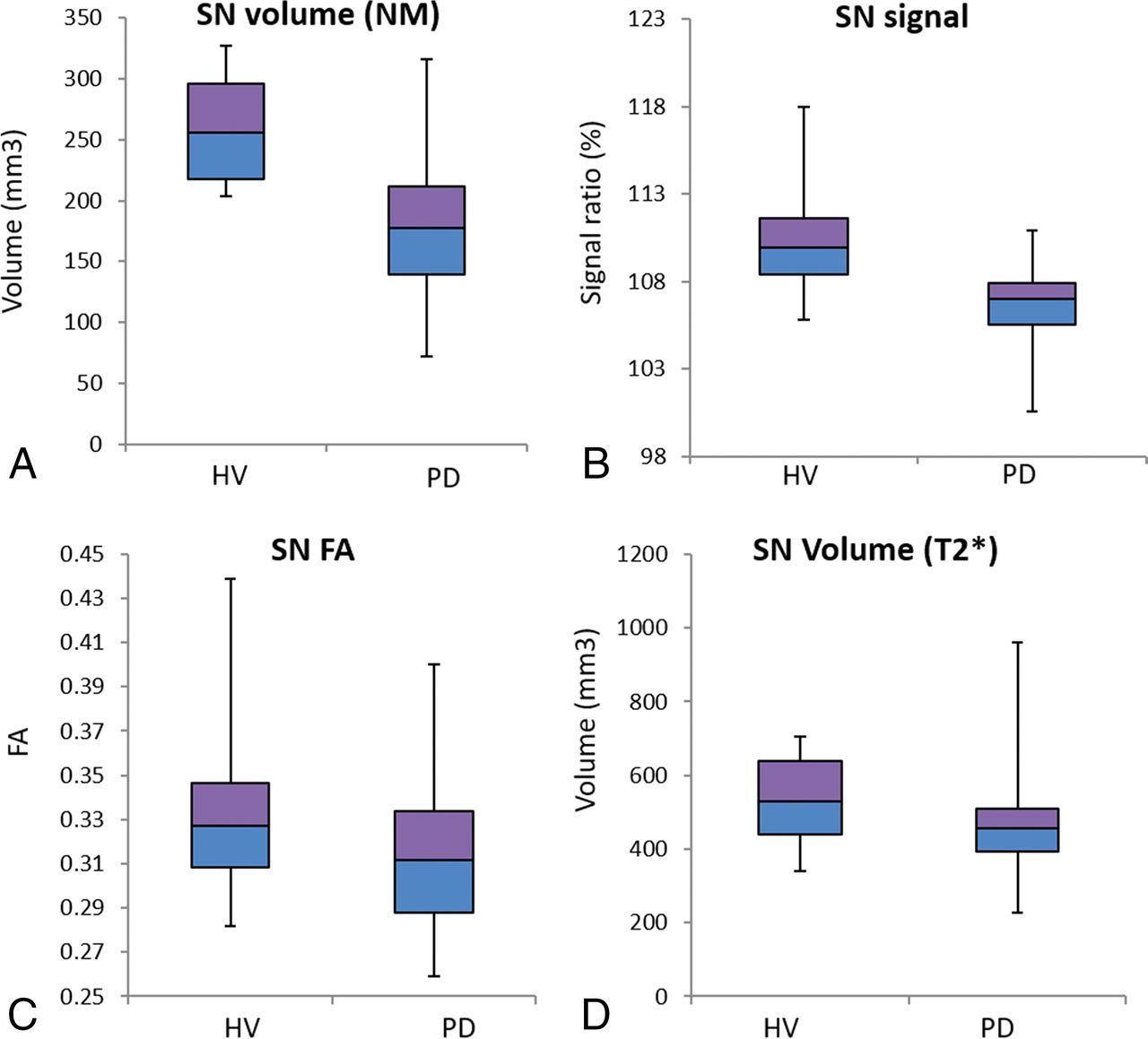
  • Fig 2.
    • Download figure
    • Open in new tab
    • Download powerpoint
    Fig 2.

    Boxplot of the 3T NM SN volume (cubic millimeters) (A) and signal intensity ratio (B) (calculated by normalizing the mean signal of the SN in each slice to the signal in the background region), the 3T NM FA (C), and the 7T T2*WI SN volume (D) shows a reduction in all measures in patients with PD compared with HVs.

  • Fig 3.
    • Download figure
    • Open in new tab
    • Download powerpoint
    Fig 3.

    Receiver operating characteristic curves of the 3T NM volume, the 3T NM signal intensity ratio, the 3T NM FA, the 7T T2* volume (A), and the combination of the first 3 biomarkers for differentiating patients with PD from HVs (B).

Tables

  • Figures
    • View popup
    Table 1:

    Quantitative measurements in the SNa

    SidePDHV
    3T NM ROI
        Volume
            L184.41 ± 68.84261.45 ± 47.68b
            R176.91 ± 62.89258.89 ± 46.62b
            Mean LR180.66 ± 63.63260.17 ± 42.87b
        Signal
            L106.74 ± 2.64110.21 ± 2.55b
            R106.61 ± 3.32110.46 ± 2.82b
            Mean LR110.56 ± 2.62110.33 ± 2.40b
        FA
            L0.32 ± 0.040.34 ± 0.04c
            R0.31 ± 0.040.33 ± 0.04
            Mean LR0.31 ± 0.030.33 ± 0.04c
        AD
            L0.68 ± 0.090.59 ± 0.10d
            R0.70 ± 0.080.62 ± 0.10c
            Mean LR0.69 ± 0.080.60 ± 0.10c
        RD
            L0.44 ± 0.060.38 ± 0.07d
            R0.46 ± 0.060.40 ± 0.07c
            Mean LR0.45 ± 0.060.39 ± 0.07c
        MD
            L0.52 ± 0.070.45 ± 0.08d
            R0.54 ± 0.060.48 ± 0.08c
            Mean LR0.53 ± 0.110.46 ± 0.08c
        R2*
            L35.91 ± 4.9536.66 ± 2.84
            R36.10 ± 6.6436.54 ± 2.80
            Mean LR35.65 ± 6.4636.60 ± 2.64
    3T T2 ROI
        FA
            L0.39 ± 0.060.42 ± 0.06
            R0.37 ± 0.060.40 ± 0.05
            Mean LR0.38 ± 0.060.40 ± 0.05
        AD
            L0.50 ± 0.090.45 ± 0.10
            R0.53 ± 0.100.49 ± 0.11
            Mean LR0.52 ± 0.090.47 ± 0.10
        RD
            L0.30 ± 0.060.26 ± 0.10c
            R0.33 ± 0.060.29 ± 0.06c
            Mean LR0.32 ± 0.060.28 ± 0.0c
        MD
            L0.37 ± 0.070.32 ± 0.07c
            R0.40 ± 0.070.35 ± 0.08c
            Mean LR0.38 ± 0.070.34 ± 0.08c
        R2*
            L28.47 ± 4.1730.21 ± 3.23
            R28.91 ± 3.9829.93 ± 2.87
            Mean LR28.69 ± 3.9730.07 ± 2.88
    7T T2*WI ROI
        T2WI volume
            L459.75 ± 120.74541.14 ± 118.80c
            R468.11 ± 130.00523.01 ± 121.85c
            Mean LR463.93 ± 122.83532.07 ± 118.33c
    • Note:—L indicates left; R, right; LR, average of left and right.

    • ↵a Quantitative measurements in the substantia nigra (mean and SD) are presented.

    • ↵b Significant differences between patients with PD and HVs are indicated using P < .001.

    • ↵c Significant differences between patients with PD and HVs are indicated using P < .05.

    • ↵d Significant differences between patients with PD and HVs are indicated using P < .005.

    • View popup
    Table 2:

    Diagnostic accuracy of quantitative and qualitative (visual) assessments

    SensitivitySpecificityPPVNPVAUC (95% CI)Diagnostic AccuracyCutoff
    Quantitative measurements
        3T NM signal0.880.80.880.80.88 (0.79–0.97)0.86110.35
        Volume10.720.8610.86 (0.76–0.95)0.89200.02
        7T T2*WI volume0.650.720.820.540.69 (0.53–0.84)0.68498.70
    Diffusion measures
        FA 3T NM0.750.530.750.550.68 (0.54–0.82)0.680.33
        3T T2WI0.650.700.80.520.64 (0.5–0.79)0.660.37
        AD 3T NM0.70.650.780.540.73 (0.59–0.87)0.680.64
        3T T2WI0.610.750.810.520.65 (0.49–0.80)0.660.5
        RD 3T NM0.640.750.820.540.72 (0.58–0.86)0.670.43
        3T T2WI0.720.60.760.550.68 (0.52–0.83)0.670.29
        MD 3T NM0.670.750.830.560.72 (0.58–0.87)0.70.51
        3T T2WI0.640.750.820.540.67 (0.53–0.82)0.670.38
    Combined measurements
        NM signal and volume + FA (3T NM)0.920.950.970.860.96 (0.9–1)0.934.4
    Qualitative evaluation
        NM at 3T0.830.90.940.9–0.86–
        DNH at 7T
            Method 10.970.80.90.94–0.91–
            Method 20.940.850.920.9–0.91–
    • Note:—AUC indicates area under the curve; NPV, negative predictive value; PPV, positive predictive value.

PreviousNext
Back to top

In this issue

American Journal of Neuroradiology: 39 (8)
American Journal of Neuroradiology
Vol. 39, Issue 8
1 Aug 2018
  • Table of Contents
  • Index by author
  • Complete Issue (PDF)
Advertisement
Print
Download PDF
Email Article

Thank you for your interest in spreading the word on American Journal of Neuroradiology.

NOTE: We only request your email address so that the person you are recommending the page to knows that you wanted them to see it, and that it is not junk mail. We do not capture any email address.

Enter multiple addresses on separate lines or separate them with commas.
Comparative Study of MRI Biomarkers in the Substantia Nigra to Discriminate Idiopathic Parkinson Disease
(Your Name) has sent you a message from American Journal of Neuroradiology
(Your Name) thought you would like to see the American Journal of Neuroradiology web site.
CAPTCHA
This question is for testing whether or not you are a human visitor and to prevent automated spam submissions.
Cite this article
N. Pyatigorskaya, B. Magnin, M. Mongin, L. Yahia-Cherif, R. Valabregue, D. Arnaldi, C. Ewenczyk, C. Poupon, M. Vidailhet, S. Lehéricy
Comparative Study of MRI Biomarkers in the Substantia Nigra to Discriminate Idiopathic Parkinson Disease
American Journal of Neuroradiology Aug 2018, 39 (8) 1460-1467; DOI: 10.3174/ajnr.A5702

Citation Manager Formats

  • BibTeX
  • Bookends
  • EasyBib
  • EndNote (tagged)
  • EndNote 8 (xml)
  • Medlars
  • Mendeley
  • Papers
  • RefWorks Tagged
  • Ref Manager
  • RIS
  • Zotero
0 Responses
Respond to this article
Share
Bookmark this article
Comparative Study of MRI Biomarkers in the Substantia Nigra to Discriminate Idiopathic Parkinson Disease
N. Pyatigorskaya, B. Magnin, M. Mongin, L. Yahia-Cherif, R. Valabregue, D. Arnaldi, C. Ewenczyk, C. Poupon, M. Vidailhet, S. Lehéricy
American Journal of Neuroradiology Aug 2018, 39 (8) 1460-1467; DOI: 10.3174/ajnr.A5702
del.icio.us logo Twitter logo Facebook logo Mendeley logo
  • Tweet Widget
  • Facebook Like
  • Google Plus One
Purchase

Jump to section

  • Article
    • Abstract
    • ABBREVIATIONS:
    • Materials and Methods
    • Results
    • Discussion
    • Conclusions
    • Footnotes
    • References
  • Figures & Data
  • Info & Metrics
  • Responses
  • References
  • PDF

Related Articles

  • No related articles found.
  • PubMed
  • Google Scholar

Cited By...

  • Nigral volume loss in prodromal, early, and moderate Parkinsons disease
  • In vivo detection of substantia nigra and locus coeruleus volume loss in Parkinsons disease using neuromelanin-sensitive MRI: Replication in two cohorts
  • Multi view based imaging genetics analysis on Parkinson disease
  • Parkinson Disease Propagation Using MRI Biomarkers and Partial Least Squares Path Modeling
  • Crossref
  • Google Scholar

This article has not yet been cited by articles in journals that are participating in Crossref Cited-by Linking.

More in this TOC Section

  • Diagnostic Neuroradiology of Monoclonal Antibodies
  • Clinical Outcomes After Chiari I Decompression
  • Segmentation of Brain Metastases with BLAST
Show more Adult Brain

Similar Articles

Advertisement

Indexed Content

  • Current Issue
  • Accepted Manuscripts
  • Article Preview
  • Past Issues
  • Editorials
  • Editor's Choice
  • Fellows' Journal Club
  • Letters to the Editor
  • Video Articles

Cases

  • Case Collection
  • Archive - Case of the Week
  • Archive - Case of the Month
  • Archive - Classic Case

More from AJNR

  • Trainee Corner
  • Imaging Protocols
  • MRI Safety Corner
  • Book Reviews

Multimedia

  • AJNR Podcasts
  • AJNR Scantastics

Resources

  • Turnaround Time
  • Submit a Manuscript
  • Submit a Video Article
  • Submit an eLetter to the Editor/Response
  • Manuscript Submission Guidelines
  • Statistical Tips
  • Fast Publishing of Accepted Manuscripts
  • Graphical Abstract Preparation
  • Imaging Protocol Submission
  • Evidence-Based Medicine Level Guide
  • Publishing Checklists
  • Author Policies
  • Become a Reviewer/Academy of Reviewers
  • News and Updates

About Us

  • About AJNR
  • Editorial Board
  • Editorial Board Alumni
  • Alerts
  • Permissions
  • Not an AJNR Subscriber? Join Now
  • Advertise with Us
  • Librarian Resources
  • Feedback
  • Terms and Conditions
  • AJNR Editorial Board Alumni

American Society of Neuroradiology

  • Not an ASNR Member? Join Now

© 2025 by the American Society of Neuroradiology All rights, including for text and data mining, AI training, and similar technologies, are reserved.
Print ISSN: 0195-6108 Online ISSN: 1936-959X

Powered by HighWire