
Article Figures & Data
Figures
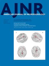
- Fig 1.
Magnified lesion views taken from axial slices of patients with RRMS enrolled in our study. CVS lesions are shown for each brain region: infratentorial (A), juxtacortical (B), periventricular (C), and subcortical lesions (D). For the lesions in the periventricular and subcortical regions, a hypointense rim is observed around the lesion on the IEV-SWI. Arrows point to select lesions and the central vessels running through them. Magnified panels range from 3.0 to 4.0 cm.
- Fig 2.
Examples of juxtacortical, periventricular, and subcortical lesions for HC participants. Arrows identify central veins running though the body of WML. IEV-SWI allows the visualization of CVS submillimeter vessels enabling accurate definition of %PVWML. Magnified panels range from 2.0 to 3.0 cm.
- Fig 3.
Average %PVWMLPk presented for the RRMS and HC groups in the LL lesion pool (A), NC lesion pool (B), and SV lesion pool (C). A large number of WML with central veins are observed to have multiple central veins. The removal of lesions with multiple central veins results in a wider spread in the data. This yields a reduced diagnostic value of %PVWML for the NC lesion pool (C). The average %PVWML is found to be significantly different between the RRMS and HC groups for all lesion pools as per the Mann-Whitney U test (P < .001).
- Fig 4.
The diagnostic accuracy of the Average %PVWMLPk, as analyzed by the receiver operating characteristic test, presented for the pool of all the lesions of >3 mm (LL pool), the pool of nonconfluent lesions of >3 mm (NC pool), and the pool of nonconfluent lesions of >3 mm with a single central vessel (SV pool).


Tables
Control Subjects Patients with RRMS No. of subjects 18 17 No. of women 15 11 No. of men 3 7 Age (yr)b 37.4 ± 5.8 (26–46) 39.4 ± 5.4 (26–46) EDSS NA 2.2 ± 1.6 (0–6) EDSS to scan time gap (days) NA 297 ± 49 LL Lesion Pool NC Lesion Pool SV Lesion Pool Bias ± SD 95% LoA Bias ± SD 95% LoA Bias ± SD 95% LoA R1, R2 4.4 ± 8.5 −12.3:21.1 4.0 ± 10.1 −23.7:15.7 −12.7 ± 20.6 −53:27.7 R1, R3 (1) 16.4 ± 9.8 −2.9:35.6 −12.5 ± 3.3 −19.0:−6.0 −22.3 ± 12.6 −47:2.4 R1, R3 (2) 1.9 ± 6.7 −11.3:15.1 −12.7 ± 3.1 −18.7:−6.6 −22.9 ± 11.6 −45.6:−0.2 R2, R3 (1) 12 ± 11.5 −10.5:34.5 −8.5 ± 7.6 −23.4:6.3 −9.6 ± 8.6 −26.4:7.2 R2, R3 (2) 2.4 ± 11.1 −24.2:19.3 −8.7 ± 7.8 −23.8:6.5 −10.3 ± 9.4 −28.6:8.1 R3 (1), R3 (2) −14.5 ± 3.8 −21.9:−7.1 −0.1 ± 0.2 −0.6:0.3 −0.6 ± 1.1 −2.8:1.5 Note:—R1 indicates reader 1; R2, reader 2; R3 (1), reader 3, first assessment; R3 (2) reader 3, second assessment; LoA, limits of agreement.
- Table 3:
Location-specific and averaged %PVWML (averaged over the 3 readers) are presented for each of the lesion poolsa
RRMS HC At Each Locationb Averagec At Each Locationb Averagec LL lesion pool Average %PVWMLinfra 10 ± 18 55 ± 14 0 ± 0 5 ± 6 Average %PVWMLjuxta 59 ± 31 3 ± 8 Average %PVWMLperi 68 ± 35 4 ± 10 Average %PVWMLsubcort 82 ± 16 12 ± 17 NC lesion pool Average %PVWMLinfra 3 ± 8 91 ± 15 0 ± 0 18 ± 23 Average %PVWMLjuxta 50 ± 34 3 ± 8 Average %PVWMLperi 47 ± 31 1 ± 6 Average %PVWMLsubcort 84 ± 17 16 ± 21 SV lesion pool Average %PVWMLinfra 2 ± 6 76 ± 24 0 ± 0 17 ± 23 Average %PVWMLjuxta 52 ± 39 3 ± 8 Average %PVWMLperi 20 ± 23 0 ± 0 Average %PVWMLsubcort 78 ± 22 16 ± 21 LL Lesion Pool NC Lesion Pool SV Lesion Pool Infratentorial region Threshold; sensitivity, 95% CI >13%; 29%, 10%–56% >13%; 12%, 2%–36% >13%; 6%, 0%–29% Specificity, 95% CI 100%, 82%–100% 100%, 82%–100% 100%, 82%–100% AUC 0.65 0.56 0.53 Juxtacortical region Threshold; sensitivity, 95% CI >19%; 82%, 57%–96% >7%; 82%, 57%–96%, >29%; 65%, 38%–86% Specificity, 95% CI 89%, 65%–99% 89%, 65%–99% 100%, 82%–100% AUC 0.93 0.89 0.86 Periventricular region Threshold; sensitivity, 95% CI >13%; 88%, 64%–99% >13%; 82%, 57%–96% >13%; 53%, 28%–77% Specificity, 95% CI 83%, 59%–96% 94%, 73%–100% 100%, 82%–100% AUC 0.93 0.90 0.77 Subcortical region Threshold; sensitivity, 95% CI >51%; 94%, 71%–100% >61%; 94%, 71%–100% >61%; 82%, 57%–96% Specificity, 95% CI 100%, 82%–100% 100%, 82%–100% 100%, 82%–100% AUC 0.99 0.99 0.96 Averaged results (over brain volume) Threshold; sensitivity, 95% CI >30%; 94%, 71%–100% >67%; 94%, 71%–100% >66%; 77%, 50%–93% Specificity, 95% CI 100%, 82%–100% 100%, 82%–100% 100%, 82%–100% AUC 0.99 0.99 0.95 Note:—AUC indicates area under the receiver operating characteristic curve.


Jump to section
Related Articles
Cited By...
- Central Vein Sign in Multiple Sclerosis: A Comparison Study of the Diagnostic Performance of 3T versus 7T MRI
- Value of 3T Susceptibility-Weighted Imaging in the Diagnosis of Multiple Sclerosis
- The Central Vein Sign in Radiologically Isolated Syndrome
- Gadolinium-Enhanced Susceptibility-Weighted Imaging in Multiple Sclerosis: Optimizing the Recognition of Active Plaques for Different MR Imaging Sequences