Advertisement

Main menu

  • Home
  • Content
    • Current Issue
    • Accepted Manuscripts
    • Article Preview
    • Past Issue Archive
    • Video Articles
    • AJNR Case Collection
    • Case of the Week Archive
    • Case of the Month Archive
    • Classic Case Archive
  • Special Collections
    • AJNR Awards
    • Low-Field MRI
    • Alzheimer Disease
    • ASNR Foundation Special Collection
    • Photon-Counting CT
    • View All
  • Multimedia
    • AJNR Podcasts
    • AJNR SCANtastic
    • Trainee Corner
    • MRI Safety Corner
    • Imaging Protocols
  • For Authors
    • Submit a Manuscript
    • Submit a Video Article
    • Submit an eLetter to the Editor/Response
    • Manuscript Submission Guidelines
    • Statistical Tips
    • Fast Publishing of Accepted Manuscripts
    • Graphical Abstract Preparation
    • Imaging Protocol Submission
    • Author Policies
  • About Us
    • About AJNR
    • Editorial Board
    • Editorial Board Alumni
  • More
    • Become a Reviewer/Academy of Reviewers
    • Subscribers
    • Permissions
    • Alerts
    • Feedback
    • Advertisers
    • ASNR Home

User menu

  • Alerts
  • Log in

Search

  • Advanced search
American Journal of Neuroradiology

American Journal of Neuroradiology

ASHNR American Society of Functional Neuroradiology ASHNR American Society of Pediatric Neuroradiology ASSR
  • Alerts
  • Log in

Advanced Search

  • Home
  • Content
    • Current Issue
    • Accepted Manuscripts
    • Article Preview
    • Past Issue Archive
    • Video Articles
    • AJNR Case Collection
    • Case of the Week Archive
    • Case of the Month Archive
    • Classic Case Archive
  • Special Collections
    • AJNR Awards
    • Low-Field MRI
    • Alzheimer Disease
    • ASNR Foundation Special Collection
    • Photon-Counting CT
    • View All
  • Multimedia
    • AJNR Podcasts
    • AJNR SCANtastic
    • Trainee Corner
    • MRI Safety Corner
    • Imaging Protocols
  • For Authors
    • Submit a Manuscript
    • Submit a Video Article
    • Submit an eLetter to the Editor/Response
    • Manuscript Submission Guidelines
    • Statistical Tips
    • Fast Publishing of Accepted Manuscripts
    • Graphical Abstract Preparation
    • Imaging Protocol Submission
    • Author Policies
  • About Us
    • About AJNR
    • Editorial Board
    • Editorial Board Alumni
  • More
    • Become a Reviewer/Academy of Reviewers
    • Subscribers
    • Permissions
    • Alerts
    • Feedback
    • Advertisers
    • ASNR Home
  • Follow AJNR on Twitter
  • Visit AJNR on Facebook
  • Follow AJNR on Instagram
  • Join AJNR on LinkedIn
  • RSS Feeds

AJNR Awards, New Junior Editors, and more. Read the latest AJNR updates

Research ArticleAdult Brain

Evaluation of Thick-Slab Overlapping MIP Images of Contrast-Enhanced 3D T1-Weighted CUBE for Detection of Intracranial Metastases: A Pilot Study for Comparison of Lesion Detection, Interpretation Time, and Sensitivity with Nonoverlapping CUBE MIP, CUBE, and Inversion-Recovery-Prepared Fast-Spoiled Gradient Recalled Brain Volume

B.C. Yoon, A.F. Saad, P. Rezaii, M. Wintermark, G. Zaharchuk and M. Iv
American Journal of Neuroradiology September 2018, 39 (9) 1635-1642; DOI: https://doi.org/10.3174/ajnr.A5747
B.C. Yoon
aFrom the Department of Radiology, Division of Neuroimaging and Neurointervention, Stanford University, Stanford, California.
  • Find this author on Google Scholar
  • Find this author on PubMed
  • Search for this author on this site
  • ORCID record for B.C. Yoon
A.F. Saad
aFrom the Department of Radiology, Division of Neuroimaging and Neurointervention, Stanford University, Stanford, California.
  • Find this author on Google Scholar
  • Find this author on PubMed
  • Search for this author on this site
  • ORCID record for A.F. Saad
P. Rezaii
aFrom the Department of Radiology, Division of Neuroimaging and Neurointervention, Stanford University, Stanford, California.
  • Find this author on Google Scholar
  • Find this author on PubMed
  • Search for this author on this site
  • ORCID record for P. Rezaii
M. Wintermark
aFrom the Department of Radiology, Division of Neuroimaging and Neurointervention, Stanford University, Stanford, California.
  • Find this author on Google Scholar
  • Find this author on PubMed
  • Search for this author on this site
  • ORCID record for M. Wintermark
G. Zaharchuk
aFrom the Department of Radiology, Division of Neuroimaging and Neurointervention, Stanford University, Stanford, California.
  • Find this author on Google Scholar
  • Find this author on PubMed
  • Search for this author on this site
  • ORCID record for G. Zaharchuk
M. Iv
aFrom the Department of Radiology, Division of Neuroimaging and Neurointervention, Stanford University, Stanford, California.
  • Find this author on Google Scholar
  • Find this author on PubMed
  • Search for this author on this site
  • ORCID record for M. Iv
  • Article
  • Figures & Data
  • Info & Metrics
  • Responses
  • References
  • PDF
Loading

Article Figures & Data

Figures

  • Tables
  • Fig 1.
    • Download figure
    • Open in new tab
    • Download powerpoint
    Fig 1.

    Total number of brain metastases detected. No significant difference was found among the total number of metastases detected using CUBE, nC-MIP, oC-MIP, and IR-FSPGR-BRAVO (P = .062) using 1-way ANOVA with a Bonferroni adjustment. The orange line denotes the total number of ground truth lesions (n = 308).

  • Fig 2.
    • Download figure
    • Open in new tab
    • Download powerpoint
    Fig 2.

    Mean time for interpretation. A, Both readers had significantly reduced interpretation time using nC-MIP and oC-MIP compared with CUBE and IR-FSPGR-BRAVO, saving at least 50 seconds per case (on average). B, The use of nC-MIP and oC-MIP with the option to cross-reference an equivocal lesion to the source images (nC-MIP+XR and oC-MIP+XR, respectively) did not result in a significant change in interpretation time compared with the use of nC-MIP or oC-MIP alone. However, time for interpretation for all CUBE MIPs was significantly reduced compared with CUBE. Error bars represent the SD. One-way ANOVA with a Bonferroni adjustment. Triple asterisks indicate P < .001; ns, no significance.
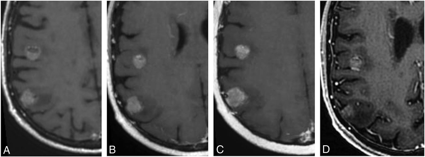
  • Fig 3.
    • Download figure
    • Open in new tab
    • Download powerpoint
    Fig 3.

    Enhancing cerebral metastases in a 74-year-old male with metastatic tongue squamous cell carcinoma. Postcontrast T1-weighted CUBE (A), nonoverlapping CUBE MIP (nC-MIP) (B), overlapping CUBE MIP (oC-MIP) (C), and IR-FSPGR-BRAVO (D) images demonstrate enhancing metastatic lesions. The lesions appear most conspicuous with CUBE MIPs (B and C). The contrast-to-noise ratio of lesions was highest for oC-MIP (C).

  • Fig 4.
    • Download figure
    • Open in new tab
    • Download powerpoint
    Fig 4.

    Comparison between nonoverlapping and overlapping CUBE MIP. A small 3-mm metastasis in a 57-year-old woman with non-small cell lung cancer is seen across 4 different slices with oC-MIP but is only identified on 2 slices with nC-MIP.

Tables

  • Figures
    • View popup
    Table 1:

    Interrater agreement using the Cohen κ coefficient for T1-weighted CUBE, nC-MIP, oC-MIP, and IR-FSPGR-BRAVO

    Image Typeκ95% Confidence IntervalAgreement
    CUBE0.2350.024–0.447Fair
    nC-MIP0.2220.069–0.374Fair
    oC-MIP0.5980.371–0.825Moderate
    IR-FSPGR-BRAVO0.4450.290–0.599Moderate
    • View popup
    Table 2:

    CNR of the smallest and largest metastases on CUBE, nC-MIP, oC-MIP, IR-FSPGR-BRAVO, and overlapping IR-FSPGR-BRAVO MIPa

    Image TypeCNR of the Smallest Lesion (<4 mm)CNR of the Largest Lesion
    CUBE2.35 ± 2.02.49 ± 1.84
    nC-MIP1.55 ± 0.532.69 ± 0.93
    oC-MIP2.35 ± 1.643.23 ± 1.92
    IR-FSPGR-BRAVO0.62 ± 0.391.45 ± 1.14
    IR-FSPGR-BRAVO MIP0.90 ± 0.781.69 ± 1.14
    • ↵a Numbers are means.

    • View popup
    Table 3:

    Sensitivity, number of false-negatives, number of false-positives, and number of discrepant lesions (FN + FP) per case for CUBE, nC-MIP, oC-MIP, IR-FSPGR-BRAVO, and non-overlapping and overlapping CUBE MIPsa

    Reader 1Reader 2
    % SensitivityMean FNMean FPMean Discrepancy% SensitivityMean FNMean FPMean Discrepancy
    CUBE97.1 ± 14.80.08 ± 0.280.31 ± 0.630.40 ± 0.6593.0 ± 16.80.38 ± 0.700.38 ± 0.820.75 ± 0.93
    nC-MIP94.7 ± 20.40.08 ± 0.280.58 ± 0.980.65 ± 0.9690.2 ± 16.80.60 ± 0.960.71 ± 1.981.31 ± 2.00
    oC-MIP95.8 ± 15.40.19 ± 0.490.54 ± 0.920.73 ± 0.9495.8 ± 15.40.19 ± 0.500.33 ± 0.690.75 ± 0.93
    BRAVO91.5 ± 17.00.58 ± 0.990.21 ± 0.500.79 ± 0.9989.0 ± 19.50.85 ± 1.490.31 ± 0.591.17 ± 1.42
    nC-MIP + XR95.5 ± 9.80.33 ± 0.660.25 ± 0.700.58 ± 0.8791.2 ± 12.30.67 ± 0.930.29 ± 0.740.95 ± 1.01
    oC-MIP + XR96.6 ± 7.30.29 ± 0.620.21 ± 0.460.68 ± 0.9694.8 ± 12.30.38 ± 0.790.23 ± 0.470.60 ± 0.82
    • Note:—FN indicates false-negative; FP, false-positive.

    • ↵a With the option to cross-reference a lesion to the source images (nC-MIP+XR and oC-MIP+XR, respectively) for both readers. Numbers are means.

PreviousNext
Back to top

In this issue

American Journal of Neuroradiology: 39 (9)
American Journal of Neuroradiology
Vol. 39, Issue 9
1 Sep 2018
  • Table of Contents
  • Index by author
  • Complete Issue (PDF)
Advertisement
Print
Download PDF
Email Article

Thank you for your interest in spreading the word on American Journal of Neuroradiology.

NOTE: We only request your email address so that the person you are recommending the page to knows that you wanted them to see it, and that it is not junk mail. We do not capture any email address.

Enter multiple addresses on separate lines or separate them with commas.
Evaluation of Thick-Slab Overlapping MIP Images of Contrast-Enhanced 3D T1-Weighted CUBE for Detection of Intracranial Metastases: A Pilot Study for Comparison of Lesion Detection, Interpretation Time, and Sensitivity with Nonoverlapping CUBE MIP, CUBE, …
(Your Name) has sent you a message from American Journal of Neuroradiology
(Your Name) thought you would like to see the American Journal of Neuroradiology web site.
CAPTCHA
This question is for testing whether or not you are a human visitor and to prevent automated spam submissions.
Cite this article
B.C. Yoon, A.F. Saad, P. Rezaii, M. Wintermark, G. Zaharchuk, M. Iv
Evaluation of Thick-Slab Overlapping MIP Images of Contrast-Enhanced 3D T1-Weighted CUBE for Detection of Intracranial Metastases: A Pilot Study for Comparison of Lesion Detection, Interpretation Time, and Sensitivity with Nonoverlapping CUBE MIP, CUBE, and Inversion-Recovery-Prepared Fast-Spoiled Gradient Recalled Brain Volume
American Journal of Neuroradiology Sep 2018, 39 (9) 1635-1642; DOI: 10.3174/ajnr.A5747

Citation Manager Formats

  • BibTeX
  • Bookends
  • EasyBib
  • EndNote (tagged)
  • EndNote 8 (xml)
  • Medlars
  • Mendeley
  • Papers
  • RefWorks Tagged
  • Ref Manager
  • RIS
  • Zotero
0 Responses
Respond to this article
Share
Bookmark this article
Evaluation of Thick-Slab Overlapping MIP Images of Contrast-Enhanced 3D T1-Weighted CUBE for Detection of Intracranial Metastases: A Pilot Study for Comparison of Lesion Detection, Interpretation Time, and Sensitivity with Nonoverlapping CUBE MIP, CUBE, and Inversion-Recovery-Prepared Fast-Spoiled Gradient Recalled Brain Volume
B.C. Yoon, A.F. Saad, P. Rezaii, M. Wintermark, G. Zaharchuk, M. Iv
American Journal of Neuroradiology Sep 2018, 39 (9) 1635-1642; DOI: 10.3174/ajnr.A5747
del.icio.us logo Twitter logo Facebook logo Mendeley logo
  • Tweet Widget
  • Facebook Like
  • Google Plus One
Purchase

Jump to section

  • Article
    • Abstract
    • ABBREVIATIONS:
    • Materials and Methods
    • Results
    • Discussion
    • Conclusions
    • Footnotes
    • References
  • Figures & Data
  • Info & Metrics
  • Responses
  • References
  • PDF

Related Articles

  • No related articles found.
  • PubMed
  • Google Scholar

Cited By...

  • No citing articles found.
  • Crossref
  • Google Scholar

This article has not yet been cited by articles in journals that are participating in Crossref Cited-by Linking.

More in this TOC Section

  • Diagnostic Neuroradiology of Monoclonal Antibodies
  • Clinical Outcomes After Chiari I Decompression
  • Segmentation of Brain Metastases with BLAST
Show more Adult Brain

Similar Articles

Advertisement

Indexed Content

  • Current Issue
  • Accepted Manuscripts
  • Article Preview
  • Past Issues
  • Editorials
  • Editor's Choice
  • Fellows' Journal Club
  • Letters to the Editor
  • Video Articles

Cases

  • Case Collection
  • Archive - Case of the Week
  • Archive - Case of the Month
  • Archive - Classic Case

More from AJNR

  • Trainee Corner
  • Imaging Protocols
  • MRI Safety Corner
  • Book Reviews

Multimedia

  • AJNR Podcasts
  • AJNR Scantastics

Resources

  • Turnaround Time
  • Submit a Manuscript
  • Submit a Video Article
  • Submit an eLetter to the Editor/Response
  • Manuscript Submission Guidelines
  • Statistical Tips
  • Fast Publishing of Accepted Manuscripts
  • Graphical Abstract Preparation
  • Imaging Protocol Submission
  • Evidence-Based Medicine Level Guide
  • Publishing Checklists
  • Author Policies
  • Become a Reviewer/Academy of Reviewers
  • News and Updates

About Us

  • About AJNR
  • Editorial Board
  • Editorial Board Alumni
  • Alerts
  • Permissions
  • Not an AJNR Subscriber? Join Now
  • Advertise with Us
  • Librarian Resources
  • Feedback
  • Terms and Conditions
  • AJNR Editorial Board Alumni

American Society of Neuroradiology

  • Not an ASNR Member? Join Now

© 2025 by the American Society of Neuroradiology All rights, including for text and data mining, AI training, and similar technologies, are reserved.
Print ISSN: 0195-6108 Online ISSN: 1936-959X

Powered by HighWire