Advertisement

Main menu

  • Home
  • Content
    • Current Issue
    • Accepted Manuscripts
    • Article Preview
    • Past Issue Archive
    • Video Articles
    • AJNR Case Collection
    • Case of the Week Archive
    • Case of the Month Archive
    • Classic Case Archive
  • Special Collections
    • AJNR Awards
    • Low-Field MRI
    • Alzheimer Disease
    • ASNR Foundation Special Collection
    • Photon-Counting CT
    • View All
  • Multimedia
    • AJNR Podcasts
    • AJNR SCANtastic
    • Trainee Corner
    • MRI Safety Corner
    • Imaging Protocols
  • For Authors
    • Submit a Manuscript
    • Submit a Video Article
    • Submit an eLetter to the Editor/Response
    • Manuscript Submission Guidelines
    • Statistical Tips
    • Fast Publishing of Accepted Manuscripts
    • Graphical Abstract Preparation
    • Imaging Protocol Submission
    • Author Policies
  • About Us
    • About AJNR
    • Editorial Board
    • Editorial Board Alumni
  • More
    • Become a Reviewer/Academy of Reviewers
    • Subscribers
    • Permissions
    • Alerts
    • Feedback
    • Advertisers
    • ASNR Home

User menu

  • Alerts
  • Log in

Search

  • Advanced search
American Journal of Neuroradiology

American Journal of Neuroradiology

ASHNR American Society of Functional Neuroradiology ASHNR American Society of Pediatric Neuroradiology ASSR
  • Alerts
  • Log in

Advanced Search

  • Home
  • Content
    • Current Issue
    • Accepted Manuscripts
    • Article Preview
    • Past Issue Archive
    • Video Articles
    • AJNR Case Collection
    • Case of the Week Archive
    • Case of the Month Archive
    • Classic Case Archive
  • Special Collections
    • AJNR Awards
    • Low-Field MRI
    • Alzheimer Disease
    • ASNR Foundation Special Collection
    • Photon-Counting CT
    • View All
  • Multimedia
    • AJNR Podcasts
    • AJNR SCANtastic
    • Trainee Corner
    • MRI Safety Corner
    • Imaging Protocols
  • For Authors
    • Submit a Manuscript
    • Submit a Video Article
    • Submit an eLetter to the Editor/Response
    • Manuscript Submission Guidelines
    • Statistical Tips
    • Fast Publishing of Accepted Manuscripts
    • Graphical Abstract Preparation
    • Imaging Protocol Submission
    • Author Policies
  • About Us
    • About AJNR
    • Editorial Board
    • Editorial Board Alumni
  • More
    • Become a Reviewer/Academy of Reviewers
    • Subscribers
    • Permissions
    • Alerts
    • Feedback
    • Advertisers
    • ASNR Home
  • Follow AJNR on Twitter
  • Visit AJNR on Facebook
  • Follow AJNR on Instagram
  • Join AJNR on LinkedIn
  • RSS Feeds

AJNR Awards, New Junior Editors, and more. Read the latest AJNR updates

Research ArticlePediatric Neuroimaging

“Ears of the Lynx” MRI Sign Is Associated with SPG11 and SPG15 Hereditary Spastic Paraplegia

B. Pascual, S.T. de Bot, M.R. Daniels, M.C. França, C. Toro, M. Riverol, P. Hedera, M.T. Bassi, N. Bresolin, B.P. van de Warrenburg, B. Kremer, J. Nicolai, P. Charles, J. Xu, S. Singh, N.J. Patronas, S.H. Fung, M.D. Gregory and J.C. Masdeu
American Journal of Neuroradiology January 2019, 40 (1) 199-203; DOI: https://doi.org/10.3174/ajnr.A5935
B. Pascual
aFrom the Departments of Neurology (B.P., M.R.D., J.C.M.)
  • Find this author on Google Scholar
  • Find this author on PubMed
  • Search for this author on this site
  • ORCID record for B. Pascual
S.T. de Bot
dDepartment of Neurology (S.T.d.B.), Leiden University Medical Centre, Leiden, the Netherlands
  • Find this author on Google Scholar
  • Find this author on PubMed
  • Search for this author on this site
  • ORCID record for S.T. de Bot
M.R. Daniels
aFrom the Departments of Neurology (B.P., M.R.D., J.C.M.)
  • Find this author on Google Scholar
  • Find this author on PubMed
  • Search for this author on this site
  • ORCID record for M.R. Daniels
M.C. França Jr
eDepartment of Neurology (M.C.F.), University of Campinas, Campinas, Brazil
  • Find this author on Google Scholar
  • Find this author on PubMed
  • Search for this author on this site
  • ORCID record for M.C. França Jr
C. Toro
fNational Institutes of Health Intramural Research Program (C.T., N.J.P., M.D.G.), Bethesda, Maryland
  • Find this author on Google Scholar
  • Find this author on PubMed
  • Search for this author on this site
  • ORCID record for C. Toro
M. Riverol
gDepartment of Neurology (M.R.), Clínica Universidad de Navarra, Pamplona, Spain
  • Find this author on Google Scholar
  • Find this author on PubMed
  • Search for this author on this site
  • ORCID record for M. Riverol
P. Hedera
hDepartment of Neurology (P.H.), Vanderbilt University Medical Center, Nashville, Tennessee
  • Find this author on Google Scholar
  • Find this author on PubMed
  • Search for this author on this site
  • ORCID record for P. Hedera
M.T. Bassi
iLaboratory of Molecular Biology (M.T.B.), Scientific Institute Istituto di Ricovero e Cura a Carattere Scientifico E. Medea, Bosisio Parini, Lecco, Italy
  • Find this author on Google Scholar
  • Find this author on PubMed
  • Search for this author on this site
  • ORCID record for M.T. Bassi
N. Bresolin
jDepartment of Neuroscience and Mental Health (N.B.), University Hospital Policlinico Ca'Granda, University of Milan, Milan, Italy
  • Find this author on Google Scholar
  • Find this author on PubMed
  • Search for this author on this site
  • ORCID record for N. Bresolin
B.P. van de Warrenburg
kDepartment of Neurology (B.P.v.d.W.), Donders Institute for Brain, Cognition, and Behaviour, Radboud University Medical Center, Nijmegen, the Netherlands
  • Find this author on Google Scholar
  • Find this author on PubMed
  • Search for this author on this site
  • ORCID record for B.P. van de Warrenburg
B. Kremer
lDepartment of Neurology (B.K.), University Medical Center Groningen, Groningen, the Netherlands
  • Find this author on Google Scholar
  • Find this author on PubMed
  • Search for this author on this site
  • ORCID record for B. Kremer
J. Nicolai
mDepartment of Neurology (J.N.), Maastricht University Medical Centre, Maastricht, the Netherlands
  • Find this author on Google Scholar
  • Find this author on PubMed
  • Search for this author on this site
  • ORCID record for J. Nicolai
P. Charles
nDepartment of Genetics (P.C.), Hôpital Pitié-Salpêtrière, Paris, France.
  • Find this author on Google Scholar
  • Find this author on PubMed
  • Search for this author on this site
  • ORCID record for P. Charles
J. Xu
bBiostatistics (J.X.)
  • Find this author on Google Scholar
  • Find this author on PubMed
  • Search for this author on this site
  • ORCID record for J. Xu
S. Singh
cRadiology (S.S., S.H.F.), Houston Methodist Research Institute, Houston, Texas
  • Find this author on Google Scholar
  • Find this author on PubMed
  • Search for this author on this site
  • ORCID record for S. Singh
N.J. Patronas
fNational Institutes of Health Intramural Research Program (C.T., N.J.P., M.D.G.), Bethesda, Maryland
  • Find this author on Google Scholar
  • Find this author on PubMed
  • Search for this author on this site
  • ORCID record for N.J. Patronas
S.H. Fung
cRadiology (S.S., S.H.F.), Houston Methodist Research Institute, Houston, Texas
  • Find this author on Google Scholar
  • Find this author on PubMed
  • Search for this author on this site
  • ORCID record for S.H. Fung
M.D. Gregory
fNational Institutes of Health Intramural Research Program (C.T., N.J.P., M.D.G.), Bethesda, Maryland
  • Find this author on Google Scholar
  • Find this author on PubMed
  • Search for this author on this site
  • ORCID record for M.D. Gregory
J.C. Masdeu
aFrom the Departments of Neurology (B.P., M.R.D., J.C.M.)
  • Find this author on Google Scholar
  • Find this author on PubMed
  • Search for this author on this site
  • ORCID record for J.C. Masdeu
  • Article
  • Figures & Data
  • Supplemental
  • Info & Metrics
  • Responses
  • References
  • PDF
Loading

Article Figures & Data

Figures

  • Tables
  • Fig 1.
    • Download figure
    • Open in new tab
    • Download powerpoint
    Fig 1.

    Corpus callosum size similar to normal in a patient with HSP-TCC. The patient had mild cognitive impairment and a mild spastic paraparesis. She had SPG 11 pathogenetic mutations. The ear of the lynx sign in this patient is shown in Fig 2.

  • Fig 2.
    • Download figure
    • Open in new tab
    • Download powerpoint
    Fig 2.

    Ears of the lynx on MR imaging. Axial images across the anterior forceps of the corpus callosum. Note an abnormality in the region of the forceps minor of the corpus callosum, corresponding to the genu fibers, which appear dark on T1-weighted and bright on FLAIR images (arrows). Midline sagittal images from the same individuals are seen in Fig 1.
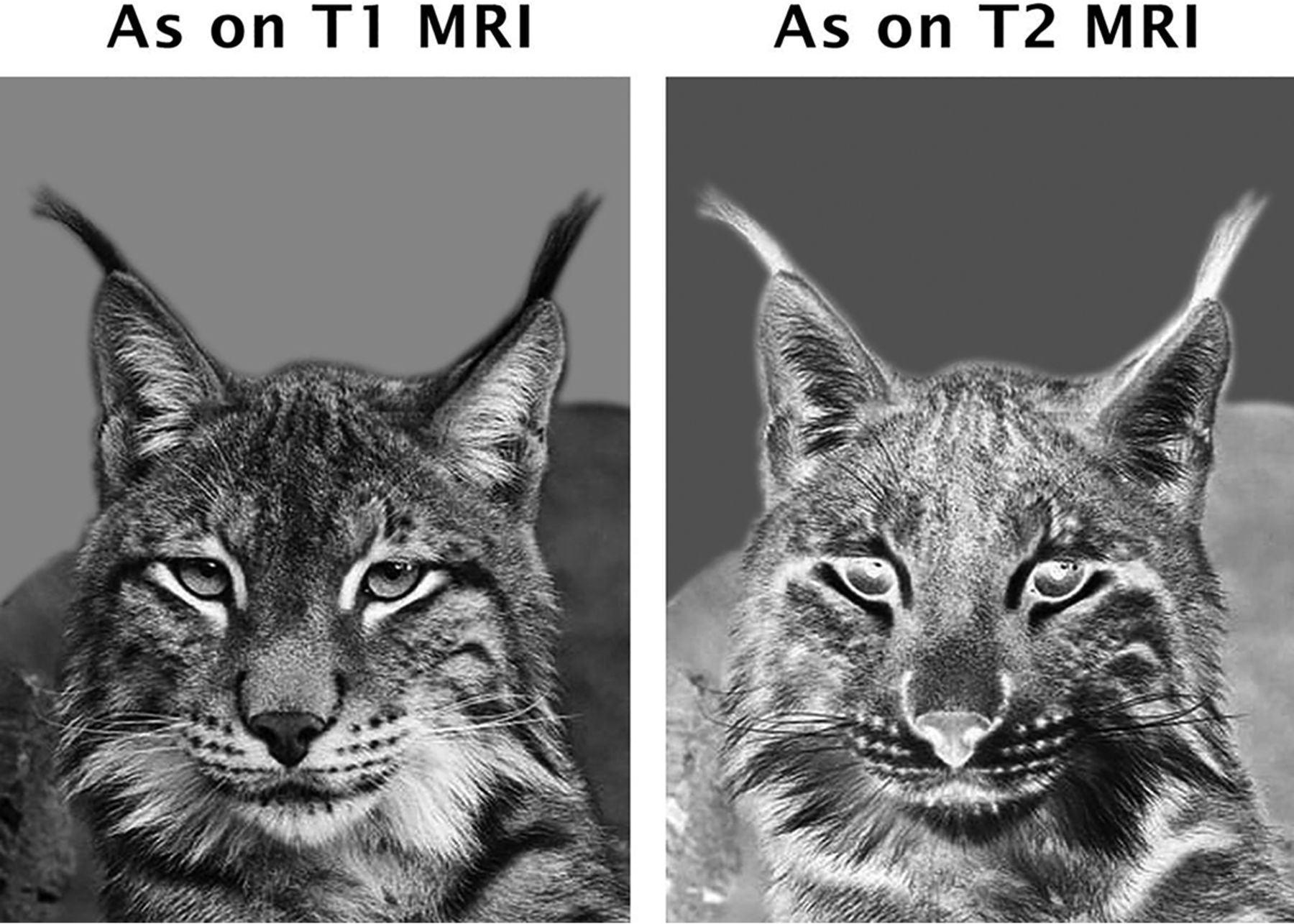
  • Fig 3.
    • Download figure
    • Open in new tab
    • Download powerpoint
    Fig 3.

    Ears of the lynx. Shown are the gray-scale and corresponding negative images of the head of a lynx. The hair tufts at the tip of the ears resemble the MR imaging finding described here. Modified with permission from an original photo taken by Aleksandar Vasic.

  • Fig 4.
    • Download figure
    • Open in new tab
    • Download powerpoint
    Fig 4.

    ROCs for T1-weighted and FLAIR images. The receiver operating characteristic curves show better discrimination for FLAIR than for T1-weighted images. For FLAIR images, the area under the ROC curve showed that the ears of the lynx sign performed in the very good-to-excellent range for a diagnostic test.

Tables

  • Figures
    • View popup
    Table 1:

    Demographics

    Diagnosis
    SPG11 (n = 31) or SPG15 (n = 3) MutationsMSHealthy Control
    Number343434
    Sex, female/male16:1816:1816:18
    Mean age (SD) at MRI (yr)24.6 (7.8)25.0 (6.8)24.6 (7.5)
    • View popup
    Table 2:

    Interrater reliability (Cohen κ)

    Cohen κ for FLAIR (95% CI)Cohen κ for T1-Weighted (95% CI)
    Rater 1 vs 20.74 (0.60–0.88)0.51 (0.32–0.71)
    Rater 2 vs 30.74 (0.60–0.88)0.42 (0.24–0.63)
    Rater 3 vs 40.77 (0.63–0.91)0.75 (0.54–0.96)
    Rater 1 vs 30.66 (0.51–0.82)0.59 (0.38–0.80)
    Rater 2 vs 40.71 (0.56–0.86)0.40 (0.20–0.60)
    Rater 1 vs 40.73 (0.59–0.87)0.62 (0.42–0.83)
    All raters0.72 (0.61–0.82)0.53 (0.35–0.69)
    • View popup
    Table 3:

    ROC results with sensitivity and specificity for FLAIR and T1-weighted images

    ROC Results
    AUC (95% CI)Sensitivity (95% CI)Specificity (95% CI)
    Sensitivity and specificity for FLAIR images
        Rater 193.9 (89.4–98.5)97.0 (84.2–99.9)90.9 (81.3–96.6)
        Rater 287.1 (79.7–94.5)81.8 (64.5–93.0)92.4 (83.2–97.5)
        Rater 387.1 (79.6–94.6)78.8 (61.1–91.0)95.5 (87.3–99.1)
        Rater 489.4 (82.3–96.5)78.8 (61.1–91.0)100 (94.6–100)
    Sensitivity and specificity for T1-weighted images
        Rater 184.8 (76.1–93.5)71.4 (51.3–86.8)98.2 (90.4–100)
        Rater 281.3 (72.0–90.5)75.0 (55.1–89.3)87.5 (75.9–94.8)
        Rater 368.8 (59.4–78.1)39.3 (21.5–59.4)98.2 (90.4–100)
        Rater 469.6 (60.4–78.9)39.3 (21.5–59.4)100 (93.6–100)
PreviousNext
Back to top

In this issue

American Journal of Neuroradiology: 40 (1)
American Journal of Neuroradiology
Vol. 40, Issue 1
1 Jan 2019
  • Table of Contents
  • Index by author
  • Complete Issue (PDF)
Advertisement
Print
Download PDF
Email Article

Thank you for your interest in spreading the word on American Journal of Neuroradiology.

NOTE: We only request your email address so that the person you are recommending the page to knows that you wanted them to see it, and that it is not junk mail. We do not capture any email address.

Enter multiple addresses on separate lines or separate them with commas.
“Ears of the Lynx” MRI Sign Is Associated with SPG11 and SPG15 Hereditary Spastic Paraplegia
(Your Name) has sent you a message from American Journal of Neuroradiology
(Your Name) thought you would like to see the American Journal of Neuroradiology web site.
CAPTCHA
This question is for testing whether or not you are a human visitor and to prevent automated spam submissions.
Cite this article
B. Pascual, S.T. de Bot, M.R. Daniels, M.C. França, C. Toro, M. Riverol, P. Hedera, M.T. Bassi, N. Bresolin, B.P. van de Warrenburg, B. Kremer, J. Nicolai, P. Charles, J. Xu, S. Singh, N.J. Patronas, S.H. Fung, M.D. Gregory, J.C. Masdeu
“Ears of the Lynx” MRI Sign Is Associated with SPG11 and SPG15 Hereditary Spastic Paraplegia
American Journal of Neuroradiology Jan 2019, 40 (1) 199-203; DOI: 10.3174/ajnr.A5935

Citation Manager Formats

  • BibTeX
  • Bookends
  • EasyBib
  • EndNote (tagged)
  • EndNote 8 (xml)
  • Medlars
  • Mendeley
  • Papers
  • RefWorks Tagged
  • Ref Manager
  • RIS
  • Zotero
0 Responses
Respond to this article
Share
Bookmark this article
“Ears of the Lynx” MRI Sign Is Associated with SPG11 and SPG15 Hereditary Spastic Paraplegia
B. Pascual, S.T. de Bot, M.R. Daniels, M.C. França, C. Toro, M. Riverol, P. Hedera, M.T. Bassi, N. Bresolin, B.P. van de Warrenburg, B. Kremer, J. Nicolai, P. Charles, J. Xu, S. Singh, N.J. Patronas, S.H. Fung, M.D. Gregory, J.C. Masdeu
American Journal of Neuroradiology Jan 2019, 40 (1) 199-203; DOI: 10.3174/ajnr.A5935
del.icio.us logo Twitter logo Facebook logo Mendeley logo
  • Tweet Widget
  • Facebook Like
  • Google Plus One
Purchase

Jump to section

  • Article
    • Abstract
    • ABBREVIATIONS:
    • Materials and Methods
    • Results
    • Discussion
    • Conclusions
    • Acknowledgment
    • Footnotes
    • References
  • Figures & Data
  • Supplemental
  • Info & Metrics
  • Responses
  • References
  • PDF

Related Articles

  • PubMed
  • Google Scholar

Cited By...

  • 'Ear of the lynx sign: hereditary spastic paraplegia (HSP) type 11
  • Clinical and neurogenetic characterisation of autosomal recessive RBL2-associated progressive neurodevelopmental disorder
  • Systematic Analysis of Brain MRI Findings in Adaptor Protein Complex 4-Associated Hereditary Spastic Paraplegia
  • 'Ears of the Lynx sign: an important and useful MRI clue for diagnosis of hereditary spastic paraplegia (HSP) caused by mutation in SPG 15 gene
  • Crossref
  • Google Scholar

This article has not yet been cited by articles in journals that are participating in Crossref Cited-by Linking.

More in this TOC Section

  • FRACTURE MR in Congenital Vertebral Anomalies
  • Comparing MRI Perfusion in Pediatric Brain Tumors
  • Sodium MRI in Pediatric Brain Tumors
Show more Pediatric Neuroimaging

Similar Articles

Advertisement

Indexed Content

  • Current Issue
  • Accepted Manuscripts
  • Article Preview
  • Past Issues
  • Editorials
  • Editor's Choice
  • Fellows' Journal Club
  • Letters to the Editor
  • Video Articles

Cases

  • Case Collection
  • Archive - Case of the Week
  • Archive - Case of the Month
  • Archive - Classic Case

More from AJNR

  • Trainee Corner
  • Imaging Protocols
  • MRI Safety Corner
  • Book Reviews

Multimedia

  • AJNR Podcasts
  • AJNR Scantastics

Resources

  • Turnaround Time
  • Submit a Manuscript
  • Submit a Video Article
  • Submit an eLetter to the Editor/Response
  • Manuscript Submission Guidelines
  • Statistical Tips
  • Fast Publishing of Accepted Manuscripts
  • Graphical Abstract Preparation
  • Imaging Protocol Submission
  • Evidence-Based Medicine Level Guide
  • Publishing Checklists
  • Author Policies
  • Become a Reviewer/Academy of Reviewers
  • News and Updates

About Us

  • About AJNR
  • Editorial Board
  • Editorial Board Alumni
  • Alerts
  • Permissions
  • Not an AJNR Subscriber? Join Now
  • Advertise with Us
  • Librarian Resources
  • Feedback
  • Terms and Conditions
  • AJNR Editorial Board Alumni

American Society of Neuroradiology

  • Not an ASNR Member? Join Now

© 2025 by the American Society of Neuroradiology All rights, including for text and data mining, AI training, and similar technologies, are reserved.
Print ISSN: 0195-6108 Online ISSN: 1936-959X

Powered by HighWire