Advertisement

Main menu

  • Home
  • Content
    • Current Issue
    • Accepted Manuscripts
    • Article Preview
    • Past Issue Archive
    • AJNR Case Collection
    • Case of the Week Archive
    • Classic Case Archive
    • Case of the Month Archive
  • Special Collections
    • Spinal CSF Leak Articles (Jan 2020-June 2024)
    • 2024 AJNR Journal Awards
    • Most Impactful AJNR Articles
  • Multimedia
    • AJNR Podcast
    • AJNR Scantastics
    • Video Articles
  • For Authors
    • Submit a Manuscript
    • Author Policies
    • Fast publishing of Accepted Manuscripts
    • Graphical Abstract Preparation
    • Manuscript Submission Guidelines
    • Imaging Protocol Submission
    • Submit a Case for the Case Collection
  • About Us
    • About AJNR
    • Editorial Board
  • More
    • Become a Reviewer/Academy of Reviewers
    • Subscribers
    • Permissions
    • Alerts
    • Feedback
    • Advertisers
    • ASNR Home
  • Other Publications
    • ajnr

User menu

  • Alerts
  • Log in

Search

  • Advanced search
American Journal of Neuroradiology

American Journal of Neuroradiology

ASHNR American Society of Functional Neuroradiology ASHNR American Society of Pediatric Neuroradiology ASSR
  • Alerts
  • Log in

Advanced Search

  • Home
  • Content
    • Current Issue
    • Accepted Manuscripts
    • Article Preview
    • Past Issue Archive
    • AJNR Case Collection
    • Case of the Week Archive
    • Classic Case Archive
    • Case of the Month Archive
  • Special Collections
    • Spinal CSF Leak Articles (Jan 2020-June 2024)
    • 2024 AJNR Journal Awards
    • Most Impactful AJNR Articles
  • Multimedia
    • AJNR Podcast
    • AJNR Scantastics
    • Video Articles
  • For Authors
    • Submit a Manuscript
    • Author Policies
    • Fast publishing of Accepted Manuscripts
    • Graphical Abstract Preparation
    • Manuscript Submission Guidelines
    • Imaging Protocol Submission
    • Submit a Case for the Case Collection
  • About Us
    • About AJNR
    • Editorial Board
  • More
    • Become a Reviewer/Academy of Reviewers
    • Subscribers
    • Permissions
    • Alerts
    • Feedback
    • Advertisers
    • ASNR Home
  • Follow AJNR on Twitter
  • Visit AJNR on Facebook
  • Follow AJNR on Instagram
  • Join AJNR on LinkedIn
  • RSS Feeds

Welcome to the new AJNR, Updated Hall of Fame, and more. Read the full announcements.


AJNR is seeking candidates for the position of Associate Section Editor, AJNR Case Collection. Read the full announcement.


Getting new auth cookie, if you see this message a lot, tell someone!
Review ArticleHead & Neck
Open Access

RESISTing the Need to Quantify: Putting Qualitative FDG-PET/CT Tumor Response Assessment Criteria into Daily Practice

J.G. Peacock, C.T. Christensen and K.P. Banks
American Journal of Neuroradiology December 2019, 40 (12) 1978-1986; DOI: https://doi.org/10.3174/ajnr.A6294
J.G. Peacock
aFrom the Department of Radiology (J.G.P., K.P.B.), Brooke Army Medical Center, San Antonio, Texas
  • Find this author on Google Scholar
  • Find this author on PubMed
  • Search for this author on this site
  • ORCID record for J.G. Peacock
C.T. Christensen
bDepartment of Radiology (C.T.C.), Wilford Hall Ambulatory Surgical Center, San Antonio, Texas
  • Find this author on Google Scholar
  • Find this author on PubMed
  • Search for this author on this site
  • ORCID record for C.T. Christensen
K.P. Banks
aFrom the Department of Radiology (J.G.P., K.P.B.), Brooke Army Medical Center, San Antonio, Texas
cDepartment of Radiology (K.P.B.), Uniformed Services University of the Health Sciences, Bethesda, Maryland
  • Find this author on Google Scholar
  • Find this author on PubMed
  • Search for this author on this site
  • ORCID record for K.P. Banks
  • Article
  • Figures & Data
  • Info & Metrics
  • Responses
  • References
  • PDF
Loading

Article Figures & Data

Figures

  • Tables
  • Fig 1.
    • Download figure
    • Open in new tab
    • Download powerpoint
    Fig 1.

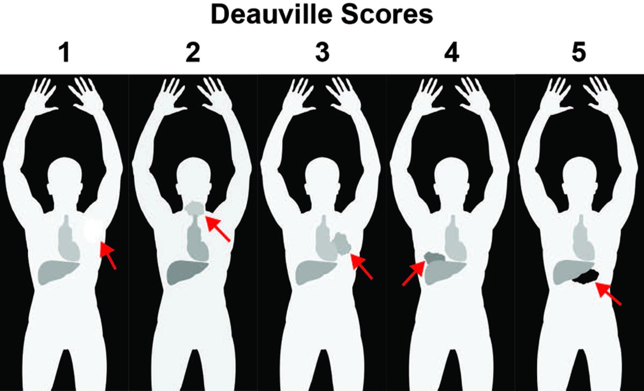
    The D5PS scores of lesions qualitatively based on the FDG uptake relative to the mediastinal blood pool (MBP) and hepatic parenchymal FDG activity. The figure demonstrates hypothetical masses (arrows) and their FDG uptake relative to the MBP and liver activity. D5PS score of 1 for a left axillary mass with FDG uptake no greater than background activity. D5PS score of 2 for cervical mass with FDG uptake above background but less than MBP or liver. D5PS score of 3 for hilar mass with FDG uptake greater than MBP but less than or equal to the hepatic activity. A D5PS score of 4 for a mass in right lung base with FDG uptake greater than both MBP and liver. A D5PS score of 5 for a midabdominal mass with FDG uptake markedly greater than that of the liver.

  • Fig 2.
    • Download figure
    • Open in new tab
    • Download powerpoint
    Fig 2.

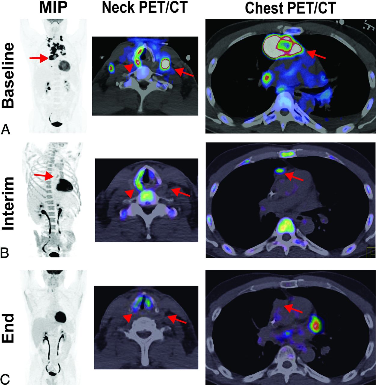
    A partial metabolic response and CMR based on Lugano criteria. A 47-year-old man with diffuse large B-cell lymphoma demonstrates (A) baseline intense FDG avidity in mediastinal (MIP and PET/CT of the chest [arrows denote a large pericardial lymph node conglomerate]) and bilateral levels III and IV lymphadenopathy (PET/CT of the neck [arrow denotes a large level III node]), a D5PS score of 5. Incidentally, on the PET/CT of the neck, the arrowhead denotes a dysfunctional right vocal cord, likely due to disease impacting the ipsilateral recurrent laryngeal nerve. B, Interim imaging demonstrates reduced but persistent FDG uptake (2 times greater than the liver) in the retrosternal, pericardial mass (MIP and PET/CT of the chest [arrows]), consistent with a D5PS score of 5 but a Lugano designation of a partial metabolic response. There was interval resolution of the cervical lymphadenopathy. C, End-of-treatment imaging shows a residual pericardial mass on CT (arrow), without FDG uptake above background, consistent with a D5PS score of 1 and a Lugano designation of CMR.

  • Fig 3.
    • Download figure
    • Open in new tab
    • Download powerpoint
    Fig 3.

    A posttreatment D5PS score of 5 but partial metabolic response based on Lugano criteria. Baseline imaging demonstrates diffuse lymphadenopathy, including cervical levels II–IV, axillary, mediastinal, abdominal periaortic/pericaval, and right greater than left iliac chains, with intense FDG avidity (arrows), consistent with a D5PS score of 5. Interim imaging during treatment demonstrates significantly reduced size and FDG avidity of the cervical, axillary, and mediastinal lymphadenopathy (arrows), now predominantly limited to the periaortic/pericaval regions and right greater than left iliac chains. The lymph nodes still have FDG avidity markedly greater than mediastinal blood pool and liver, consistent with a D5PS score of 5, but the reduction in size, number, and intensity results in a Lugano designation of a partial metabolic response.

  • Fig 4.
    • Download figure
    • Open in new tab
    • Download powerpoint
    Fig 4.

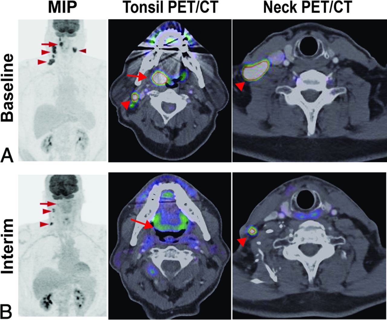
    Hopkins criteria score of 5 in right tonsillar squamous cell carcinoma, consistent with residual tumor. A 71-year-old man with right tonsillar squamous cell carcinoma. MIP from baseline PET/CT demonstrates a Hopkins criteria score of 5 in the primary right tonsillar tumor (arrow) and right greater than left level II–IV cervical lymph nodes (arrowheads). Examination after radiation and chemotherapy shows resolution of the right tonsillar FDG uptake (arrow) and some of the cervical lymph nodes but persistent, focal, and intense FDG uptake in 2 ipsilateral level III and IV lymph nodes (arrowheads); a Hopkins criteria score of 5 is consistent with residual tumor.
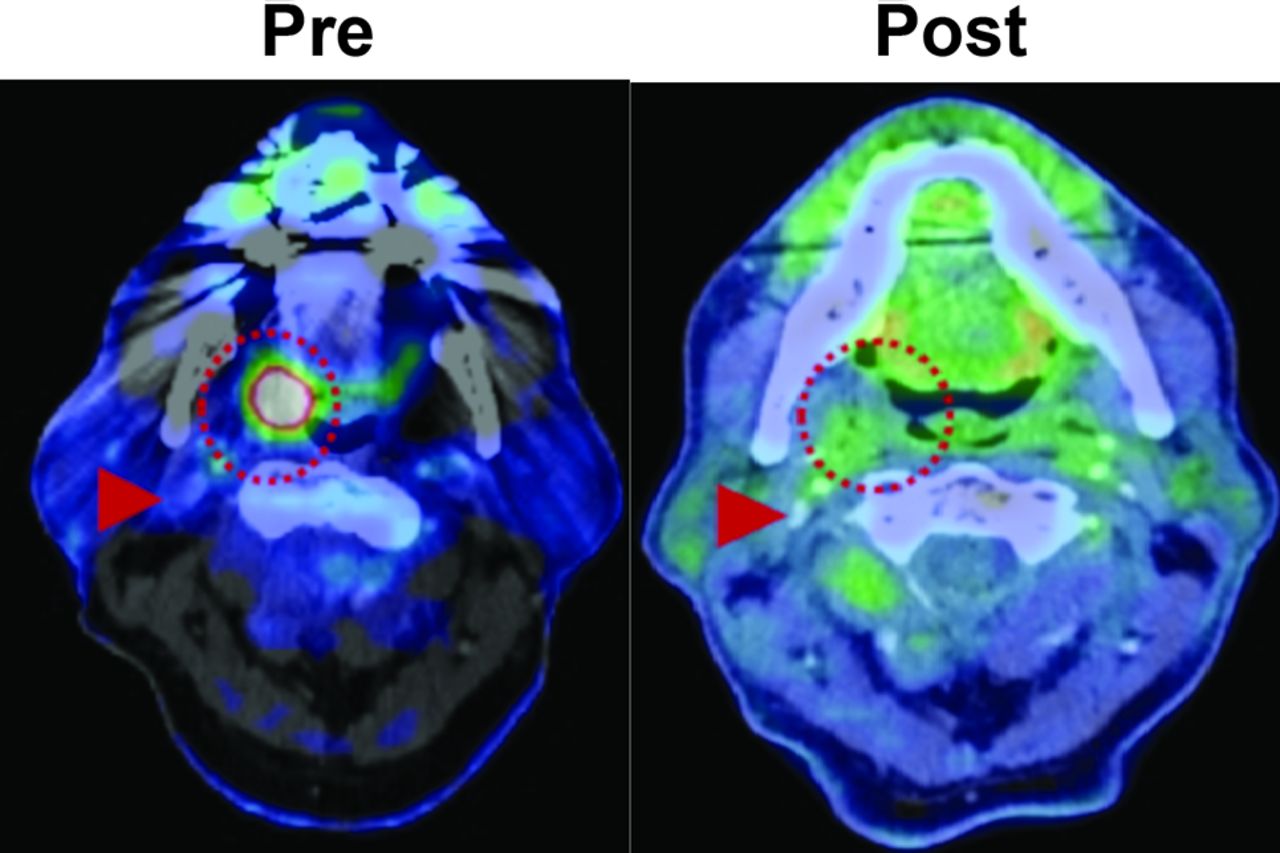
  • Fig 5.
    • Download figure
    • Open in new tab
    • Download powerpoint
    Fig 5.

    Hopkins criteria score of 2 and NI-RADS score of 1 in right tonsillar squamous cell carcinoma, consistent with a CMR. A 71-year-old man with an intensely FDG-avid right tonsillar squamous cell carcinoma (dotted circle) on pretreatment PET/CT. After treatment, the mass has resolved, with FDG uptake in the region just above the adjacent right internal jugular vein (arrowhead) but similar to the surrounding oropharyngeal soft tissues, consistent with a Hopkins criteria score of 2 and an NI-RADS score of 1 at the primary site. By using Hopkins or NI-RADs criteria, the findings are consistent with an overall CMR.

  • Fig 6.
    • Download figure
    • Open in new tab
    • Download powerpoint
    Fig 6.

    An NI-RADS score of 2a in left tonsillar squamous cell carcinoma (SCCa) after chemoradiation, consistent with a Hopkins criteria score of 3 and posttreatment inflammation. A 63-year-old man with a history of left tonsillar SCCa. A, Baseline oblique MIP shows markedly hypermetabolic primary left tonsillar SCCa (arrow). After completion of chemoradiation therapy, repeated FDG PET/CT was obtained. B, Oblique MIP and axial PET/CT show residual hypermetabolic mucosal activity of moderate intensity (arrow) throughout the tonsil bed. The findings are compatible with NI-RADS 2a. Linked management recommendations in the NI-RADS criteria suggest correlation with direct visualization given that such a finding typically represents non-neoplastic FDG uptake. As a comparison, the FDG uptake is greater than the liver and much greater than the internal jugular vein, this is consistent with a Hopkins criteria score of 3. The findings suggest benign posttreatment inflammation. Lesions with this score can be false-negatives and demonstrate intermediate overall survival and progression-free survival compared with scores of 1–2 and 4–5. Due to the false-negative probability, these lesions should be biopsied before altering the treatment plan.

Tables

  • Figures
    • View popup
    Table 1:

    Lugano classification for CT-based lymphoma responsea

    TechniqueComplete ResponsePartial ResponseNo ResponsePD
    CTComplete disappearance of disease or decrease in the lymph node long axis to <1.5 cmMultiple: ≥ 50% decrease in SPD of up to 6 lesions. Single: ≥ 50% decrease in PPDNo criteria for PD and <50% decrease in SPD of up to 6 lesions.(1) New or increased lymphadenopathy (long axis >1.5 cm): new nodes, ≥ 50% increase in PPD, or >0.5 cm increase in diameter for ≤2 cm or >1.0 cm for >2 cm; (2) splenic volume increase: with splenomegaly, >50% increase in length, or without splenomegaly, ≥2 cm increase in length; (3) new or larger lesions; (4) recurrent previously resolved lesions; (5) new extranodal lesion >1 cm (or <1 cm if unequivocally lymphoma)
    FDG-PET/CTD5PS score of 1, 2, or 3 in disease sites, with or without residual massD5PS score of 4 or 5, with reduced uptake compared with baselineD5PS score of 4 or 5, with no signficant change in FDG uptakeD5PS score of 4 or 5, with increased uptake intensity compared with baseline and/or new FDG-avid foci
    • Note:—SPD indicates sum of the product of the diameters (PPD).

    • ↵a For non-FDG avid lymphomas, the following CT-based measurement criteria are used to determine tumor response classification; adapted from Ref. 15.

    • View popup
    Table 2:

    D5PSa

    D5PS ScorebLesion FDG Uptake
    1At or below background
    2Less than or equal to mediastinal blood pool
    3Greater than or equal to mediastinal, but less than hepatic blood pool
    4Greater than hepatic blood pool
    5Markedly greater than hepatic blood pool
    XDesignator for nonlymphomatous lesion
    • ↵a Adapted from Ref. 23.

    • ↵b The D5PS score is used in the Lugano classification for grading lymphoma response.

    • View popup
    Table 3:

    Hopkins criteria scores for head and neck cancersa

    Hopkins Criteria ScorebLesion FDG UptakeTherapy Response
    1Focal uptake less than IJVCMR
    2Focal uptake greater than IJV, but less than liverLikely CMR
    3Diffuse uptake greater than IJV and liverLikely posttreatment inflammation
    4Focal uptake greater than liverLikely residual tumor
    5Focal uptake markedly greater than liverResidual tumor
    • Note:—IJV indicates internal jugular vein.

    • ↵a Adapted from Ref. 16.

    • ↵b Five-point scale Hopkins criteria scores used to classify head and neck cancer response after treatment; new lesions are considered to be PD.

    • View popup
    Table 4:

    NI-RADS risk stratification guidelinesa

    DescriptionCategoryImaging FindingsManagement
    Incomplete0New baseline study or unavailable previous imagingPrevious imaging
    No evidence of recurrence1Posttreatment changes without mass or abnormal FDG uptakeRoutine surveillance
    Low suspicion (superficial)2aEnhancement without FDG uptake or mild/moderate FDG uptake without a massDirect visual inspection
    Low suspicion (deep)2bEnhancement without FDG uptake or mild/moderate FDG uptake without a massShort imaging follow-up
    High suspicion3New or enlarged mass or lymph node, enhancement, and intense FDG uptakeBiopsy
    Definitive recurrence4Pathologically proved or definite progressionClinical management
    • ↵a The NI-RADS risk stratification guidelines grade head and neck tumor treatment response; adapted from Ref. 26.

PreviousNext
Back to top

In this issue

American Journal of Neuroradiology: 40 (12)
American Journal of Neuroradiology
Vol. 40, Issue 12
1 Dec 2019
  • Table of Contents
  • Index by author
  • Complete Issue (PDF)
Advertisement
Print
Download PDF
Email Article

Thank you for your interest in spreading the word on American Journal of Neuroradiology.

NOTE: We only request your email address so that the person you are recommending the page to knows that you wanted them to see it, and that it is not junk mail. We do not capture any email address.

Enter multiple addresses on separate lines or separate them with commas.
RESISTing the Need to Quantify: Putting Qualitative FDG-PET/CT Tumor Response Assessment Criteria into Daily Practice
(Your Name) has sent you a message from American Journal of Neuroradiology
(Your Name) thought you would like to see the American Journal of Neuroradiology web site.
CAPTCHA
This question is for testing whether or not you are a human visitor and to prevent automated spam submissions.
Cite this article
J.G. Peacock, C.T. Christensen, K.P. Banks
RESISTing the Need to Quantify: Putting Qualitative FDG-PET/CT Tumor Response Assessment Criteria into Daily Practice
American Journal of Neuroradiology Dec 2019, 40 (12) 1978-1986; DOI: 10.3174/ajnr.A6294

Citation Manager Formats

  • BibTeX
  • Bookends
  • EasyBib
  • EndNote (tagged)
  • EndNote 8 (xml)
  • Medlars
  • Mendeley
  • Papers
  • RefWorks Tagged
  • Ref Manager
  • RIS
  • Zotero
0 Responses
Respond to this article
Share
Bookmark this article
RESISTing the Need to Quantify: Putting Qualitative FDG-PET/CT Tumor Response Assessment Criteria into Daily Practice
J.G. Peacock, C.T. Christensen, K.P. Banks
American Journal of Neuroradiology Dec 2019, 40 (12) 1978-1986; DOI: 10.3174/ajnr.A6294
del.icio.us logo Twitter logo Facebook logo Mendeley logo
  • Tweet Widget
  • Facebook Like
  • Google Plus One
Purchase

Jump to section

  • Article
    • Abstract
    • ABBREVIATIONS:
    • INTRODUCTION
    • THE LUGANO CLASSIFICATION FOR LYMPHOMA TREATMENT RESPONSE EVALUATION
    • HOPKINS CRITERIA FOR HEAD AND NECK TUMOR RESPONSE EVALUATION
    • NI-RADS GUIDELINES FOR HEAD AND NECK TUMORS
    • CONCLUSIONS
    • Footnotes
    • References
  • Figures & Data
  • Info & Metrics
  • Responses
  • References
  • PDF

Related Articles

  • No related articles found.
  • PubMed
  • Google Scholar

Cited By...

  • 18F-FDG PET/CT Prediction of Treatment Outcomes in Human Papillomavirus-Positive, Locally Advanced Oropharyngeal Cancer Patients Receiving Deintensified Therapy: Results from NRG-HN002
  • First-in-Humans Evaluation of Safety and Dosimetry of 64Cu-LLP2A for PET Imaging
  • Crossref (3)
  • Google Scholar

This article has been cited by the following articles in journals that are participating in Crossref Cited-by Linking.

  • First-in-Humans Evaluation of Safety and Dosimetry of64Cu-LLP2A for PET Imaging
    Richard Laforest, Anchal Ghai, Tyler J. Fraum, Reiko Oyama, Jennifer Frye, Helen Kaemmerer, Greg Gaehle, Tom Voller, Cedric Mpoy, Buck E. Rogers, Mark Fiala, Kooresh I. Shoghi, Samuel Achilefu, Michael Rettig, Ravi Vij, John F. DiPersio, Sally Schwarz, Monica Shokeen, Farrokh Dehdashti
    Journal of Nuclear Medicine 2023 64 2
  • Improved non-invasive positron emission tomographic imaging of chemotherapy-induced tumor cell death using Zirconium-89-labeled APOMAB®
    Vasilios Liapis, William Tieu, Stacey E. Rudd, Paul S. Donnelly, Nicole L. Wittwer, Michael P. Brown, Alexander H. Staudacher
    EJNMMI Radiopharmacy and Chemistry 2020 5 1
  • 18F-FDG PET/CT Prediction of Treatment Outcomes in Human Papillomavirus–Positive, Locally Advanced Oropharyngeal Cancer Patients Receiving Deintensified Therapy: Results from NRG-HN002
    Rathan M. Subramaniam, Lyudmila DeMora, Min Yao, Sue S. Yom, Maura Gillison, Jimmy J. Caudell, John Waldron, Ping Xia, Christine H. Chung, Minh Tam Truong, Michelle Echevarria, Jason W. Chan, Jessica L. Geiger, Loren Mell, Samantha Seaward, Wade L. Thorstad, Jonathan Jay Beitler, Khalil Sultanem, Diagjin Blakaj, Quynh-Thu Le
    Journal of Nuclear Medicine 2023 64 3

More in this TOC Section

  • Peritumoral Signal in Vestibular Schwannomas
  • Chondrosarcoma vs Synovial Chondromatosis: Imaging
  • NI-RADS for HEAD&NECK Cancer Recurrence
Show more HEAD & NECK

Similar Articles

Advertisement

Indexed Content

  • Current Issue
  • Accepted Manuscripts
  • Article Preview
  • Past Issues
  • Editorials
  • Editors Choice
  • Fellow Journal Club
  • Letters to the Editor

Cases

  • Case Collection
  • Archive - Case of the Week
  • Archive - Case of the Month
  • Archive - Classic Case

Special Collections

  • Special Collections

Resources

  • News and Updates
  • Turn around Times
  • Submit a Manuscript
  • Author Policies
  • Manuscript Submission Guidelines
  • Evidence-Based Medicine Level Guide
  • Publishing Checklists
  • Graphical Abstract Preparation
  • Imaging Protocol Submission
  • Submit a Case
  • Become a Reviewer/Academy of Reviewers
  • Get Peer Review Credit from Publons

Multimedia

  • AJNR Podcast
  • AJNR SCANtastic
  • Video Articles

About Us

  • About AJNR
  • Editorial Board
  • Not an AJNR Subscriber? Join Now
  • Alerts
  • Feedback
  • Advertise with us
  • Librarian Resources
  • Permissions
  • Terms and Conditions

American Society of Neuroradiology

  • Not an ASNR Member? Join Now

© 2025 by the American Society of Neuroradiology All rights, including for text and data mining, AI training, and similar technologies, are reserved.
Print ISSN: 0195-6108 Online ISSN: 1936-959X

Powered by HighWire