Advertisement

Main menu

  • Home
  • Content
    • Current Issue
    • Accepted Manuscripts
    • Article Preview
    • Past Issue Archive
    • Video Articles
    • AJNR Case Collection
    • Case of the Week Archive
    • Case of the Month Archive
    • Classic Case Archive
  • Special Collections
    • AJNR Awards
    • Low-Field MRI
    • Alzheimer Disease
    • ASNR Foundation Special Collection
    • Photon-Counting CT
    • View All
  • Multimedia
    • AJNR Podcasts
    • AJNR SCANtastic
    • Trainee Corner
    • MRI Safety Corner
    • Imaging Protocols
  • For Authors
    • Submit a Manuscript
    • Submit a Video Article
    • Submit an eLetter to the Editor/Response
    • Manuscript Submission Guidelines
    • Statistical Tips
    • Fast Publishing of Accepted Manuscripts
    • Graphical Abstract Preparation
    • Imaging Protocol Submission
    • Author Policies
  • About Us
    • About AJNR
    • Editorial Board
    • Editorial Board Alumni
  • More
    • Become a Reviewer/Academy of Reviewers
    • Subscribers
    • Permissions
    • Alerts
    • Feedback
    • Advertisers
    • ASNR Home

User menu

  • Alerts
  • Log in

Search

  • Advanced search
American Journal of Neuroradiology

American Journal of Neuroradiology

ASHNR American Society of Functional Neuroradiology ASHNR American Society of Pediatric Neuroradiology ASSR
  • Alerts
  • Log in

Advanced Search

  • Home
  • Content
    • Current Issue
    • Accepted Manuscripts
    • Article Preview
    • Past Issue Archive
    • Video Articles
    • AJNR Case Collection
    • Case of the Week Archive
    • Case of the Month Archive
    • Classic Case Archive
  • Special Collections
    • AJNR Awards
    • Low-Field MRI
    • Alzheimer Disease
    • ASNR Foundation Special Collection
    • Photon-Counting CT
    • View All
  • Multimedia
    • AJNR Podcasts
    • AJNR SCANtastic
    • Trainee Corner
    • MRI Safety Corner
    • Imaging Protocols
  • For Authors
    • Submit a Manuscript
    • Submit a Video Article
    • Submit an eLetter to the Editor/Response
    • Manuscript Submission Guidelines
    • Statistical Tips
    • Fast Publishing of Accepted Manuscripts
    • Graphical Abstract Preparation
    • Imaging Protocol Submission
    • Author Policies
  • About Us
    • About AJNR
    • Editorial Board
    • Editorial Board Alumni
  • More
    • Become a Reviewer/Academy of Reviewers
    • Subscribers
    • Permissions
    • Alerts
    • Feedback
    • Advertisers
    • ASNR Home
  • Follow AJNR on Twitter
  • Visit AJNR on Facebook
  • Follow AJNR on Instagram
  • Join AJNR on LinkedIn
  • RSS Feeds

AJNR Awards, New Junior Editors, and more. Read the latest AJNR updates

Research ArticlePediatric Neuroimaging

Volumetric MRI Study of the Brain in Fetuses with Intrauterine Cytomegalovirus Infection and Its Correlation to Neurodevelopmental Outcome

A. Grinberg, E. Katorza, D. Hoffman, R. Ber, A. Mayer and S. Lipitz
American Journal of Neuroradiology February 2019, 40 (2) 353-358; DOI: https://doi.org/10.3174/ajnr.A5948
A. Grinberg
aFrom the Antenatal Diagnostic Unit (A.G., E.K., D.H., R.B., S.L.), Department of Obstetrics and Gynecology
cSackler School of Medicine (A.G., E.K., D.H., R.B., A.M., S.L.), Tel Aviv University, Tel Aviv, Israel.
  • Find this author on Google Scholar
  • Find this author on PubMed
  • Search for this author on this site
  • ORCID record for A. Grinberg
E. Katorza
aFrom the Antenatal Diagnostic Unit (A.G., E.K., D.H., R.B., S.L.), Department of Obstetrics and Gynecology
cSackler School of Medicine (A.G., E.K., D.H., R.B., A.M., S.L.), Tel Aviv University, Tel Aviv, Israel.
  • Find this author on Google Scholar
  • Find this author on PubMed
  • Search for this author on this site
  • ORCID record for E. Katorza
D. Hoffman
aFrom the Antenatal Diagnostic Unit (A.G., E.K., D.H., R.B., S.L.), Department of Obstetrics and Gynecology
cSackler School of Medicine (A.G., E.K., D.H., R.B., A.M., S.L.), Tel Aviv University, Tel Aviv, Israel.
  • Find this author on Google Scholar
  • Find this author on PubMed
  • Search for this author on this site
  • ORCID record for D. Hoffman
R. Ber
aFrom the Antenatal Diagnostic Unit (A.G., E.K., D.H., R.B., S.L.), Department of Obstetrics and Gynecology
cSackler School of Medicine (A.G., E.K., D.H., R.B., A.M., S.L.), Tel Aviv University, Tel Aviv, Israel.
  • Find this author on Google Scholar
  • Find this author on PubMed
  • Search for this author on this site
  • ORCID record for R. Ber
A. Mayer
bDepartment of Diagnostic Radiology (A.M.), Chaim Sheba Medical Center, Tel-Hashomer, Israel
cSackler School of Medicine (A.G., E.K., D.H., R.B., A.M., S.L.), Tel Aviv University, Tel Aviv, Israel.
  • Find this author on Google Scholar
  • Find this author on PubMed
  • Search for this author on this site
  • ORCID record for A. Mayer
S. Lipitz
aFrom the Antenatal Diagnostic Unit (A.G., E.K., D.H., R.B., S.L.), Department of Obstetrics and Gynecology
cSackler School of Medicine (A.G., E.K., D.H., R.B., A.M., S.L.), Tel Aviv University, Tel Aviv, Israel.
  • Find this author on Google Scholar
  • Find this author on PubMed
  • Search for this author on this site
  • ORCID record for S. Lipitz
  • Article
  • Figures & Data
  • Info & Metrics
  • Responses
  • References
  • PDF
Loading

Article Figures & Data

Figures

  • Tables
  • Fig 1.
    • Download figure
    • Open in new tab
    • Download powerpoint
    Fig 1.

    Representative slices of structure boundaries; A, STV and CV. B, REBV and LEBV.
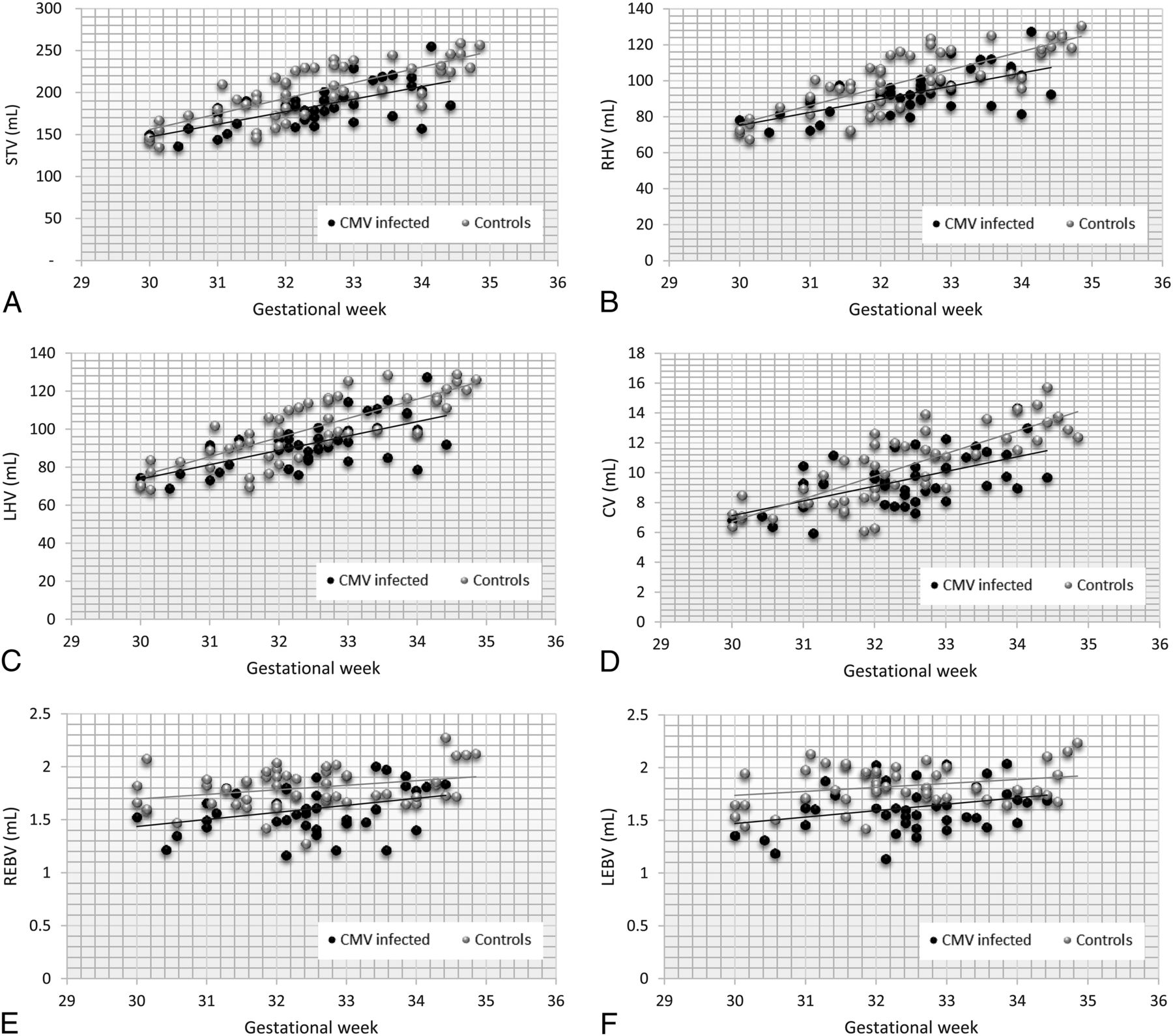
  • Fig 2.
    • Download figure
    • Open in new tab
    • Download powerpoint
    Fig 2.

    Scatterplots of volumetric measurements according to the gestational age of the CMV-infected group and the noninfected controls. Simple linear regression lines are shown for each set of data. A, STV. B, RHV. C, LHV. D, CV. E, REBV. F, LEBV.

Tables

  • Figures
    • View popup
    Table 1:

    Means/medians of brain volume measurements for CMV-infected and control groupsa

    Volume (mL)CMV-Infected Fetuses (n = 42)Control Fetuses (n = 50)P
    STV184.7 (24.2)200.9 (32.6).008b
    RHV93.2 (11.8)100.2 (17.1).022b
    LHV92.4 (12.5)99.5 (17.1).02b
    CV9.5 (1.7)10.3 (2.2).084
    REBV1.58 (1.46–1.79)1.82 (1.66–1.92)<.001b
    LEBV1.61 (1.47–1.76)1.79 (1.71–1.96)<.001b
    • ↵a STV, RHV, LHV, CV are presented as mean (SD); REBV and LEBV are presented as median (interquartile range).

    • ↵b Significant.

    • View popup
    Table 2:

    Brain volumes of CMV-infected fetuses demonstrated as z scoresa

    VolumesZ ScorePb
    STV−1.26 (−1.71 to −0.31)<.001
    RHV−1.06 (−1.65 to −0.15)<.001
    LHV−1.08 (−1.6 to −0.14)<.001
    CV−1.11 (−1.8 to −0.5)<.001
    REBV−1.13 (−2.14 to −0.28).001
    LEBV−1.64 (−2.58 to −0.26)<.001
    • ↵a Data are presented as median and interquartile range.

    • ↵b All P values are significant.

PreviousNext
Back to top

In this issue

American Journal of Neuroradiology: 40 (2)
American Journal of Neuroradiology
Vol. 40, Issue 2
1 Feb 2019
  • Table of Contents
  • Index by author
  • Complete Issue (PDF)
Advertisement
Print
Download PDF
Email Article

Thank you for your interest in spreading the word on American Journal of Neuroradiology.

NOTE: We only request your email address so that the person you are recommending the page to knows that you wanted them to see it, and that it is not junk mail. We do not capture any email address.

Enter multiple addresses on separate lines or separate them with commas.
Volumetric MRI Study of the Brain in Fetuses with Intrauterine Cytomegalovirus Infection and Its Correlation to Neurodevelopmental Outcome
(Your Name) has sent you a message from American Journal of Neuroradiology
(Your Name) thought you would like to see the American Journal of Neuroradiology web site.
CAPTCHA
This question is for testing whether or not you are a human visitor and to prevent automated spam submissions.
Cite this article
A. Grinberg, E. Katorza, D. Hoffman, R. Ber, A. Mayer, S. Lipitz
Volumetric MRI Study of the Brain in Fetuses with Intrauterine Cytomegalovirus Infection and Its Correlation to Neurodevelopmental Outcome
American Journal of Neuroradiology Feb 2019, 40 (2) 353-358; DOI: 10.3174/ajnr.A5948

Citation Manager Formats

  • BibTeX
  • Bookends
  • EasyBib
  • EndNote (tagged)
  • EndNote 8 (xml)
  • Medlars
  • Mendeley
  • Papers
  • RefWorks Tagged
  • Ref Manager
  • RIS
  • Zotero
0 Responses
Respond to this article
Share
Bookmark this article
Volumetric MRI Study of the Brain in Fetuses with Intrauterine Cytomegalovirus Infection and Its Correlation to Neurodevelopmental Outcome
A. Grinberg, E. Katorza, D. Hoffman, R. Ber, A. Mayer, S. Lipitz
American Journal of Neuroradiology Feb 2019, 40 (2) 353-358; DOI: 10.3174/ajnr.A5948
del.icio.us logo Twitter logo Facebook logo Mendeley logo
  • Tweet Widget
  • Facebook Like
  • Google Plus One
Purchase

Jump to section

  • Article
    • Abstract
    • ABBREVIATIONS:
    • Materials and Methods
    • Results
    • Discussion
    • Conclusions
    • Footnotes
    • References
  • Figures & Data
  • Info & Metrics
  • Responses
  • References
  • PDF

Related Articles

  • No related articles found.
  • PubMed
  • Google Scholar

Cited By...

  • Correlation between 2D and 3D Fetal Brain MRI Biometry and Neurodevelopmental Outcomes in Fetuses with Suspected Microcephaly and Macrocephaly
  • Normal Human Brain Growth
  • Crossref
  • Google Scholar

This article has not yet been cited by articles in journals that are participating in Crossref Cited-by Linking.

More in this TOC Section

  • FRACTURE MR in Congenital Vertebral Anomalies
  • Comparing MRI Perfusion in Pediatric Brain Tumors
  • Sodium MRI in Pediatric Brain Tumors
Show more Pediatric Neuroimaging

Similar Articles

Advertisement

Indexed Content

  • Current Issue
  • Accepted Manuscripts
  • Article Preview
  • Past Issues
  • Editorials
  • Editor's Choice
  • Fellows' Journal Club
  • Letters to the Editor
  • Video Articles

Cases

  • Case Collection
  • Archive - Case of the Week
  • Archive - Case of the Month
  • Archive - Classic Case

More from AJNR

  • Trainee Corner
  • Imaging Protocols
  • MRI Safety Corner
  • Book Reviews

Multimedia

  • AJNR Podcasts
  • AJNR Scantastics

Resources

  • Turnaround Time
  • Submit a Manuscript
  • Submit a Video Article
  • Submit an eLetter to the Editor/Response
  • Manuscript Submission Guidelines
  • Statistical Tips
  • Fast Publishing of Accepted Manuscripts
  • Graphical Abstract Preparation
  • Imaging Protocol Submission
  • Evidence-Based Medicine Level Guide
  • Publishing Checklists
  • Author Policies
  • Become a Reviewer/Academy of Reviewers
  • News and Updates

About Us

  • About AJNR
  • Editorial Board
  • Editorial Board Alumni
  • Alerts
  • Permissions
  • Not an AJNR Subscriber? Join Now
  • Advertise with Us
  • Librarian Resources
  • Feedback
  • Terms and Conditions
  • AJNR Editorial Board Alumni

American Society of Neuroradiology

  • Not an ASNR Member? Join Now

© 2025 by the American Society of Neuroradiology All rights, including for text and data mining, AI training, and similar technologies, are reserved.
Print ISSN: 0195-6108 Online ISSN: 1936-959X

Powered by HighWire