
Article Figures & Data
Figures
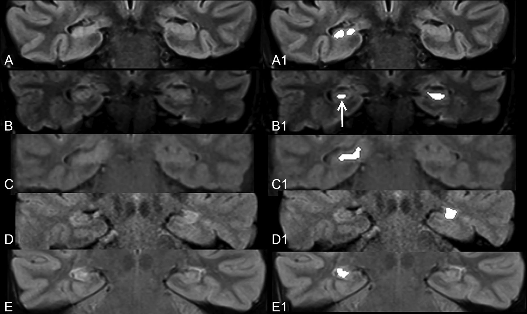
- Fig 1.
Coronal FLAIR images (A–E) across the medial temporal lobes in patients with pathologically confirmed right-sided (A, C, and E) and left-sided (B and D) MTS. Corresponding images following image processing (A1–E1) demonstrate confluent areas of marked exaggeration of signal intensity of the diseased hippocampi. Note a similar exaggeration of signal intensity to a smaller extent in the right hippocampus (arrow) in B1, presumably indicating bilateral disease.
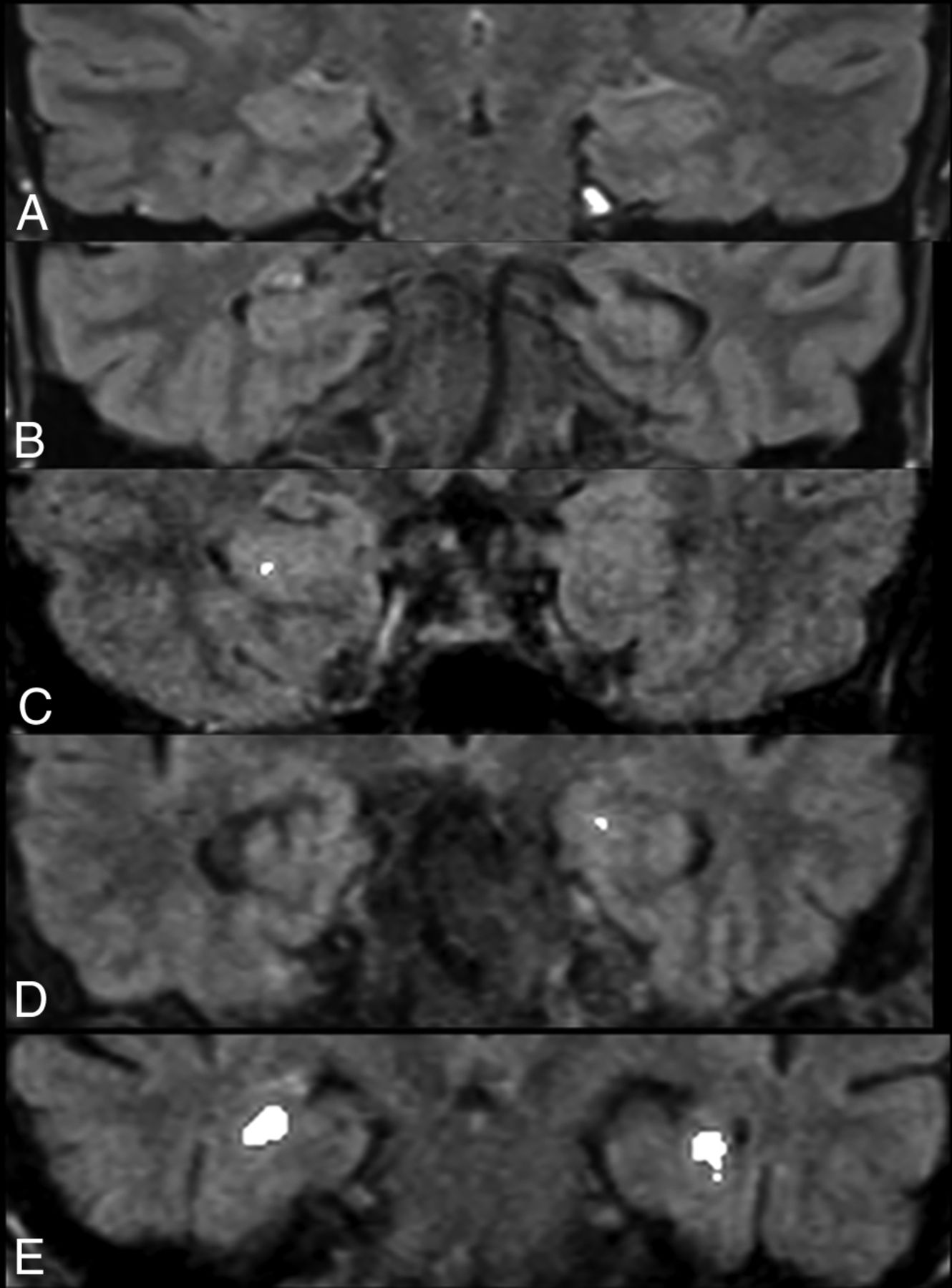
- Fig 2.
Representative postprocessed coronal FLAIR images across the medial temporal lobes of 5 controls without seizures. Processing did not result in any alteration of hippocampal signal (A and B) for most control hippocampi. While punctate (C and D) foci of signal exaggeration were noted in some control hippocampi, false-positive confluent regions of increased intensity mimicking MTS (E) were observed in the bilateral hippocampi of 1 (2%) control.


Tables
Subjects Controls Mean age (range) (yr) 34.6 (18–58) 42.8 (21–79) Men 24 13 Women 27 38 Selection source Pathology database RIS search and review Inclusion criteria Age older than 18 yr Age older than 18 yr Pathologically proven MTS Brain and orbital MRI Coronal FLAIR images extending across hippocampi Exclusion criteria Preoperative coronal FLAIR through hippocampi not available Seizures
Known or imaging suspicion of neurologic disease known to affect hippocampus
Reported hippocampal abnormalitiesIndication for MRI Refractory epilepsy Diplopia, cranial neuropathy, visual dysfunction, optic neuropathy, papilledema, retrobulbar pain, ptosis, ocular findings MR imaging magnet strength (No.) 3T (36) 1.5T (15) 3T (6) 1.5T (45) Coronal 2D FLAIR parameters Thickness (mm) 3 3 FOV (mm) 180 × 180 166 × 190 TR (ms) 9000–11,000 9000–10,000 TE (ms) 80–110 80–110 TI (ms) 2500 2500 Matrix 256 × 256 256 × 224 Note:—RIS indicates radiology information system.
- Table 2:
Effect of image processing on signal intensity ratings for subjects with MTS, right-sided control hippocampi, and left-sided control hippocampi on a 5-point scalea
Median Minimum 25th Percentile 75th Percentile Maximum P Subjects 4.00 1.00 1.00 2.00 4.00 <.001 Subjects (processed) 5.00 1.00 1.00 2.00 5.00 Controls (R) 1.00 1.00 1.00 1.50 3.50 <.01 Controls (R, processed) 1.00 1.00 1.00 1.00 5.00 Controls (L) 1.00 1.00 1.00 1.50 2.00 .03 Controls (L, processed) 1.00 1.00 1.00 1.00 5.00 Note:—R indicates right; L, left.
↵a 1, Definitely normal; 2, probably normal; 3, possibly normal; 4, probably abnormal; 5, definitely abnormal.
- Table 3:
Diagnostic performance of 6 blinded readers in detection of MTS-related hippocampal signal alteration before and after image processing with a proprietary algorithm
Reader Sensitivity Specificity PPV NPV Accuracy Before processing R1 72.55 98.04 97.37 78.13 0.85 R2 60.78 98.04 96.88 71.43 0.79 R3 43.14 100.00 100.00 63.75 0.72 R4 58.82 92.16 88.24 69.12 0.75 R5 86.27 80.39 81.48 85.42 0.83 R6 66.67 100.00 100.00 75.00 0.83 Mean (SD) 64.71 (14.46) 94.77 (7.61) 93.99 (7.50) 73.81 (7.53) 0.80 (0.05) After Processing R1 74.51 96.08 95.00 79.03 0.85 R2 78.43 98.04 97.56 81.97 0.88 R3 72.55 98.04 97.37 78.13 0.85 R4 78.43 92.16 90.91 81.03 0.85 R5 72.55 98.04 97.37 78.13 0.85 R6 74.51 98.04 97.44 79.37 0.86 Mean (SD) 75.16 (2.68) 96.73 (2.37) 95.94 (2.65) 79.61 (1.57) 0.86 (0.01) Note:—R1 and R2 indicate radiology residents; R3 and R4, neuroradiology fellows; R5 and R6, attending neuroradiologists; PPV, positive predictive value; NPV, negative predictive value.


Jump to section
Related Articles
Cited By...
- No citing articles found.