
Article Figures & Data
Figures
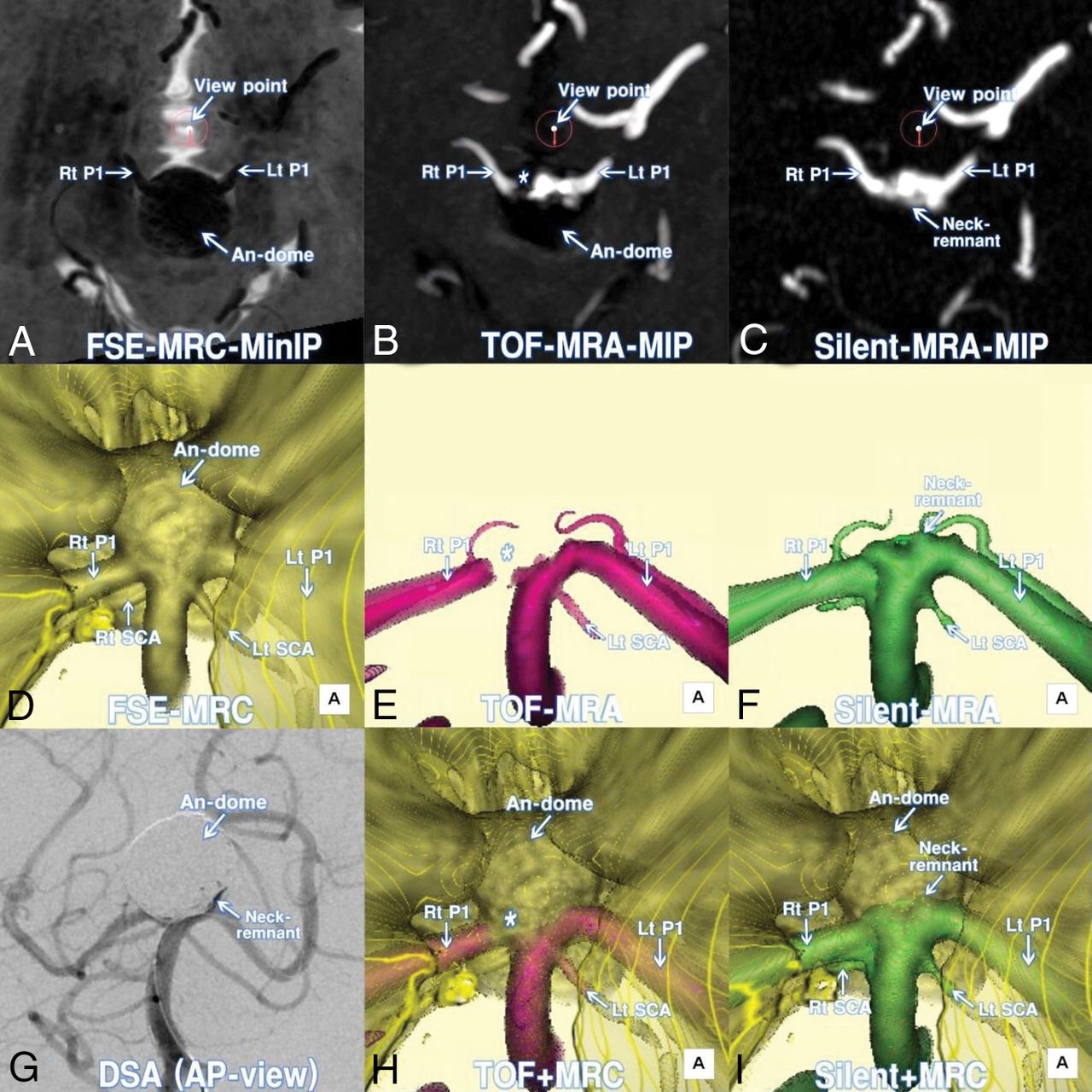
- Fig 1.
A 69-year-old woman (patient 10) treated by coiling for a large unruptured top of the basilar artery (BA-top) aneurysm. A, Axial minimum intensity projection (MinIP) image of postcoiling FSE-MRC. The encircled arrow indicates the viewing position for the following 3D images. B, Axial MIP image of postcoiling TOF-MRA. C, Axial MIP image of postcoiling silent MRA. D, Postcoiling 3D FSE-MRC image. E, Postcoiling 3D TOF-MRA image. Note the gap (asterisk) of the parent artery and irregularity of the neck. F, Postcoiling 3D silent MRA image. Parent arteries and the coiled neck are clearly visualized. G, DSA image at the completion of coiling. Note that the configuration of the neck complex with remnants and parent arteries is depicted with the shadow of the coiled dome. H, A 3D multifusion image of postcoiling TOF-MRA and FSE-MRC. I, A 3D multifusion image of postcoiling silent MRA and FSE-MRC. Note that the coiled neck complex with remnants is clearly visualized together with the coiled dome and adjacent brain parenchyma. P1 indicates the first segment of posterior cerebral artery; Lt, left; Rt, right; AP, anteroposterior; An, aneurysm.
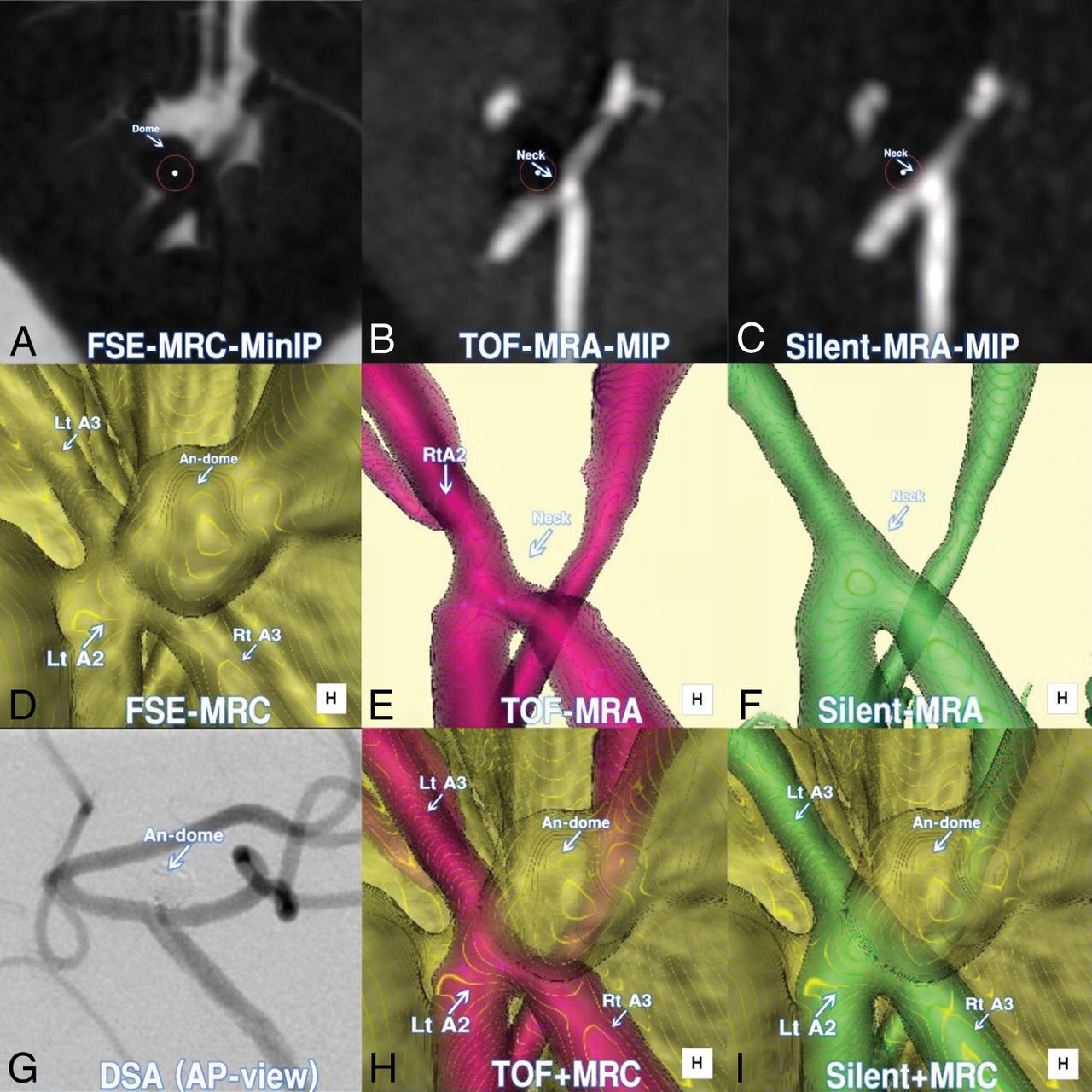
- Fig 2.
A 78-year-old woman (patient 6) treated by coiling for an unruptured distal anterior cerebral artery (A2–A3) aneurysm. A, Axial minimum intensity projection (MinIP) image of postcoiling FSE-MRC. B, Axial MIP image of postcoiling TOF-MRA. C, Axial MIP image of postcoiling silent MRA. D, Postcoiling 3D FSE-MRC image. E, Postcoiling 3D TOF-MRA image. Note the defects of the parent artery and the neck. F, Postcoiling 3D silent MRA image. Note that the parent arteries and coiled neck without remnants are clearly visualized. G, DSA image at the completion of coiling. Note that the configuration of the neck complex without remnants is depicted with the shadow of the coiled dome. H, A 3D multifusion image of postcoiling TOF-MRA and FSE-MRC. I, A 3D multifusion image of postcoiling silent MRA and FSE-MRC. Note the complete occlusion of the neck without remnants together with the coiled dome. A2 indicates the second segment of the anterior cerebral artery; A3, the third segment of the anterior cerebral artery; An, aneurysm; Lt, left; Rt, right; AP, anteroposterior.
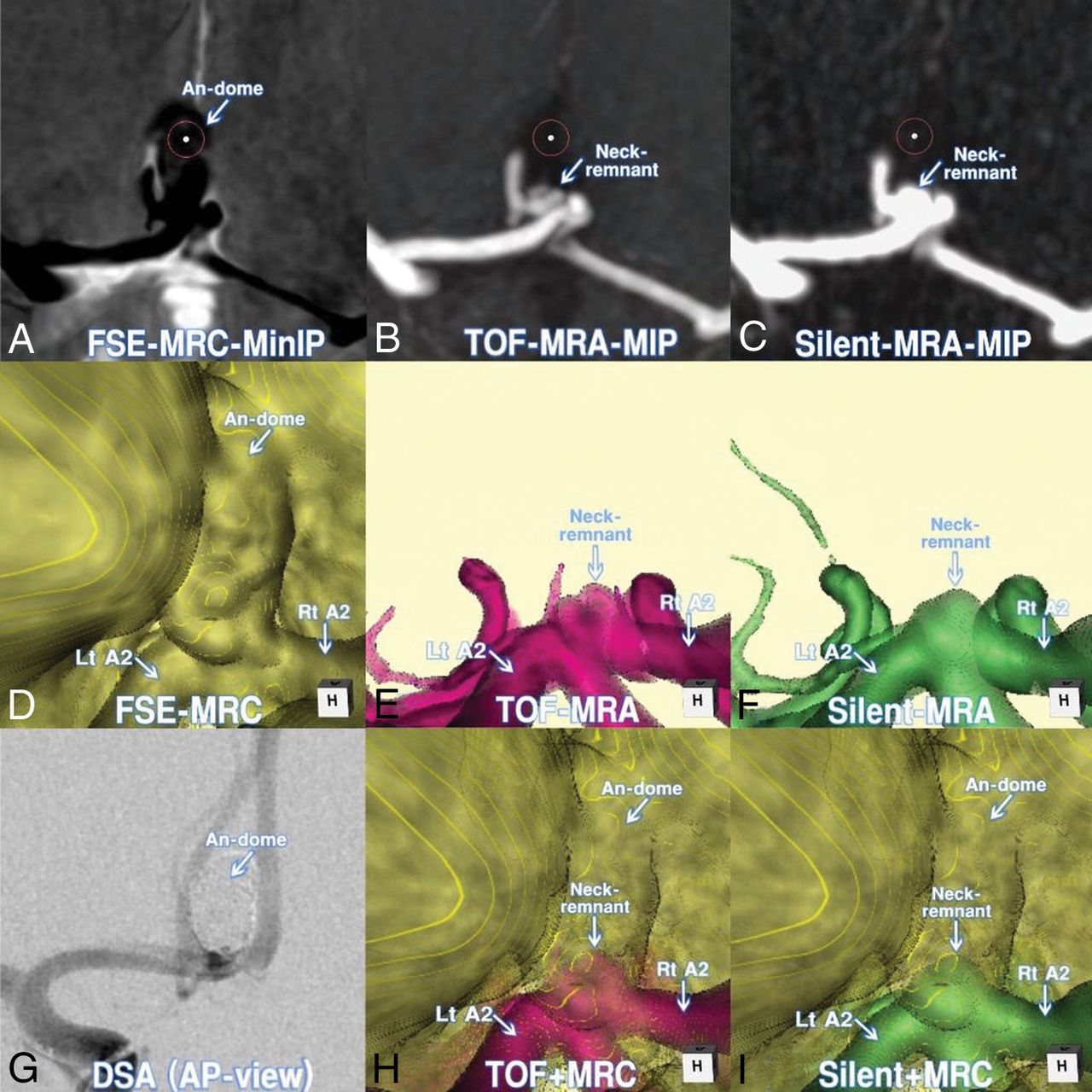
- Fig 3.
A 58-year-old man (patient 3) treated by endovascular coiling for an unruptured AComA aneurysm. A, Axial minimum intensity projection (MinIP) image of postcoiling FSE-MRC. B, Axial MIP image of postcoiling TOF-MRA. C, Axial MIP image of postcoiling silent MRA. D, Postcoiling 3D FSE-MRC image. E, Postcoiling 3D TOF-MRA image. Note the minor defects at the parent artery and the neck. F, Postcoiling 3D silent MRA image. Note that the parent arteries and coiled neck are clearly visualized. G, DSA image at the completion of coiling. Note the configuration of the neck complex with the shadow of the coiled dome. H, A 3D multifusion image of postcoiling TOF-MRA and FSE-MRC. I, A 3D multifusion image of postcoiling silent MRA and FSE-MRC. Note the clear visualization of the coiled neck complex with remnants, together with the coiled dome and adjacent brain parenchyma. Rt indicates right; Lt, left; AP, anteroposterior.


Jump to section
Related Articles
Cited By...
- No citing articles found.