Advertisement

Main menu

  • Home
  • Content
    • Current Issue
    • Accepted Manuscripts
    • Article Preview
    • Past Issue Archive
    • AJNR Case Collection
    • Case of the Week Archive
    • Classic Case Archive
    • Case of the Month Archive
  • Special Collections
    • Spinal CSF Leak Articles (Jan 2020-June 2024)
    • 2024 AJNR Journal Awards
    • Most Impactful AJNR Articles
  • Multimedia
    • AJNR Podcast
    • AJNR Scantastics
    • Video Articles
  • For Authors
    • Submit a Manuscript
    • Author Policies
    • Fast publishing of Accepted Manuscripts
    • Graphical Abstract Preparation
    • Manuscript Submission Guidelines
    • Imaging Protocol Submission
    • Submit a Case for the Case Collection
  • About Us
    • About AJNR
    • Editorial Board
  • More
    • Become a Reviewer/Academy of Reviewers
    • Subscribers
    • Permissions
    • Alerts
    • Feedback
    • Advertisers
    • ASNR Home
  • Other Publications
    • ajnr

User menu

  • Alerts
  • Log in

Search

  • Advanced search
American Journal of Neuroradiology

American Journal of Neuroradiology

ASHNR American Society of Functional Neuroradiology ASHNR American Society of Pediatric Neuroradiology ASSR
  • Alerts
  • Log in

Advanced Search

  • Home
  • Content
    • Current Issue
    • Accepted Manuscripts
    • Article Preview
    • Past Issue Archive
    • AJNR Case Collection
    • Case of the Week Archive
    • Classic Case Archive
    • Case of the Month Archive
  • Special Collections
    • Spinal CSF Leak Articles (Jan 2020-June 2024)
    • 2024 AJNR Journal Awards
    • Most Impactful AJNR Articles
  • Multimedia
    • AJNR Podcast
    • AJNR Scantastics
    • Video Articles
  • For Authors
    • Submit a Manuscript
    • Author Policies
    • Fast publishing of Accepted Manuscripts
    • Graphical Abstract Preparation
    • Manuscript Submission Guidelines
    • Imaging Protocol Submission
    • Submit a Case for the Case Collection
  • About Us
    • About AJNR
    • Editorial Board
  • More
    • Become a Reviewer/Academy of Reviewers
    • Subscribers
    • Permissions
    • Alerts
    • Feedback
    • Advertisers
    • ASNR Home
  • Follow AJNR on Twitter
  • Visit AJNR on Facebook
  • Follow AJNR on Instagram
  • Join AJNR on LinkedIn
  • RSS Feeds

Welcome to the new AJNR, Updated Hall of Fame, and more. Read the full announcements.


AJNR is seeking candidates for the position of Associate Section Editor, AJNR Case Collection. Read the full announcement.


Table of Contents

June 01, 2019; Volume 40,Issue 6

Perspectives

  • You have access
    Perspectives
    Azza Elgendy
    American Journal of Neuroradiology June 2019, 40 (6) 927; DOI: https://doi.org/10.3174/ajnr.P0066

Review Article

  • Adult Brain
    Open Access
    Texture Analysis in Cerebral Gliomas: A Review of the Literature
    N. Soni, S. Priya and G. Bathla
    American Journal of Neuroradiology June 2019, 40 (6) 928-934; DOI: https://doi.org/10.3174/ajnr.A6075

Social Media Vignette

  • You have access
    The Continued Rise in Professional Use of Social Media at Scientific Meetings: An Analysis of Twitter Use during the ASNR 2018 Annual Meeting
    G. D'Anna, M.M. Chen, J.L. McCarty, A. Radmanesh and A.L. Kotsenas
    American Journal of Neuroradiology June 2019, 40 (6) 935-937; DOI: https://doi.org/10.3174/ajnr.A6064

General Contents

  • EDITOR'S CHOICEAdult Brain
    Open Access
    Ensemble of Convolutional Neural Networks Improves Automated Segmentation of Acute Ischemic Lesions Using Multiparametric Diffusion-Weighted MRI
    S. Winzeck, S.J.T. Mocking, R. Bezerra, M.J.R.J. Bouts, E.C. McIntosh, I. Diwan, P. Garg, A. Chutinet, W.T. Kimberly, W.A. Copen, P.W. Schaefer, H. Ay, A.B. Singhal, K. Kamnitsas, B. Glocker, A.G. Sorensen and O. Wu
    American Journal of Neuroradiology June 2019, 40 (6) 938-945; DOI: https://doi.org/10.3174/ajnr.A6077

    Convolutional neural networks were trained on combinations of DWI, ADC, and low b-value-weighted images from 116 subjects. The performances of the networks (measured by the Dice score, sensitivity, and precision) were compared with one another and with ensembles of 5 networks. An ensemble of convolutional neural networks trained on DWI, ADC, and low b-value-weighted images produced the most accurate acute infarct segmentation over individual networks. Automated volumes correlated with manually measured volumes for the independent cohort.

  • EDITOR'S CHOICEAdult Brain
    Open Access
    A Novel Collateral Imaging Method Derived from Time-Resolved Dynamic Contrast-Enhanced MR Angiography in Acute Ischemic Stroke: A Pilot Study
    H.G. Roh, E.Y. Kim, I.S. Kim, H.J. Lee, J.J. Park, S.B. Lee, J.W. Choi, Y.S. Jeon, M. Park, S.U. Kim and H.J. Kim
    American Journal of Neuroradiology June 2019, 40 (6) 946-953; DOI: https://doi.org/10.3174/ajnr.A6068

    The purpose of this study was to introduce a multiphase MRA collateral map derived from time-resolved dynamic contrast-enhanced MRA and to verify the value of the multiphase MRA collateral map in acute ischemic stroke by comparing it with the multiphase collateral imaging method (MRP collateral map) derived from dynamic susceptibility contrast-enhanced MR perfusion. The authors generated collateral maps using dynamic signals from dynamic contrast-enhanced MRA and DSC-MRP in 67 patients using a Matlab-based in-house program and graded the collateral scores of the multiphase MRA collateral map and the MRP collateral map independently. Interobserver reliabilities and intermethod agreement between both collateral maps for collateral grading were tested. The interobserver reliabilities forcollateral grading using multiphase MRA or MRP collateral maps were excellent. They conclude that the dynamic signals of dynamic contrast-enhanced MRA can generate multiphasecollateral images and show the possibility of the multiphase MRA collateral map as a useful collateral imaging method in acute ischemic stroke.

  • Adult Brain
    Open Access
    Diagnostic Accuracy of High-Resolution Black-Blood MRI in the Evaluation of Intracranial Large-Vessel Arterial Occlusions
    A.S. Al-Smadi, R.N. Abdalla, A.H. Elmokadem, A. Shaibani, M.C. Hurley, M.B. Potts, B.S. Jahromi, T.J. Carroll and S.A. Ansari
    American Journal of Neuroradiology June 2019, 40 (6) 954-959; DOI: https://doi.org/10.3174/ajnr.A6065
  • FELLOWS' JOURNAL CLUBAdult Brain
    Open Access
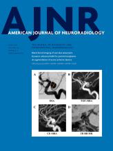
    Surveillance of Unruptured Intracranial Saccular Aneurysms Using Noncontrast 3D-Black-Blood MRI: Comparison of 3D-TOF and Contrast-Enhanced MRA with 3D-DSA
    C. Zhu, X. Wang, L. Eisenmenger, B. Tian, Q. Liu, A.J. Degnan, C. Hess, D. Saloner and J. Lu
    American Journal of Neuroradiology June 2019, 40 (6) 960-966; DOI: https://doi.org/10.3174/ajnr.A6080

    Sixty-four patients with 68 saccular unruptured intracranial aneurysms were recruited. Patients underwent 3T MR imaging with 3D-TOF-MRA, 3D black-blood MR imaging, and contrast-enhanced MRA, and they underwent 3D rotational angiography within 2 weeks. The neck, width, and height of the unruptured intracranial aneurysms were measured by 2 radiologists independently on 3D rotational angiography and 3 MR imaging sequences. 3D black-blood MR imaging demonstrates the best agreement with DSA, with the smallest limits of agreement and measurement error. 3D-TOF-MRA had the largest limits of agreement and measurement error. The authors conclude that 3D black-blood MR imaging achieves better accuracy for aneurysm size measurements compared with 3D-TOF, using 3D rotational angiography as a criterion standard.

  • Adult Brain
    Open Access
    Baseline and Evolutionary Radiologic Features in Sporadic, Hemorrhagic Brain Cavernous Malformations
    K.D. Flemming, S. Kumar, G. Lanzino and W. Brinjikji
    American Journal of Neuroradiology June 2019, 40 (6) 967-972; DOI: https://doi.org/10.3174/ajnr.A6076
  • Adult Brain
    You have access
    The Occipital Emissary Vein: A Possible Marker for Pseudotumor Cerebri
    A. Hedjoudje, A. Piveteau, C. Gonzalez-Campo, A. Moghekar, P. Gailloud and D. San Millán
    American Journal of Neuroradiology June 2019, 40 (6) 973-978; DOI: https://doi.org/10.3174/ajnr.A6061
  • Adult Brain
    Open Access
    Association between Tumor Acidity and Hypervascularity in Human Gliomas Using pH-Weighted Amine Chemical Exchange Saturation Transfer Echo-Planar Imaging and Dynamic Susceptibility Contrast Perfusion MRI at 3T
    Y.-L. Wang, J. Yao, A. Chakhoyan, C. Raymond, N. Salamon, L.M. Liau, P.L. Nghiemphu, A. Lai, W.B. Pope, N. Nguyen, M. Ji, T.F. Cloughesy and B.M. Ellingson
    American Journal of Neuroradiology June 2019, 40 (6) 979-986; DOI: https://doi.org/10.3174/ajnr.A6063
  • Adult Brain
    Open Access
    Quantitative Susceptibility Mapping of Time-Dependent Susceptibility Changes in Multiple Sclerosis Lesions
    S. Zhang, T.D. Nguyen, S.M. Hurtado Rúa, U.W. Kaunzner, S. Pandya, I. Kovanlikaya, P. Spincemaille, Y. Wang and S.A. Gauthier
    American Journal of Neuroradiology June 2019, 40 (6) 987-993; DOI: https://doi.org/10.3174/ajnr.A6071
  • EDITOR'S CHOICEAdult Brain
    You have access
    Susceptibility-Weighted MR Imaging Hypointense Rim in Progressive Multifocal Leukoencephalopathy: The End Point of Neuroinflammation and a Potential Outcome Predictor
    M.M Thurnher, J. Boban, A. Rieger and E. Gelpi
    American Journal of Neuroradiology June 2019, 40 (6) 994-1000; DOI: https://doi.org/10.3174/ajnr.A6072

    This retrospective study included 18 patients with a definite diagnosis of progressive multifocal leukoencephalopathy. Ten patients were HIV-positive, 3 patients had natalizumab-associated PML, 1 patient had multiple myeloma, 3 patients had a history of lymphoma, and 1 was diagnosed with acute myeloid leukemia. Patients were divided into short- (up to 12 months) and long-term (>12 months) survivors. A total of 93 initial and follow-up MR imaging examinations were reviewed. On SWI, the presence and development of a hypointense rim at the periphery of the PML lesions were noted. A postmortem histologic examination was performed in 2 patients: A rim formed in one, and in one, there was no rim. A total of 73 progressive multifocal leukoencephalopathy lesions were observed. In 13 (72.2%) patients, a well-defined thin, linear, hypointense rim at the periphery of the lesion toward the cortical side was present, while in 5 (27.8%) patients, it was completely absent. All 11 long-term survivors and 2 short-term survivors presented with a prominent SWI-hypointense rim. The thin, uniformly linear, gyriform SWI-hypointense rim in the paralesional U-fibers in patients with definite PML might represent an end point stage of the neuroinflammatory process in long-term survivors.

  • Interventional
    Open Access
    Thrombectomy with Conscious Sedation Compared with General Anesthesia: A DEFUSE 3 Analysis
    C.J. Powers, D. Dornbos, M. Mlynash, D. Gulati, M. Torbey, S.M. Nimjee, M.G. Lansberg, G.W. Albers and M.P. Marks
    American Journal of Neuroradiology June 2019, 40 (6) 1001-1005; DOI: https://doi.org/10.3174/ajnr.A6059
  • FELLOWS' JOURNAL CLUBInterventional
    You have access
    First-Line Sofia Aspiration Thrombectomy Approach within the Endovascular Treatment of Ischemic Stroke Multicentric Registry: Efficacy, Safety, and Predictive Factors of Success
    G. Marnat, X. Barreau, L. Detraz, R. Bourcier, B. Gory, A. Sgreccia, F. Gariel, J. Berge, P. Menegon, M. Kyheng, J. Labreuche, A. Consoli, R. Blanc and B. Lapergue on behalf of the ETIS Investigators
    American Journal of Neuroradiology June 2019, 40 (6) 1006-1012; DOI: https://doi.org/10.3174/ajnr.A6074

    The authors performed a retrospective analysis of the prospectively maintained Endovascular Treatment of Ischemic Stroke multicentric registry. Data from consecutive patients who benefited from thrombectomy with a first-line Sofia approach between January 2013 and April 2018 were studied. We excluded other first-line approaches (stent retriever or combined aspiration and stent retriever) and extracranial occlusions. During the study period, 296 patients were treated. Mean age and initial NIHSS score were, respectively, 69.5 years and 16. Successful reperfusion, defined by the modified TICI 2b/3, was obtained in 86.1%. Complete reperfusion (modified TICI 3) was obtained in 41.2%. A first-pass effect was achieved in 24.2%. A rescue stent retriever approach was required in 29.7%. The first-line contact aspiration approach appeared safe and efficient with Sofia catheters. These devices achieved very high reperfusion rates with a low requirement for stent retriever rescue therapy, especially for M1 occlusions.

  • Interventional
    You have access
    Current Hospital Demographics of Subarachnoid Hemorrhage Based on CT Angiography and 3D Rotational Angiography in a Neurosurgical Center
    S.B.T. van Rooij, R.S. Bechan, W.J. van Rooij and M.E. Sprengers
    American Journal of Neuroradiology June 2019, 40 (6) 1013-1017; DOI: https://doi.org/10.3174/ajnr.A6060
  • Interventional
    Open Access
    Histologic and Biomolecular Similarities in Healing between Aneurysms and Cutaneous Skin Wounds
    J.R. Ayers-Ringler, Z. Khashim, Y.-H. Ding, D.F. Kallmes and R. Kadirvel
    American Journal of Neuroradiology June 2019, 40 (6) 1018-1021; DOI: https://doi.org/10.3174/ajnr.A6046
  • Extracranial Vascular
    You have access
    An Ultrasonographic Multiparametric Carotid Plaque Risk Index Associated with Cerebrovascular Symptomatology: A Study Comparing Color Doppler Imaging and Contrast-Enhanced Ultrasonography
    V. Rafailidis, I. Chryssogonidis, C. Xerras, E. Grisan, G.-A. Cheimariotis, T. Tegos, D. Rafailidis, P.S. Sidhu and A. Charitanti-Kouridou
    American Journal of Neuroradiology June 2019, 40 (6) 1022-1028; DOI: https://doi.org/10.3174/ajnr.A6056
  • Head & Neck
    You have access
    Dynamic Contrast-Enhanced MRI to Differentiate Parotid Neoplasms Using Golden-Angle Radial Sparse Parallel Imaging
    J.L. Mogen, K.T. Block, N.K. Bansal, J.T. Patrie, S. Mukherjee, E. Zan, M. Hagiwara, G.M. Fatterpekar and S.H. Patel
    American Journal of Neuroradiology June 2019, 40 (6) 1029-1036; DOI: https://doi.org/10.3174/ajnr.A6055
  • Head & Neck
    You have access
    Localization of Parotid Gland Tumors in Relation to the Intraparotid Facial Nerve on 3D Double-Echo Steady-State with Water Excitation Sequence
    H. Fujii, A. Fujita, H. Kanazawa, E. Sung, O. Sakai and H. Sugimoto
    American Journal of Neuroradiology June 2019, 40 (6) 1037-1042; DOI: https://doi.org/10.3174/ajnr.A6078
  • Head & Neck
    You have access
    Evaluating the Utility of a Postprocessing Algorithm for MRI Evaluation of Optic Neuritis
    L. Stunkel, A. Sharma, M.S. Parsons, A. Salter and G.P. Van Stavern
    American Journal of Neuroradiology June 2019, 40 (6) 1043-1048; DOI: https://doi.org/10.3174/ajnr.A6057
  • Head & Neck
    You have access
    A Scoring System for Prediction of Cervical Lymph Node Metastasis in Patients with Head and Neck Squamous Cell Carcinoma
    M.S. Chung, Y.J. Choi, S.O. Kim, Y.S. Lee, J.Y. Hong, J.H. Lee and J.H. Baek
    American Journal of Neuroradiology June 2019, 40 (6) 1049-1054; DOI: https://doi.org/10.3174/ajnr.A6066
  • FELLOWS' JOURNAL CLUBHead & Neck
    You have access
    Readout-Segmented Echo-Planar DWI for the Detection of Cholesteatomas: Correlation with Surgical Validation
    N. Fischer, V.H. Schartinger, D. Dejaco, J. Schmutzhard, H. Riechelmann, M. Plaikner and B. Henninger
    American Journal of Neuroradiology June 2019, 40 (6) 1055-1059; DOI: https://doi.org/10.3174/ajnr.A6079

    Readout-segmented echo-planar (RESOLVE)-DWI is a new alternative technique for obtaining DWI with high quality, delivering sharp images at high spatial resolution and reduced slice thickness. Fifty patients with chronic otitis media who underwent MR imaging before an operation of the middle ear were included. The MR imaging protocol consisted of axial and coronal readout-segmented echo-planar DWI with b-values of 0 and 1000 s/mm2 and 3-mm slice thickness. The readout segmented echo-planar diffusion-weighted images were fused with standard T2-weighted sequences for better anatomic assignment. Readout-segmented echo-planar DWI detected 22 of the 25 cases of surgically proved cholesteatoma. It has an accuracy of 92%, a sensitivity of 88%, a specificity of 96%, a positive predictive value of 96%, and a negativepredictive value of 89%. Readout-segmented echo-planar DWI is a promising and reliable MR imaging sequence for the detection and exclusion of cholesteatoma.

  • Pediatrics
    Open Access
    Radiologic and Pathologic Features of the Transmantle Sign in Focal Cortical Dysplasia: The T1 Signal Is Useful for Differentiating Subtypes
    Yukio Kimura, A. Shioya, Y. Saito, Y. Oitani, Y. Shigemoto, E. Morimoto, F. Suzuki, N. Ikegaya, Yuiko Kimura, K. Iijima, Y. Takayama, M. Iwasaki, M. Sasaki and N. Sato
    American Journal of Neuroradiology June 2019, 40 (6) 1060-1066; DOI: https://doi.org/10.3174/ajnr.A6067
  • Pediatrics
    You have access
    Radiation Dose and Image Quality in Pediatric Neck CT
    S.V. Tipnis, W.J. Rieter, D. Patel, S.T. Stalcup, M.G. Matheus and M.V. Spampinato
    American Journal of Neuroradiology June 2019, 40 (6) 1067-1073; DOI: https://doi.org/10.3174/ajnr.A6073
  • Spine
    Open Access
    Deep Learning–Based Automatic Segmentation of Lumbosacral Nerves on CT for Spinal Intervention: A Translational Study
    G. Fan, H. Liu, Z. Wu, Y. Li, C. Feng, D. Wang, J. Luo, W.M. Wells and S. He
    American Journal of Neuroradiology June 2019, 40 (6) 1074-1081; DOI: https://doi.org/10.3174/ajnr.A6070

Online Features

  • Letters

    • LETTER
      You have access
      Vessel Wall Imaging of Unruptured Intracranial Aneurysms: Ready for Prime Time? Not so Fast!
      V.T. Lehman and W. Brinjikji
      American Journal of Neuroradiology June 2019, 40 (6) E26-E29; DOI: https://doi.org/10.3174/ajnr.A6048
    • LETTER
      You have access
      Patients with High-Grade Gliomas and Café-au-Lait Macules: Is Neurofibromatosis Type 1 the Only Diagnosis?
      L. Guerrini-Rousseau, M. Suerink, J. Grill, E. Legius, K. Wimmer and L. Brugières
      American Journal of Neuroradiology June 2019, 40 (6) E30-E31; DOI: https://doi.org/10.3174/ajnr.A6058
    • LETTER
      You have access
      Reply:
      N. Kadom, R.C. Castellino and D.S. Wolf
      American Journal of Neuroradiology June 2019, 40 (6) E32; DOI: https://doi.org/10.3174/ajnr.A6062

Erratum

  • Erratum
    You have access
    Erratum
    American Journal of Neuroradiology June 2019, 40 (6) E33; DOI: https://doi.org/10.3174/ajnr.A6069
Back to top
PreviousNext

In this issue

American Journal of Neuroradiology: 40 (6)
American Journal of Neuroradiology
Vol. 40, Issue 6
1 Jun 2019
  • Table of Contents
  • Index by author
  • Complete Issue (PDF)
Sign up for alerts
Advertisement

Jump to

  • Perspectives
  • Review Article
  • Social Media Vignette
  • General Contents
  • Online Features
    • Letters
  • Erratum
  • Letters
  • Most Read
  • Most Cited
Loading
Advertisement

Indexed Content

  • Current Issue
  • Accepted Manuscripts
  • Article Preview
  • Past Issues
  • Editorials
  • Editors Choice
  • Fellow Journal Club
  • Letters to the Editor

Cases

  • Case Collection
  • Archive - Case of the Week
  • Archive - Case of the Month
  • Archive - Classic Case

Special Collections

  • Special Collections

Resources

  • News and Updates
  • Turn around Times
  • Submit a Manuscript
  • Author Policies
  • Manuscript Submission Guidelines
  • Evidence-Based Medicine Level Guide
  • Publishing Checklists
  • Graphical Abstract Preparation
  • Imaging Protocol Submission
  • Submit a Case
  • Become a Reviewer/Academy of Reviewers
  • Get Peer Review Credit from Publons

Multimedia

  • AJNR Podcast
  • AJNR SCANtastic
  • Video Articles

About Us

  • About AJNR
  • Editorial Board
  • Not an AJNR Subscriber? Join Now
  • Alerts
  • Feedback
  • Advertise with us
  • Librarian Resources
  • Permissions
  • Terms and Conditions

American Society of Neuroradiology

  • Not an ASNR Member? Join Now

© 2025 by the American Society of Neuroradiology All rights, including for text and data mining, AI training, and similar technologies, are reserved.
Print ISSN: 0195-6108 Online ISSN: 1936-959X

Powered by HighWire