
Article Figures & Data
Figures
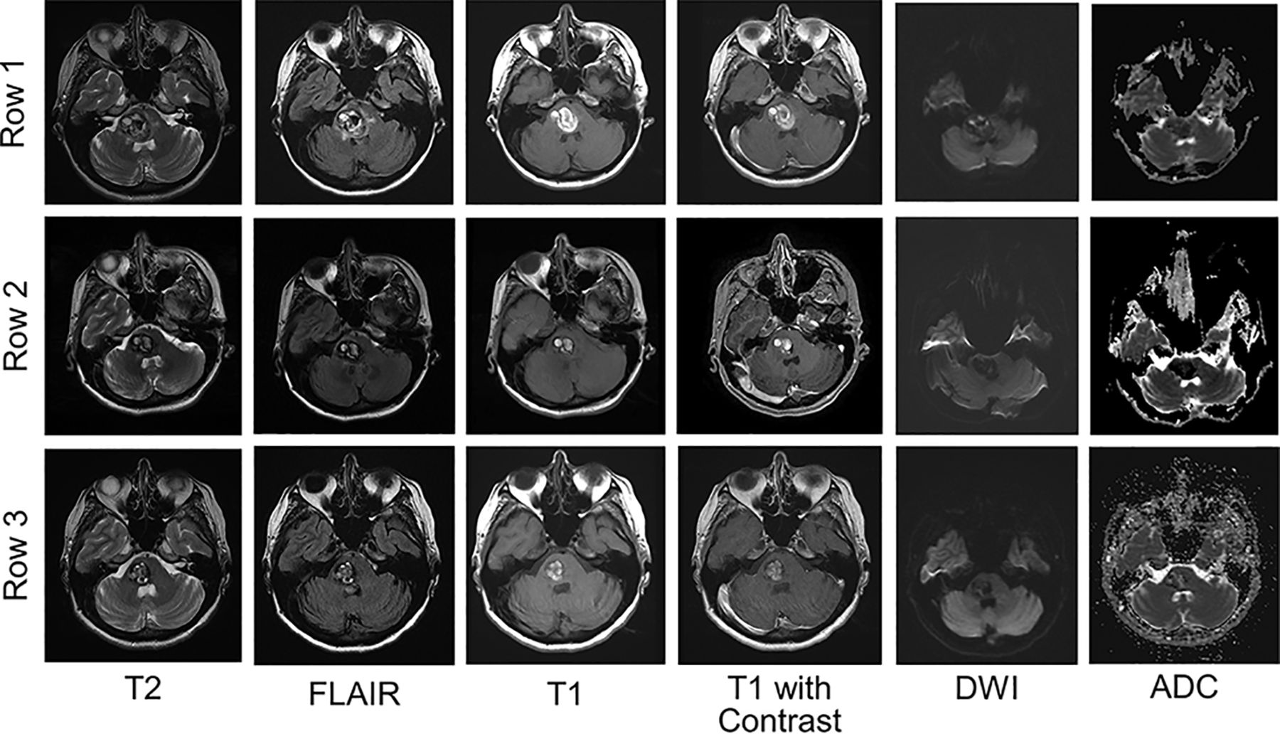
- Fig 1.
Typical evolution of a hemorrhagic CM on MR imaging with time. Row 1, A hemorrhagic CM in the pons at the time of acute focal symptoms. MR imaging demonstrates subacute hemorrhage with increased T1 and T2 signal, with surrounding edema on FLAIR. In this patient, there was no enhancement of the lesion. DWI and ADC maps demonstrate low signal intensity. Rows 2 and 3, The evolution of the lesion 1 and 8 months later, respectively. The edema is reduced by 1 month and absent at 8 months. The lesion decreases in size, but there remains an increased T1 signal. DWI remains low signal intensity throughout. The ADC map demonstrates low or mixed intensity by 8 months.
- Fig 2.
Radiologic evolution of hemorrhagic cavernous malformations. Row 1, An acute hemorrhage into a medullary cavernous malformation with surrounding FLAIR hyperintensity. There is a small developmental venous anomaly on contrast imaging. A follow-up MR imaging (row 2) was performed 3 months later. The cavernous malformation has evolved from a Zabramski type I to type III lesion. There is no persistent edema, and the ADC and DWI demonstrate hypointensity.
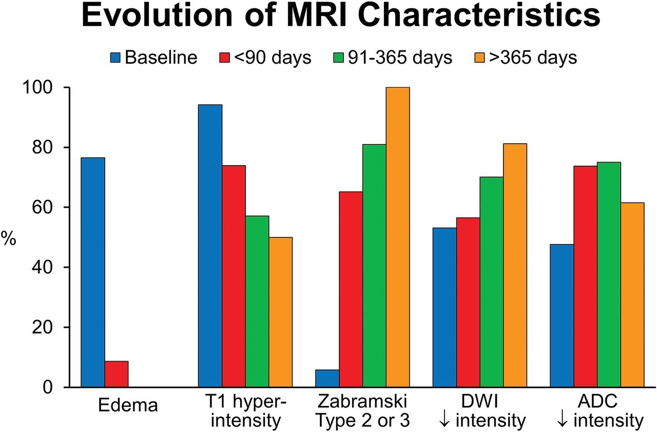
- Fig 3.
Overview of the evolutionary MR imaging changes of hemorrhagic CM with time. This graphic display shows the percentage of patients with particular MR imaging characteristics at baseline and ≤90 days, 91–365 days, and >365 days.


Tables
Clinical Information Sex 31 (60.8%) male Age at diagnosis (yr) Median, 38.3 (range, 17.7–70.5) Race 45 (90.0%) Caucasian Clinical presentation (No.) (%) Focal neurologic deficit, 37 (72.6%) Seizure, 4 (7.8%) Headache, 8 (15.6%) Spell, not seizure, 1 (2.0%) Other, 1 (2.0%) Initial MRI scan brain (No.) Symptom onset to first MRI (day) Median, 4 (range, 0–30) >1 CMa 3 (5.88%) Size (mm) Median, 12.7 (range, 4.7–34.7) Brain stem location 31 (60.8%) Location Cortical, 12 (23.5%) Supratentorial, subcortical, 6 (11.8%) Posterior fossa, 32 (62.7%) Intraventricular, 1 (2.0%) Zabramski lesion type Type I, 48 (94.2%) Type II, 3 (5.8%) DVA 21/42b (50.0%) T1 hyperintensity 48 (94.1%) Enhancement with Gd 7/42b (16.7%) Edema 39 (76.5%) DWI intensity Low, 26/49b (53.1%) Mixed, 21/49b (42.9%) High, 2/49b (4.0%) ADC map intensity Low, 19/42b (45.2%) Mixed, 23/42b (54.8%) High, 0 Perilesional high T1 signal 17/51 (33.3%) ≤90 Days 91–365 Days >365 Days No. 28 24 23 Time to follow-up MRI (day) Median, 46 (range, 12–90) Median, 159.5 (range, 92–365) Median, 1136 (range, 382-3022) Follow-up MRI demonstrated rebleed (No.) 5 3 6 T1 hyperintensity 17/23 (73.9%) 12/21 (57.1%) 8/16 (50%) T1 hyperintensity (moderate to significant) 9/17 (52.9%) 6/12 (50%) 1/8 (12.5%) Enhancement with gadolinium 6/22 (27.3%) 7/20 (35%) 9/16 (56.2%) Degree of enhancement (moderate to significant) 2/6 (33.3%) 1/7 (14.3%) 2/9 (22.2%) Edema 2/23 (8.7%) 0 0 Degree of edema (moderate to significant) 1/2 (50%) – – DWI low intensity 13/23 (56.5%) 12/17 (70.1%) 13/16 (81.2%) ADC low intensity 14/19 (73.7%) 12/16 (75.0%) 8/13 (61.5%) Change to Zabramski type II or III lesion 15/23 (65.2%) 17/21 (80.9%) 16/16 (100%) Average size change (compared with original MRI) (mm) Median, 1.4 (range, −6.5 to +1.7) Median, 2.05 (range, −24.7 to +1.1) Median, −3.3 (range, −9.7 to +8.9) Note:—– indicates no data.
↵a Denominators noted are based on the availability of the particular MRI sequence.
≤365 Days >365 Days No. 37 23 Median time to MRI (day) 101 (range, 5–263) 1136 (range, 382–3022) No. with recurrent hemorrhage 8 6 T1 hyperintensity 19/32 (59.4%) 8/16 (50.0%) T1 hyperintensity (moderate to significant) 10/19 (56.2%) 1/9 (11.1%) Enhancement with gadolinium 8/31 (25.8%) 9/16 (56.2%) Edema 2/32 (6.2%) 0 DWI low intensity 20/29 (68.9%) 13/16 (81.2%) ADC low intensity 19/28 (67.9%) 8/13 (61.5%) Change to Zabramski type II or III lesions 27/32 (84.4%) 16/16 (100%) Average size change (mm) (range) Median, −1.4 (−24.7 to +1.1) Median, −3.3 (−9.7 to +8.9) ↵a Denominators noted are based on the availability of the particular MRI sequence.


Jump to section
Related Articles
Cited By...
- No citing articles found.