Advertisement

Main menu

  • Home
  • Content
    • Current Issue
    • Accepted Manuscripts
    • Article Preview
    • Past Issue Archive
    • AJNR Case Collection
    • Case of the Week Archive
    • Classic Case Archive
    • Case of the Month Archive
  • Special Collections
    • Spinal CSF Leak Articles (Jan 2020-June 2024)
    • 2024 AJNR Journal Awards
    • Most Impactful AJNR Articles
  • Multimedia
    • AJNR Podcast
    • AJNR Scantastics
    • Video Articles
  • For Authors
    • Submit a Manuscript
    • Author Policies
    • Fast publishing of Accepted Manuscripts
    • Graphical Abstract Preparation
    • Manuscript Submission Guidelines
    • Imaging Protocol Submission
    • Submit a Case for the Case Collection
  • About Us
    • About AJNR
    • Editorial Board
  • More
    • Become a Reviewer/Academy of Reviewers
    • Subscribers
    • Permissions
    • Alerts
    • Feedback
    • Advertisers
    • ASNR Home
  • Other Publications
    • ajnr

User menu

  • Alerts
  • Log in

Search

  • Advanced search
American Journal of Neuroradiology

American Journal of Neuroradiology

ASHNR American Society of Functional Neuroradiology ASHNR American Society of Pediatric Neuroradiology ASSR
  • Alerts
  • Log in

Advanced Search

  • Home
  • Content
    • Current Issue
    • Accepted Manuscripts
    • Article Preview
    • Past Issue Archive
    • AJNR Case Collection
    • Case of the Week Archive
    • Classic Case Archive
    • Case of the Month Archive
  • Special Collections
    • Spinal CSF Leak Articles (Jan 2020-June 2024)
    • 2024 AJNR Journal Awards
    • Most Impactful AJNR Articles
  • Multimedia
    • AJNR Podcast
    • AJNR Scantastics
    • Video Articles
  • For Authors
    • Submit a Manuscript
    • Author Policies
    • Fast publishing of Accepted Manuscripts
    • Graphical Abstract Preparation
    • Manuscript Submission Guidelines
    • Imaging Protocol Submission
    • Submit a Case for the Case Collection
  • About Us
    • About AJNR
    • Editorial Board
  • More
    • Become a Reviewer/Academy of Reviewers
    • Subscribers
    • Permissions
    • Alerts
    • Feedback
    • Advertisers
    • ASNR Home
  • Follow AJNR on Twitter
  • Visit AJNR on Facebook
  • Follow AJNR on Instagram
  • Join AJNR on LinkedIn
  • RSS Feeds

Welcome to the new AJNR, Updated Hall of Fame, and more. Read the full announcements.


AJNR is seeking candidates for the position of Associate Section Editor, AJNR Case Collection. Read the full announcement.


Research ArticleSpine

Quantification of DTI in the Pediatric Spinal Cord: Application to Clinical Evaluation in a Healthy Patient Population

B.B. Reynolds, S. By, Q.R. Weinberg, A.A. Witt, A.T. Newton, H.R. Feiler, B. Ramkorun, D.B. Clayton, P. Couture, J.E. Martus, M. Adams, J.C. Wellons, S.A. Smith and A. Bhatia
American Journal of Neuroradiology July 2019, 40 (7) 1236-1241; DOI: https://doi.org/10.3174/ajnr.A6104
B.B. Reynolds
fInstitute of Imaging Science (B.B.R., S.B., Q.R.W., A.A.W., A.T.N., H.R.F., B.R., S.A.S., A.B.), Vanderbilt University, Nashville, Tennessee
  • Find this author on Google Scholar
  • Find this author on PubMed
  • Search for this author on this site
  • ORCID record for B.B. Reynolds
S. By
fInstitute of Imaging Science (B.B.R., S.B., Q.R.W., A.A.W., A.T.N., H.R.F., B.R., S.A.S., A.B.), Vanderbilt University, Nashville, Tennessee
  • Find this author on Google Scholar
  • Find this author on PubMed
  • Search for this author on this site
  • ORCID record for S. By
Q.R. Weinberg
fInstitute of Imaging Science (B.B.R., S.B., Q.R.W., A.A.W., A.T.N., H.R.F., B.R., S.A.S., A.B.), Vanderbilt University, Nashville, Tennessee
  • Find this author on Google Scholar
  • Find this author on PubMed
  • Search for this author on this site
  • ORCID record for Q.R. Weinberg
A.A. Witt
fInstitute of Imaging Science (B.B.R., S.B., Q.R.W., A.A.W., A.T.N., H.R.F., B.R., S.A.S., A.B.), Vanderbilt University, Nashville, Tennessee
  • Find this author on Google Scholar
  • Find this author on PubMed
  • Search for this author on this site
  • ORCID record for A.A. Witt
A.T. Newton
aFrom the Department of Radiology and Radiological Sciences (A.T.N., P.C., S.A.S., A.B.)
cPediatrics (A.T.N.)
fInstitute of Imaging Science (B.B.R., S.B., Q.R.W., A.A.W., A.T.N., H.R.F., B.R., S.A.S., A.B.), Vanderbilt University, Nashville, Tennessee
  • Find this author on Google Scholar
  • Find this author on PubMed
  • Search for this author on this site
  • ORCID record for A.T. Newton
H.R. Feiler
fInstitute of Imaging Science (B.B.R., S.B., Q.R.W., A.A.W., A.T.N., H.R.F., B.R., S.A.S., A.B.), Vanderbilt University, Nashville, Tennessee
  • Find this author on Google Scholar
  • Find this author on PubMed
  • Search for this author on this site
  • ORCID record for H.R. Feiler
B. Ramkorun
fInstitute of Imaging Science (B.B.R., S.B., Q.R.W., A.A.W., A.T.N., H.R.F., B.R., S.A.S., A.B.), Vanderbilt University, Nashville, Tennessee
  • Find this author on Google Scholar
  • Find this author on PubMed
  • Search for this author on this site
  • ORCID record for B. Ramkorun
D.B. Clayton
bUrology (D.B.C., M.A.)
  • Find this author on Google Scholar
  • Find this author on PubMed
  • Search for this author on this site
  • ORCID record for D.B. Clayton
P. Couture
aFrom the Department of Radiology and Radiological Sciences (A.T.N., P.C., S.A.S., A.B.)
  • Find this author on Google Scholar
  • Find this author on PubMed
  • Search for this author on this site
  • ORCID record for P. Couture
J.E. Martus
dDivision of Pediatric Orthopaedics (J.E.M.), Monroe Carell Jr. Children's Hospital at Vanderbilt, Nashville, Tennessee
  • Find this author on Google Scholar
  • Find this author on PubMed
  • Search for this author on this site
  • ORCID record for J.E. Martus
M. Adams
bUrology (D.B.C., M.A.)
  • Find this author on Google Scholar
  • Find this author on PubMed
  • Search for this author on this site
  • ORCID record for M. Adams
J.C. Wellons III
aFrom the Department of Radiology and Radiological Sciences (A.T.N., P.C., S.A.S., A.B.)
gDepartment of Ophthalmology (S.A.S., J.C.W. III), Vanderbilt University Medical Center, Nashville, Tennessee.
  • Find this author on Google Scholar
  • Find this author on PubMed
  • Search for this author on this site
  • ORCID record for J.C. Wellons III
S.A. Smith
aFrom the Department of Radiology and Radiological Sciences (A.T.N., P.C., S.A.S., A.B.)
eDepartment of Biomedical Engineering (S.A.S.)
fInstitute of Imaging Science (B.B.R., S.B., Q.R.W., A.A.W., A.T.N., H.R.F., B.R., S.A.S., A.B.), Vanderbilt University, Nashville, Tennessee
gDepartment of Ophthalmology (S.A.S., J.C.W. III), Vanderbilt University Medical Center, Nashville, Tennessee.
  • Find this author on Google Scholar
  • Find this author on PubMed
  • Search for this author on this site
  • ORCID record for S.A. Smith
A. Bhatia
aFrom the Department of Radiology and Radiological Sciences (A.T.N., P.C., S.A.S., A.B.)
fInstitute of Imaging Science (B.B.R., S.B., Q.R.W., A.A.W., A.T.N., H.R.F., B.R., S.A.S., A.B.), Vanderbilt University, Nashville, Tennessee
  • Find this author on Google Scholar
  • Find this author on PubMed
  • Search for this author on this site
  • ORCID record for A. Bhatia
  • Article
  • Figures & Data
  • Info & Metrics
  • Responses
  • References
  • PDF
Loading

Article Figures & Data

Figures

  • Tables
  • Fig 1.
    • Download figure
    • Open in new tab
    • Download powerpoint
    Fig 1.

    Diffusion images and metrics. A, Patient's sagittal T2-weighted image with overlay shows where 2 different diffusion imaging volumes (A1 and A2) were acquired and to which region they were assigned. B, Sample DTI-derived maps for each region for the same patients as in A. B0 indicates minimally weighted diffusion image; CV, cervical; UT, upper thoracic; TL, thoracolumbar; CN, conus. Scale bars show the range of visualized values for each metric.
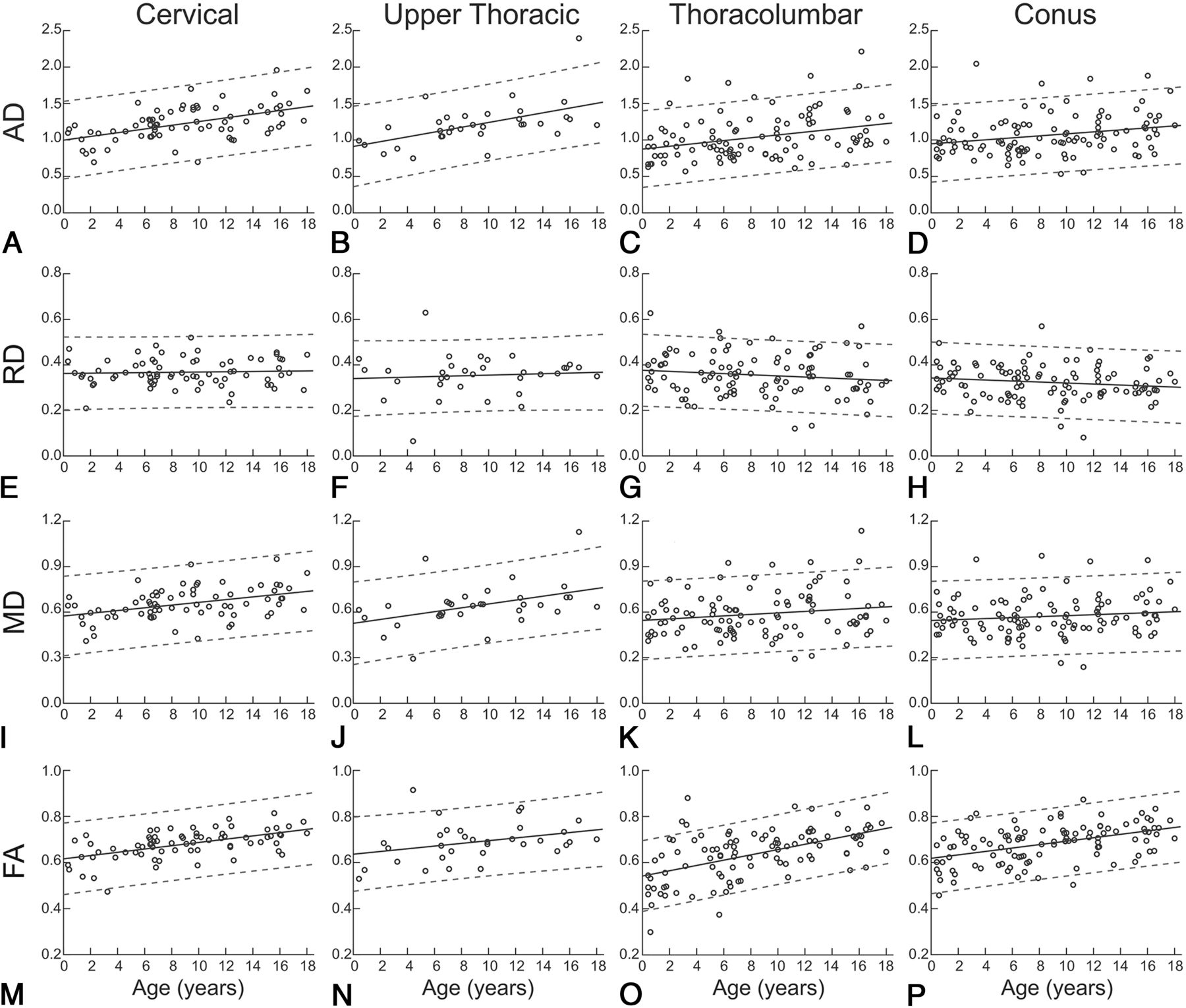
  • Fig 2.
    • Download figure
    • Open in new tab
    • Download powerpoint
    Fig 2.

    Average regional DTI indices as a function of age. Age-related trends are shown across pediatric development for each spinal cord region and diffusion-derived index. Circles show mean DTI-derived index values for individual patients, solid lines indicate the age-related trend, and dashed lines indicate the 95% confidence interval.

Tables

  • Figures
  • ANCOVA and eta statistics

    ANCOVA/FactorSum SquareddfMean SquaredFP Valueη2Partial η2
    Axial diffusivity
        Main effects
            Age3.04513.04544.26<.0010.11690.1271
            Region1.80130.6008.72<.0010.06920.0793
        Interaction
            Age × region0.28030.0931.36.260.01080.0132
        Model error20.9173040.0690.8032
        Model totals26.044
    Radial diffusivity
        Main effects
            Age0.01410.0142.24.140.00690.0073
            Region0.10130.0345.43.0010.05010.0508
        Interaction
            Age × region0.01830.0060.94.420.00870.0092
        Model error1.8903040.0060.9344
        Model totals2.023
    Mean diffusivity
        Main effects
            Age0.27010.27016.20<.0010.04690.0506
            Region0.34630.1156.93<.0010.06020.0640
        Interaction
            Age × region0.07230.0241.44.230.01250.0140
        Model error5.0613040.0170.8804
        Model totals5.749
    Fractional anisotropy
        Main effects
            Age0.56310.56396.11<.0010.22580.2402
            Region0.11530.0386.55<.0010.04620.0607
        Interaction
            Age × region0.03430.0111.94.120.01360.0187
        Model error1.7813040.0060.7143
        Model totals2.493
PreviousNext
Back to top

In this issue

American Journal of Neuroradiology: 40 (7)
American Journal of Neuroradiology
Vol. 40, Issue 7
1 Jul 2019
  • Table of Contents
  • Index by author
  • Complete Issue (PDF)
Advertisement
Print
Download PDF
Email Article

Thank you for your interest in spreading the word on American Journal of Neuroradiology.

NOTE: We only request your email address so that the person you are recommending the page to knows that you wanted them to see it, and that it is not junk mail. We do not capture any email address.

Enter multiple addresses on separate lines or separate them with commas.
Quantification of DTI in the Pediatric Spinal Cord: Application to Clinical Evaluation in a Healthy Patient Population
(Your Name) has sent you a message from American Journal of Neuroradiology
(Your Name) thought you would like to see the American Journal of Neuroradiology web site.
CAPTCHA
This question is for testing whether or not you are a human visitor and to prevent automated spam submissions.
Cite this article
B.B. Reynolds, S. By, Q.R. Weinberg, A.A. Witt, A.T. Newton, H.R. Feiler, B. Ramkorun, D.B. Clayton, P. Couture, J.E. Martus, M. Adams, J.C. Wellons, S.A. Smith, A. Bhatia
Quantification of DTI in the Pediatric Spinal Cord: Application to Clinical Evaluation in a Healthy Patient Population
American Journal of Neuroradiology Jul 2019, 40 (7) 1236-1241; DOI: 10.3174/ajnr.A6104

Citation Manager Formats

  • BibTeX
  • Bookends
  • EasyBib
  • EndNote (tagged)
  • EndNote 8 (xml)
  • Medlars
  • Mendeley
  • Papers
  • RefWorks Tagged
  • Ref Manager
  • RIS
  • Zotero
0 Responses
Respond to this article
Share
Bookmark this article
Quantification of DTI in the Pediatric Spinal Cord: Application to Clinical Evaluation in a Healthy Patient Population
B.B. Reynolds, S. By, Q.R. Weinberg, A.A. Witt, A.T. Newton, H.R. Feiler, B. Ramkorun, D.B. Clayton, P. Couture, J.E. Martus, M. Adams, J.C. Wellons, S.A. Smith, A. Bhatia
American Journal of Neuroradiology Jul 2019, 40 (7) 1236-1241; DOI: 10.3174/ajnr.A6104
del.icio.us logo Twitter logo Facebook logo Mendeley logo
  • Tweet Widget
  • Facebook Like
  • Google Plus One
Purchase

Jump to section

  • Article
    • Abstract
    • ABBREVIATIONS:
    • Materials and Methods
    • Results
    • Discussion
    • Conclusions
    • Footnotes
    • REFERENCES
  • Figures & Data
  • Info & Metrics
  • Responses
  • References
  • PDF

Related Articles

  • No related articles found.
  • PubMed
  • Google Scholar

Cited By...

  • Diffusion Kurtosis Imaging of neonatal Spinal Cord in clinical routine
  • Atlas-Based Quantification of DTI Measures in a Typically Developing Pediatric Spinal Cord
  • Crossref (8)
  • Google Scholar

This article has been cited by the following articles in journals that are participating in Crossref Cited-by Linking.

  • Cerebral White Matter Myelination and Relations to Age, Gender, and Cognition: A Selective Review
    Irina S. Buyanova, Marie Arsalidou
    Frontiers in Human Neuroscience 2021 15
  • Microstructural Changes in Compressed Cervical Spinal Cord Are Consistent With Clinical Symptoms and Symptom Duration
    Weifei Wu, Zong Yang, Tingting Zhang, Neng Ru, Fan Zhang, Bin Wu, Jie Liang
    Spine 2020 45 16
  • Atlas-Based Quantification of DTI Measures in a Typically Developing Pediatric Spinal Cord
    S. Shahrampour, B. De Leener, M. Alizadeh, D. Middleton, L. Krisa, A.E. Flanders, S.H. Faro, J. Cohen-Adad, F.B. Mohamed
    American Journal of Neuroradiology 2021 42 9
  • Correlations of diffusion tensor imaging and clinical measures with spinal cord cross-sectional area measurements in pediatric spinal cord injury patients
    Devon M. Middleton, Shiva Shahrampour, Laura Krisa, Winston Liu, Govind Nair, Steven Jacobson, Chris J. Conklin, Mahdi Alizadeh, Scott H. Faro, M. J. Mulcahey, Feroze B. Mohamed
    The Journal of Spinal Cord Medicine 2023 46 6
  • Diffusion Kurtosis Imaging of Neonatal Spinal Cord in Clinical Routine
    Rosella Trò, Monica Roascio, Domenico Tortora, Mariasavina Severino, Andrea Rossi, Julien Cohen-Adad, Marco Massimo Fato, Gabriele Arnulfo
    Frontiers in Radiology 2022 2
  • Harmonization of multi-site diffusion tensor imaging data for cervical and thoracic spinal cord at 1.5 T and 3 T using longitudinal ComBat
    Devon M. Middleton, Yutong Li, Andrew Chen, Russell Shinohara, Joshua Fisher, Laura Krisa, Mark Elliot, Scott H. Faro, John H. Woo, Adam E. Flanders, Feroze B. Mohamed
    Scientific Reports 2023 13 1
  • Functional Neuroradiology
    Kiran Shankar Talekar, Meng Law, Majda M. Thurnher, Eric D. Schwartz, Adam E. Flanders
    2023
  • Porcine Model of the Growing Spinal Cord—Changes in Diffusion Tensor Imaging Parameters
    Karolina Barbara Owsińska-Schmidt, Paulina Drobot, Anna Zimny, Marcin Adam Wrzosek
    Animals 2023 13 4

More in this TOC Section

Spine

  • Bern Score Validity for SIH
  • MP2RAGE 7T in MS Lesions of the Cervical Spine
  • Deep Learning for STIR Spine MRI Quality
Show more Spine

Pediatrics

  • Comparison of Image Quality and Radiation Dose in Pediatric Temporal Bone CT Using Photon-Counting Detector CT and Energy-Integrating Detector CT
  • SyMRI & MR Fingerprinting in Brainstem Myelination
  • Pons&Vermis Localization on Fetal MRI Using U-Net
Show more Pediatrics

Similar Articles

Advertisement

Indexed Content

  • Current Issue
  • Accepted Manuscripts
  • Article Preview
  • Past Issues
  • Editorials
  • Editors Choice
  • Fellow Journal Club
  • Letters to the Editor

Cases

  • Case Collection
  • Archive - Case of the Week
  • Archive - Case of the Month
  • Archive - Classic Case

Special Collections

  • Special Collections

Resources

  • News and Updates
  • Turn around Times
  • Submit a Manuscript
  • Author Policies
  • Manuscript Submission Guidelines
  • Evidence-Based Medicine Level Guide
  • Publishing Checklists
  • Graphical Abstract Preparation
  • Imaging Protocol Submission
  • Submit a Case
  • Become a Reviewer/Academy of Reviewers
  • Get Peer Review Credit from Publons

Multimedia

  • AJNR Podcast
  • AJNR SCANtastic
  • Video Articles

About Us

  • About AJNR
  • Editorial Board
  • Not an AJNR Subscriber? Join Now
  • Alerts
  • Feedback
  • Advertise with us
  • Librarian Resources
  • Permissions
  • Terms and Conditions

American Society of Neuroradiology

  • Not an ASNR Member? Join Now

© 2025 by the American Society of Neuroradiology All rights, including for text and data mining, AI training, and similar technologies, are reserved.
Print ISSN: 0195-6108 Online ISSN: 1936-959X

Powered by HighWire