
Article Figures & Data
Figures
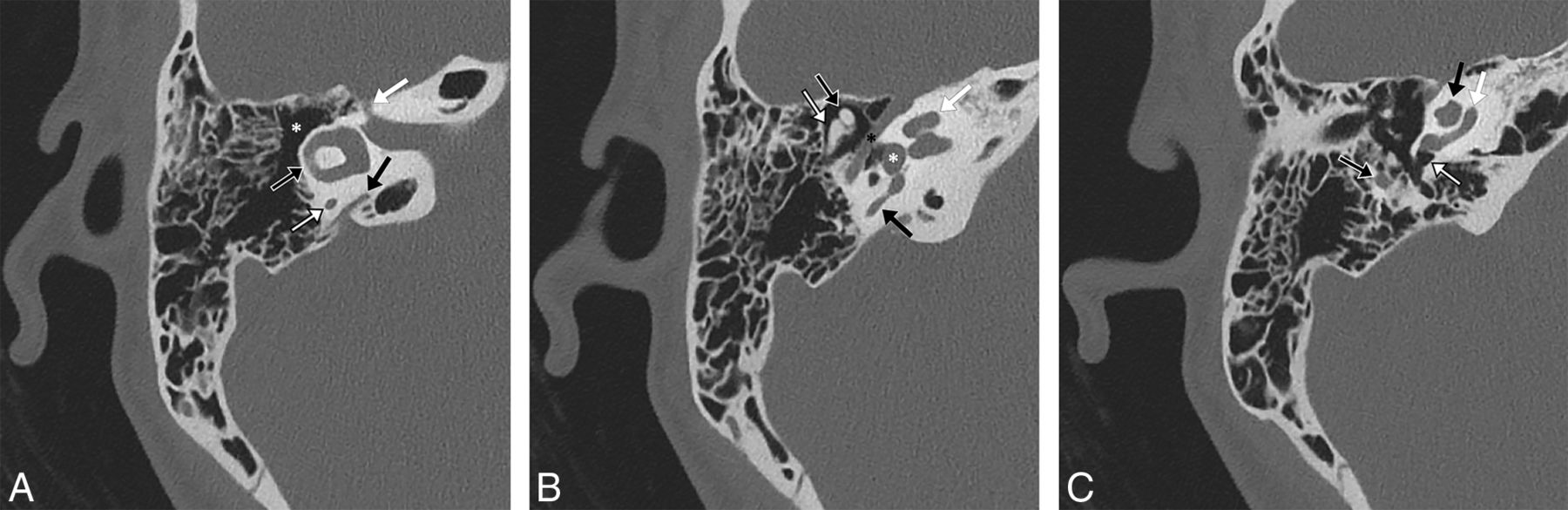
- Fig 1.
A 23-year-old woman with a normal right temporal bone. Noncontrast 0.6-mm-thick CT images in bone windows reformatted into an axial plane through the lateral semicircular canal. Major structures as seen in this plane include the following. A, at the level of the lateral semicircular canal: lateral semicircular canal (black arrow with white outline), posterior limb of the posterior semicircular canal (white arrow with black outline), vestibular aqueduct (black arrow), canal of the facial nerve labyrinthine segment (white arrow), and epitympanum/attic (asterisk). B, slightly inferior to A: the classic ice cream cone appearance of the malleus head (black arrow with white outline) and incus body (white arrow with black outline), entire length of the facial nerve tympanic segment (black asterisk), vestibule (white asterisk) at the upper aspect of the oval window, middle turn of the cochlea (white arrow), and upper limb of the posterior semicircular canal (black arrow). C, slightly inferior to B: the proximal aspect of the facial nerve mastoid segment (black arrow with white outline), round window niche (white arrow with black outline), basal turn of the cochlea (white arrow), and lower portions of the apical and middle turns of the cochlea (black arrow).
- Fig 2.
A 55-year-old woman with a normal left temporal bone. Sagittal noncontrast CT images in bone windows at the same location demonstrate the method of measuring the axial plane error angle. A, The angle between a line drawn by a radiologist at a PACS workstation through the anterior and posterior limbs of the lateral semicircular canal and a line drawn parallel to the plane chosen by the automation software (dotted line), which, in this case, is very close to the acquisition plane, is 16°. B, The angle between a line drawn by a radiologist at a PACS workstation through the anterior and posterior limbs of the lateral semicircular canal and a line drawn parallel to the plane selected by the technologist (dotted line) at the CT console is 4°.
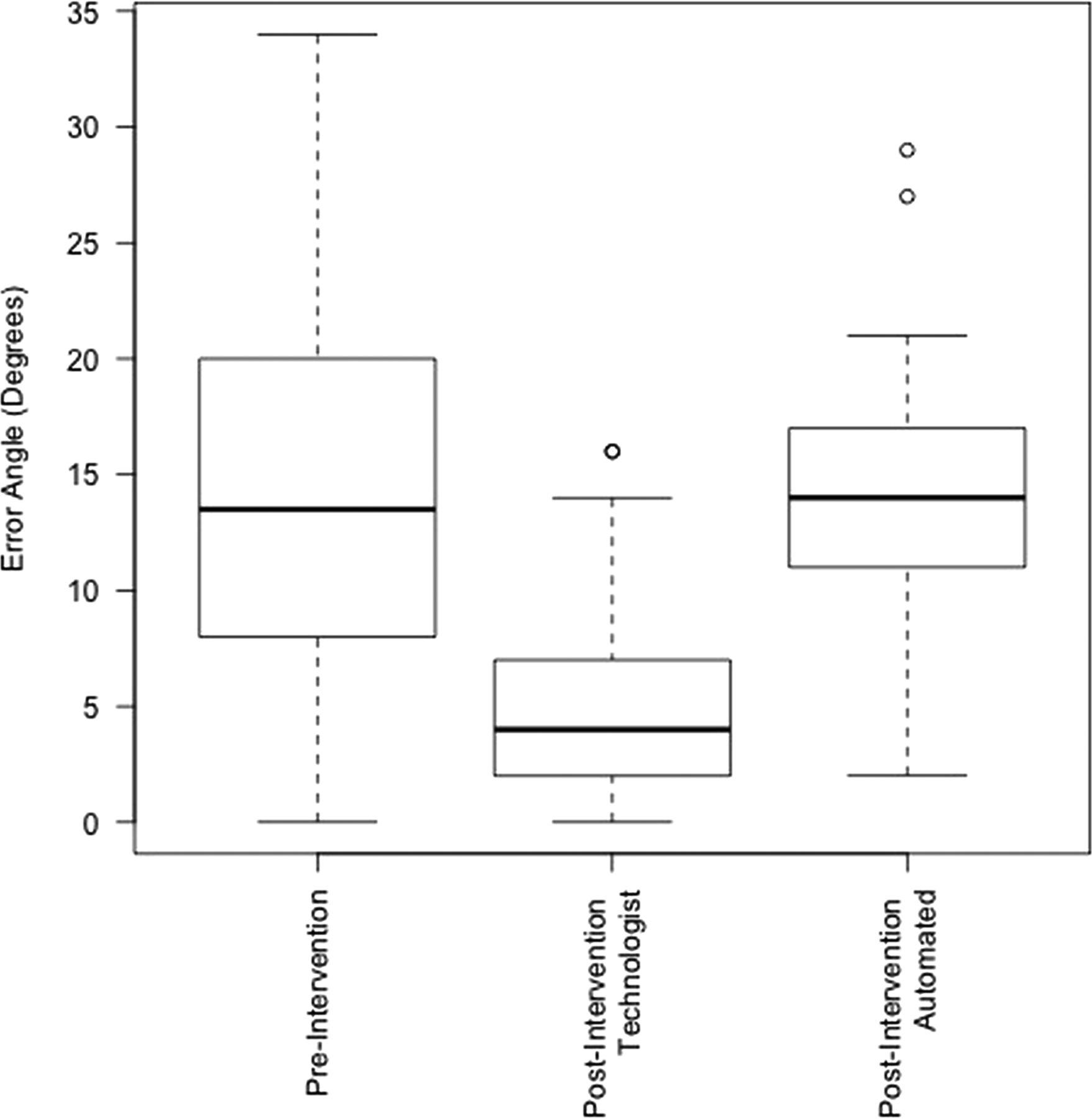
- Fig 3.
Boxplots depicting the difference in angles between the plane of the lateral semicircular canal and the plane of the temporal bone images in the preintervention group (image plane = acquisition plane), postintervention technologist group (image plane = plane as reformatted by a technologist in the clinical setting), and postintervention automated group (image plane = reformatted by commercially available syngo.via software).
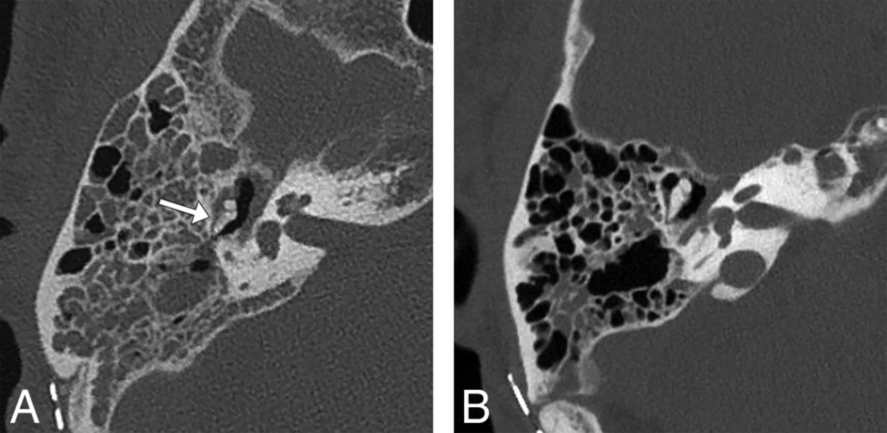
- Fig 4.
A 64-year-old man status post resection of a right vestibular schwannoma. Right temporal bone noncontrast CT images in bone windows demonstrate a possible perception error when images are reconstructed in random planes. A, Axial image in the acquisition plane obtained before the study intervention with a 29° error angle shows soft tissue in the Prussak space with a hazy appearance of the adjacent incus, suggesting possible erosion of the incus (arrow). B, Axial image in the plane of the lateral semicircular canal as reformatted by a CT technologist 3 months later following the study intervention shows the normal ice cream cone configuration of the malleus and incus with soft tissue in the Prussak space but no evident erosion of the incus, consistent with expected postoperative fluid in the middle ear cavity. The fluid had increased between the 2 examinations (not shown), so the finding is not a result of decreased fluid but a result of volume averaging with the incus body projected in an oblique plane.


Tables
Results of reformatting by a technologist
Total No. of Exams Total No. of Temporal Bones Axial Reformat Plane Correct (No.) (%) Coronal Reformat Plane Correct (No.) (%) Weeks 1–2 9 18 15 (83) 13 (72) Weeks 3–4 12 24 18 (75) 16 (67) Weeks 5–6 16 32 26 (81) 22 (69) Weeks 7–8 21 42 42 (100) 41 (98) Weeks 9–10 13 26 23 (88) 20 (77) Weeks 11–12 14 28 22 (79) 22 (79) Weeks 13–14 13 26 24 (92) 24 (92) Weeks 15–16 7 14 13 (93) 13 (93) Total 105 210 183 (87) 171 (81)


