
Article Figures & Data
Figures
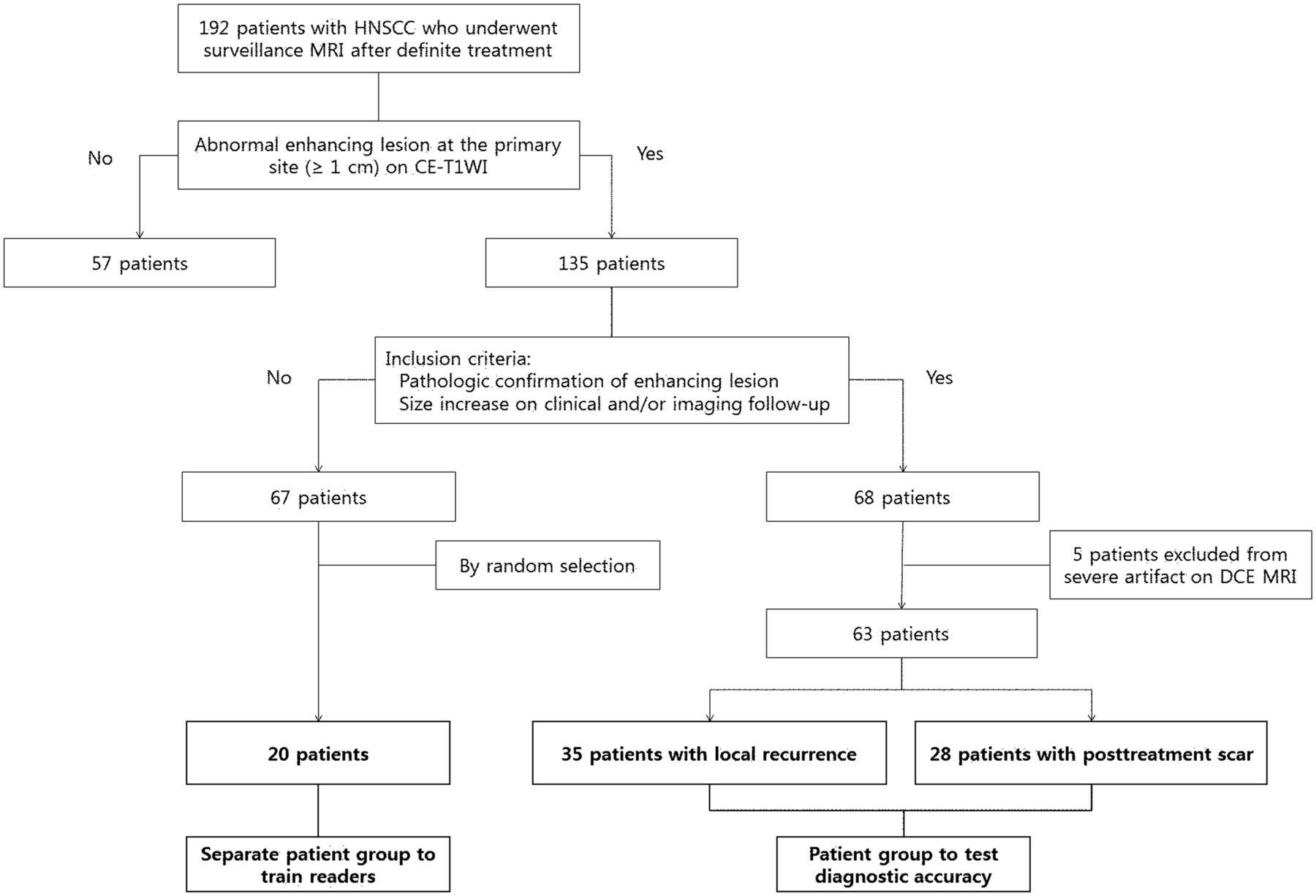
- Fig 1.
Diagram of study population enrollment.
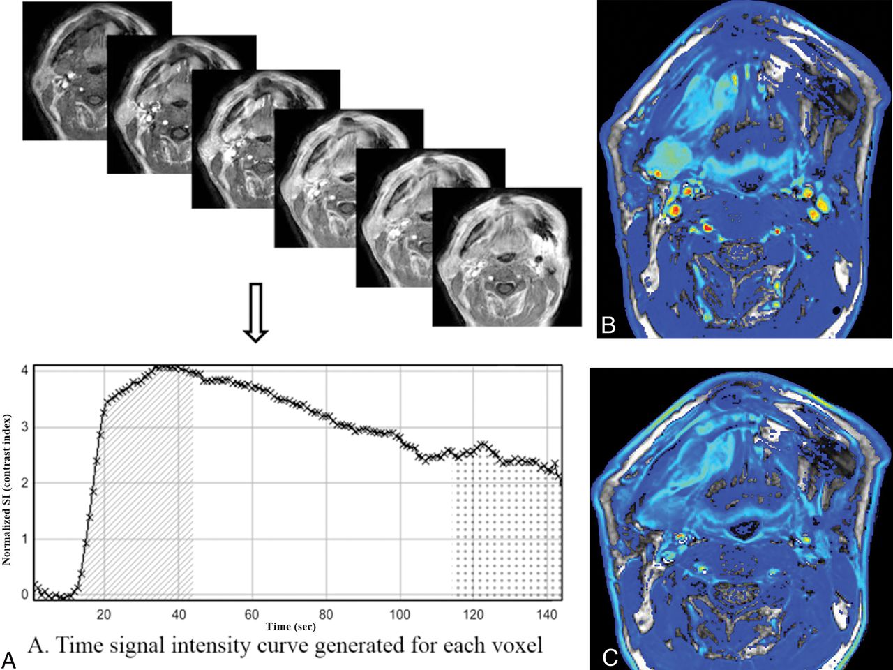
- Fig 2.
Illustration of the steps for generating color maps of initial and final 90-second AUC values using the TSI curve. A, The time course of the contrast index was plotted to obtain a TSI curve and the initial and final 90-second AUC values for each voxel. IAUC90 (diagonal pattern) and FAUC90 (dotted pattern) were defined as the trapezoidal integration of the normalized TSI curve during the initial and final 90 seconds from the onset of contrast enhancement in the voxel. B and C, Voxel-based color maps corresponding to IAUC90 and FAUC90 values were constructed in blue, green, yellow, and red.
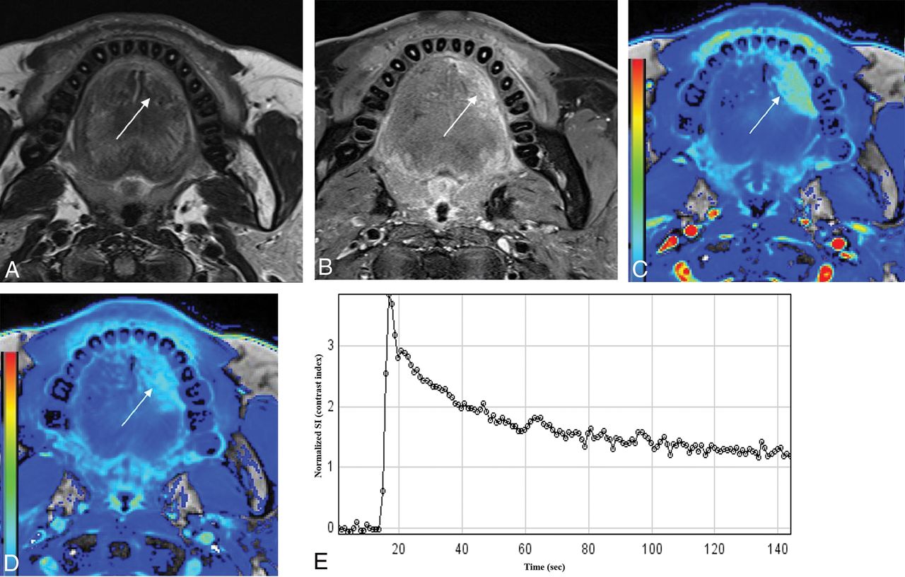
- Fig 3.
A 53-year-old man after hemiglossectomy for squamous cell carcinoma of the oral tongue 12 months previously (T2N0M0; depth of invasion, 10 mm). A and B, T2-weighted and contrast-enhanced fat-suppressed T1-weighted images show an ill-defined enhancing lesion at the operative bed of the oral tongue (arrows). C and D, IAUC90 and FAUC90 images of DCE-MR imaging show contrast washout at the corresponding area. E, The time-signal intensity curve obtained from the enhancing area using a hand-drawn ROI also shows contrast washout. This lesion was confirmed as recurrent tumor on subsequent surgical excision.
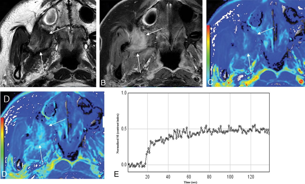
- Fig 4.
A 40-year-old female patient after wide excision for buccal cancer 36 months previously (T1N0M0). T2-weighted image shows a low signal intensity lesion in the right retroantral region (A) with heterogeneous contrast enhancement on contrast-enhanced fat-suppressed T1-weighted imaging (B) (arrows). IAUC90 (C) and FAUC90 (D) images of DCE-MR imaging show a progressive increment pattern. Time-signal intensity curve obtained at the enhancing portion also shows a progressive increment pattern (E). This lesion was confirmed as fibrosis at excisional biopsy.
- Fig 5.
A 48-year-old female patient after wide excision and flap reconstruction for right-tongue cancer 13 months previously (T1N0M0). T2-weighted image (A) shows a slightly high signal intensity lesion posterolateral to the reconstructed flap with moderate contrast enhancement on the T1-weighted image (B, arrows). IAUC90 (C) and FAUC90 (D) images of DCE-MR imaging show a prolonged enhancement pattern. The time-signal intensity curve obtained at the enhancing portion also shows a prolonged enhancement pattern (E). This lesion was confirmed as dense fibrosis at excisional biopsy.


Tables
Clinical Characteristics Separate Patient Group to Train Readers (n = 20) Patient Group to Test Diagnostic Accuracy Recurrent Tumor (n = 28) Posttreatment Change (n = 35) P Value Male/female ratio 12:8 18:10 29:6 .105 Age (mean) (yr) 55 ± 10 60 ± 11 58 ± 12 .452 Primary site Nasopharynx 5 (25) 6 (21) 9 (26) Oral cavity 6 (30) 11 (39) 11 (31) Oropharynx 4 (20) 5 (18) 5 (14) Larynx/hypopharynx 2 (10) 3 (11) 4 (11) PNS 3 (15) 3 (11) 6 (17) Treatment modality .183 OP 10 (50) 13 (46) 12 (34) CCRT 8 (45) 13 (46) 16 (45) OP + RT 2 (10) 2 (7) 7 (20) Mean time interval (range)b 28 (12–35) 27 (12–60) 24 (10–41) .518 Final diagnosis Pathologic exam 18 7 Clinical follow-up 10 28 Note:—CCRT indicates concurrent chemoradiation therapy; NA, not applicable; OP, operation; PNS, paranasal sinus; RT, radiation therapy.
↵a Numbers in parentheses are percentages except where noted.
↵b Mean time interval is expressed as months and the period between the end of treatment and MR imaging.
- Table 2:
Results of analysis of conventional MRI alone and combined interpretation of conventional and DCE-MRI for detecting local tumor recurrence
Recurrent Tumor Posttreatment Change TP FN FNR (%) TN FP FPR (%) Reader 1 Conventional MRI 21 7 25 8 27 77 Conventional and DCE-MRI 25 3 11 32 3 9 Reader 2 Conventional MRI 19 9 32 15 20 57 Conventional and DCE-MRI 23 5 18 32 3 9 Reader 3 Conventional MRI 23 5 18 10 25 71 Conventional and DCE-MRI 24 4 14 32 3 9 Note:—FN indicates false-negative; FNR, false-negative rate; FP, false-positive; FPR, false-positive rate; TN, true-negative; TP, true-positive.
- Table 3:
Diagnostic accuracies of conventional MRI alone and combined interpretation of conventional and DCE-MRI for detecting local tumor recurrencea
PPV NPV Sensitivity Specificity Accuracy Reader 1 Conventional MRI 45 (39–51)b 57 (32–80)b 79 (66–90) 22 (11–32)b 48 (35–61)b Conventional and DCE-MRI 89 (74–96)b 91 (79–97)b 89 (72–98) 91 (77–98)b 91 (80–96)b Reader 2 Conventional MRI 49 (39–58)b 63 (38–75)b 68 (54–81) 43 (25–49)b 54 (41–67)b Conventional and DCE-MRI 82 (67–91)b 91 (79–97)b 88 (70–98) 87 (71–76)b 87 (77–91)b Reader 3 Conventional MRI 48 (40–54)b 67 (42–86)b 82 (69–93) 29 (18–37)b 52 (39–65)b Conventional and DCE-MRI 89 (73–96)b 89 (76–95)b 86 (67–96) 91 (77–98)b 89 (78–95)b


