Advertisement

Main menu

  • Home
  • Content
    • Current Issue
    • Accepted Manuscripts
    • Article Preview
    • Past Issue Archive
    • AJNR Case Collection
    • Case of the Week Archive
    • Classic Case Archive
    • Case of the Month Archive
  • Special Collections
    • Spinal CSF Leak Articles (Jan 2020-June 2024)
    • 2024 AJNR Journal Awards
    • Most Impactful AJNR Articles
  • Multimedia
    • AJNR Podcast
    • AJNR Scantastics
    • Video Articles
  • For Authors
    • Submit a Manuscript
    • Author Policies
    • Fast publishing of Accepted Manuscripts
    • Graphical Abstract Preparation
    • Manuscript Submission Guidelines
    • Imaging Protocol Submission
    • Submit a Case for the Case Collection
  • About Us
    • About AJNR
    • Editorial Board
  • More
    • Become a Reviewer/Academy of Reviewers
    • Subscribers
    • Permissions
    • Alerts
    • Feedback
    • Advertisers
    • ASNR Home
  • Other Publications
    • ajnr

User menu

  • Alerts
  • Log in

Search

  • Advanced search
American Journal of Neuroradiology

American Journal of Neuroradiology

ASHNR American Society of Functional Neuroradiology ASHNR American Society of Pediatric Neuroradiology ASSR
  • Alerts
  • Log in

Advanced Search

  • Home
  • Content
    • Current Issue
    • Accepted Manuscripts
    • Article Preview
    • Past Issue Archive
    • AJNR Case Collection
    • Case of the Week Archive
    • Classic Case Archive
    • Case of the Month Archive
  • Special Collections
    • Spinal CSF Leak Articles (Jan 2020-June 2024)
    • 2024 AJNR Journal Awards
    • Most Impactful AJNR Articles
  • Multimedia
    • AJNR Podcast
    • AJNR Scantastics
    • Video Articles
  • For Authors
    • Submit a Manuscript
    • Author Policies
    • Fast publishing of Accepted Manuscripts
    • Graphical Abstract Preparation
    • Manuscript Submission Guidelines
    • Imaging Protocol Submission
    • Submit a Case for the Case Collection
  • About Us
    • About AJNR
    • Editorial Board
  • More
    • Become a Reviewer/Academy of Reviewers
    • Subscribers
    • Permissions
    • Alerts
    • Feedback
    • Advertisers
    • ASNR Home
  • Follow AJNR on Twitter
  • Visit AJNR on Facebook
  • Follow AJNR on Instagram
  • Join AJNR on LinkedIn
  • RSS Feeds

Welcome to the new AJNR, Updated Hall of Fame, and more. Read the full announcements.


AJNR is seeking candidates for the position of Associate Section Editor, AJNR Case Collection. Read the full announcement.


Research ArticlePediatrics
Open Access

Cytotoxic Lesion of the Corpus Callosum in an Adolescent with Multisystem Inflammatory Syndrome and SARS-CoV-2 Infection

J. Lin, E.C. Lawson, S. Verma, R.B. Peterson and R. Sidhu
American Journal of Neuroradiology November 2020, 41 (11) 2017-2019; DOI: https://doi.org/10.3174/ajnr.A6755
J. Lin
aFrom the Department of Pediatric Neurology (J.L., E.C.L., S.V., R.S.), Children’s Healthcare of Atlanta, Atlanta, Georgia
dDepartment of Pediatrics (J.L., S.V., R.S.), Division of Neurology, Emory University School of Medicine, Atlanta, Georgia
  • Find this author on Google Scholar
  • Find this author on PubMed
  • Search for this author on this site
  • ORCID record for J. Lin
E.C. Lawson
aFrom the Department of Pediatric Neurology (J.L., E.C.L., S.V., R.S.), Children’s Healthcare of Atlanta, Atlanta, Georgia
cDivision of Neuroradiology, Department of Neurology (E.C.L.)
  • Find this author on Google Scholar
  • Find this author on PubMed
  • Search for this author on this site
  • ORCID record for E.C. Lawson
S. Verma
aFrom the Department of Pediatric Neurology (J.L., E.C.L., S.V., R.S.), Children’s Healthcare of Atlanta, Atlanta, Georgia
dDepartment of Pediatrics (J.L., S.V., R.S.), Division of Neurology, Emory University School of Medicine, Atlanta, Georgia
  • Find this author on Google Scholar
  • Find this author on PubMed
  • Search for this author on this site
  • ORCID record for S. Verma
R.B. Peterson
bDepartment of Radiology and Imaging Sciences (R.B.P.)
  • Find this author on Google Scholar
  • Find this author on PubMed
  • Search for this author on this site
  • ORCID record for R.B. Peterson
R. Sidhu
aFrom the Department of Pediatric Neurology (J.L., E.C.L., S.V., R.S.), Children’s Healthcare of Atlanta, Atlanta, Georgia
dDepartment of Pediatrics (J.L., S.V., R.S.), Division of Neurology, Emory University School of Medicine, Atlanta, Georgia
  • Find this author on Google Scholar
  • Find this author on PubMed
  • Search for this author on this site
  • ORCID record for R. Sidhu
  • Article
  • Figures & Data
  • Info & Metrics
  • Responses
  • References
  • PDF
Loading

Abstract

SUMMARY: Multisystem inflammatory syndrome in children is a recently described complication in the late phase of Severe Acute Respiratory Syndrome coronavirus 2 (SARS-CoV-2) infection involving systemic hyperinflammation and multiorgan dysfunction. The extent of its clinical picture is actively evolving and has yet to be fully elucidated. While neurologic manifestations of SARS-CoV-2 are well-described in the adult population, reports of neurologic complications in pediatric patients with SARS-CoV-2 infection are limited. We present a pediatric patient with SARS-CoV-2 infection with development of multisystem inflammatory syndrome and acute encephalopathy causing delirium who was found to have a cytotoxic lesion of the corpus callosum on neuroimaging. Cytotoxic lesions of the corpus callosum are a well-known, typically reversible entity that can occur in a wide range of conditions, including infection, seizure, toxins, nutritional deficiencies, and Kawasaki disease. We hypothesized that the cytotoxic lesion of the corpus callosum, in the index case, was secondary to the systemic inflammation from SARS-CoV-2 infection, resulting in multisystem inflammatory syndrome in children.

ABBREVIATIONS:

CLOCC
cytotoxic lesion of the corpus callosum
COVID-19
coronavirus disease 2019
IVIG
intravenous immunoglobulin
MIS-C
multisystem inflammatory syndrome in children
PCR
polymerase chain reaction
SARS-CoV-2
Severe Acute Respiratory Syndrome coronavirus 2

Severe Acute Respiratory Syndrome coronavirus 2 (SARS-CoV-2) infection in the pediatric population is not well-understood and has, until recently, been thought of as a relatively mild disease compared with that in adults. Most reported symptoms in children are fever, respiratory tract infection, or asymptomatic infection.1 More recent reports in the pediatric population, however, describe a multisystem inflammatory syndrome in children (MIS-C).2⇓⇓⇓-6 In the adult population, neurologic manifestations of coronavirus disease 2019 (COVID-19) are being reported with increasing frequency, including seizure, Guillain-Barré syndrome, vasculitis, stroke, cranial nerve palsy, and general encephalopathy.7⇓⇓⇓-11 Cases of neurologic manifestations in pediatric patients have been limited.

We report the case of a pediatric patient positive for COVID-19 who presented with fever, vomiting, diarrhea, cough, difficulty walking, and delirium.

Case Presentation

A 13-year-old previously healthy girl, who relocated from New York to Georgia a month prior, presented to the emergency department after 3 days of fever, vomiting, diarrhea, cough, dizziness, and gait instability. The only medications given before admission were aspirin and ibuprofen. On presentation to Egleston Hospital of Children's Healthcare of Atlanta, she had a fever of 39.2°C, hypotension initially requiring pressor support, normal white blood cell count (9780 cells/µL) with increased neutrophils (88%) and lymphopenia (7%), thrombocytopenia (121,000 cells/µL), hyponatremia (128 mmol/L), elevated transaminases (aspartate aminotransferase = 292 U/L, alanine aminotransferase = 336 U/L), acute kidney injury (creatinine = 1.09 mg/dL), elevated inflammatory markers (C-reactive protein = 10.9 mg/dL, erythrocyte sedimentation rate = 63 mm/h, interleukin 6 = 65 pg/mL), and elevated cardiac markers (troponin = 18.2 ng/mL, brain natriuretic peptide = 561 pg/mL). She was admitted to the pediatric intensive care unit for presumed bacterial sepsis and started on ceftriaxone and vancomycin.

On hospital day 1, she began to have hallucinations in addition to urinary retention, prompting neurology consultation. On neurologic examination, she was agitated and combative with a variable rate of speech. She had intermittent auditory hallucinations. Her attention fluctuated. She was able to follow 1- but not 2-step directions. Cranial nerve function was normal without clinical evidence of anosmia. Muscle tone was decreased with symmetric antigravity movement of all extremities. Sensation to light touch was intact throughout. Deep tendon reflexes were 1+ with absent ankle jerks. Contrast-enhanced MR imaging of the brain (Magnetom Avanto, 1.5T; Siemens) on hospital day 1 demonstrated an abnormal focal nonenhancing lesion with increased intensity on T2-weighted sequences and restricted diffusion in the splenium of the corpus callosum (Figure). Findings on MR imaging of the spine were normal. CSF studies were unremarkable (4 white blood cells (WBC)/μL, glucose = 83 mg/dL, protein = 32 mg/dL). The electroencephalogram showed diffuse slowing without epileptiform discharges or seizures. On hospital day 2, the patient developed tachypnea requiring escalation of respiratory support to bilevel positive airway pressure, though no desaturation or hypoxemia was noted. The echocardiogram revealed depressed cardiac function with a left ventricular ejection fraction of 38%. Intravenous immunoglobulin (IVIG), 2 g/kg, was administered on hospital day 2, given concerns for atypical Kawasaki-like multisystem inflammatory state.

FIGURE.
  • Download figure
  • Open in new tab
  • Download powerpoint
FIGURE.

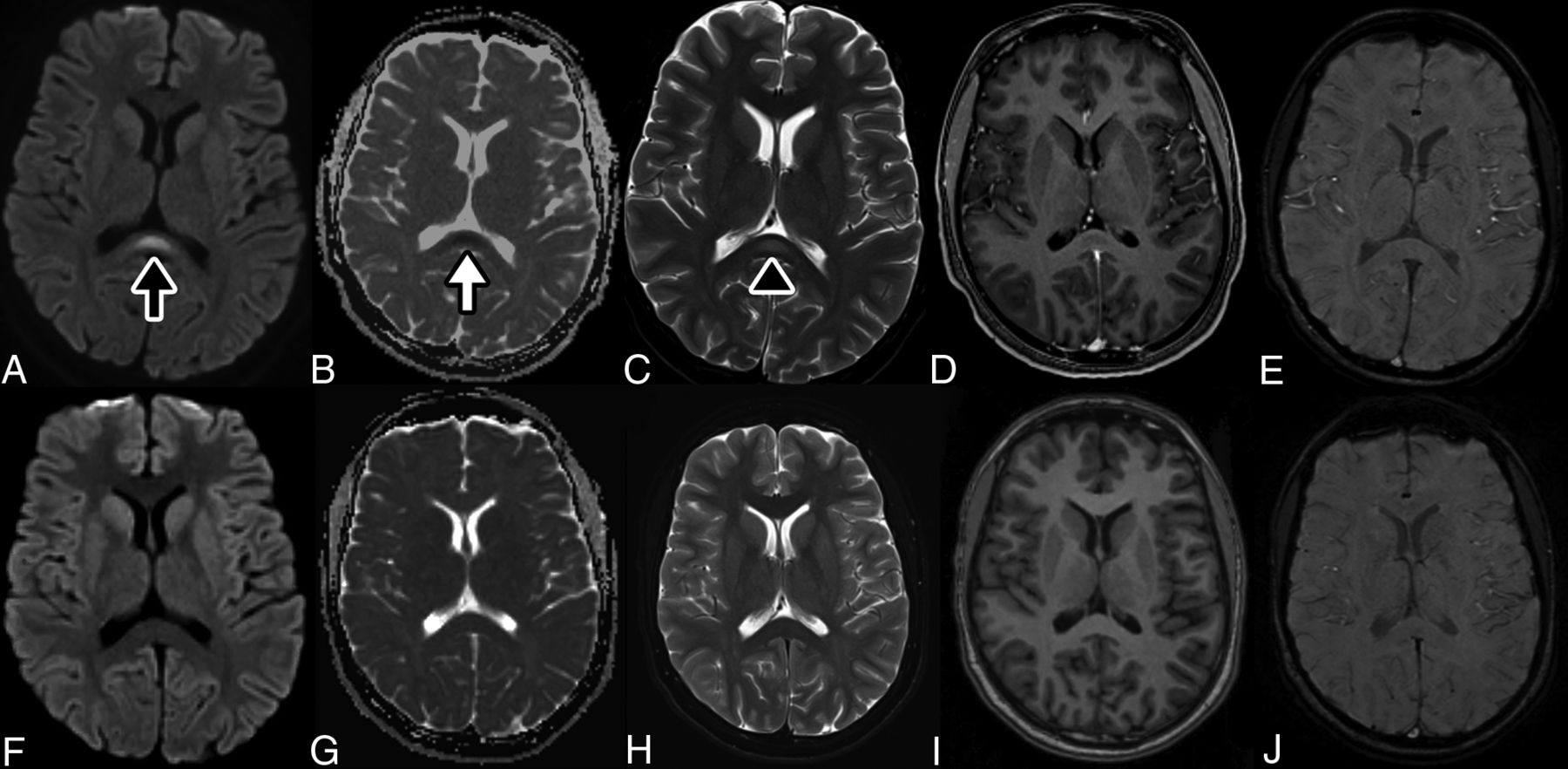
CLOCC in an adolescent with MIS-C and SARS-CoV-2.. MR image of the brain demonstrates a nonspecific focus of increased signal in the splenium of the corpus callosum on DWI sequences at b=1000 s/mm2 (black arrow, A) with associated loss of signal on apparent diffusion coefficient maps (white arrow, B) corresponding to restricted diffusion. The apparent diffusion coefficient for the lesion is 0.44 × 10⁻³ mm2/s. This lesion corresponds to a faint focus of abnormal increased signal on T2WI spin-echo sequences (black arrowhead, C). The lesion also demonstrates a lack of contrast enhancement on T1WI postcontrast thin-section image (D) and absent susceptibility, suggesting absent hemorrhage on SWI (E). The imaging findings suggest a cytotoxic lesion of the corpus callosum. Follow-up DWI (F) and ADC maps (G) with T2WI (H), T1WI (I), and SWI sequences (J) after 2.5 months demonstrate resolution of the lesion after therapy.

Extensive infectious and autoimmune investigations were performed. Respiratory viral panel and initial SARS-CoV-2 polymerase chain reaction (PCR) findings on admission were negative. The remainder of infectious evaluation was negative (CSF meningitis panel, CSF and serum arboviral panels, serum viral studies including coxsackie A9 antibody, West Nile virus antibodies, human immunodeficiency virus antigen and antibody, enterovirus PCR, parvovirus B19 PCR, adenovirus PCR, and Epstein-Barr virus PCR). The serum autoimmune encephalopathy panel had a mildly elevated glutamic acid decarboxylase antibody (0.17 nmol/L) in the setting of recent IVIG administration and was thought to likely be a false-positive. Antinuclear antibody titer was elevated to 1:320 with a negative antibody reflex panel. The remainder of autoimmune work-up had negative findings (CSF oligoclonal bands, immunoglobulin G index, CSF autoimmune encephalopathy panel, serum thyroglobulin and antithyroid peroxidase antibodies, myelin oligodendrocyte glycoprotein antibody, aquaporin-4 antibody). Given the high level of suspicion, SARS-CoV-2 PCR was repeated on hospital day 3 and found to be positive. Qualitative COVID-19 immunoglobulin G in serum drawn on hospital day 2 before IVIG administration was also positive.

During the next few days, the patient’s mental status improved gradually; however, she remained weak and tendon reflexes were persistently absent in the ankles. Creatine kinase was elevated (3600 U/L, normal = 50–275 U/L), and the electrodiagnostic study performed on hospital day 5 was unremarkable with no evidence of polyneuropathy or myopathy. On hospital day 6, respiratory support was weaned to nasal cannula, and she was able to stand unassisted. The repeat echocardiogram demonstrated return to normal cardiac function, and laboratory markers normalized. She was transferred out of the intensive care unit and discharged home 12 days following the initial presentation with follow-up in the pediatric neurology clinic scheduled. Furthermore, subsequent research laboratory studies on the CSF did not detect the presence of SARS-CoV-2 by PCR. An indirect enzyme-linked immunosorbent assay for CSF showed increased levels of immunoglobulin M (1:64) for SARS-CoV-2 S1 and E (envelope) proteins (90% sensitivity, 89% specificity; courtesy of the Dr. William Hu Laboratory, Emory University).

DISCUSSION

We describe a pediatric case of a cytotoxic lesion of the corpus callosum (CLOCC) in the setting of MIS-C due to SARS-CoV-2 infection. CLOCCs have been well-described in a wide variety of conditions, including infection, seizure, toxins, nutritional deficiencies, and Kawasaki disease.12,13 Clinically, these lesions can manifest in a wide variety of nonspecific symptoms, such as cognitive impairment, seizures, hallucinations, delirium, dysarthria, and motor weakness.12,13 Clinically, the CLOCC may have contributed to her acute encephalopathy, causing delirium with prominent auditory hallucinations. MR imaging features of CLOCCs include increased signal intensity on FLAIR sequences and decreased signal intensity on T1-weighted sequences. Diffusion is reduced, and there is no contrast enhancement.13

CLOCCs associated with classic Kawasaki disease are a well-known entity. In hyperinflammatory states, macrophage activation leads to cytokine release that subsequently results in T-cell recruitment and breakdown of the blood-brain barrier. As the cytokine cascade invades the CNS, astrocytes are triggered to release excess glutamate, precipitating further cytokine release from microglia. The corpus callosum is especially vulnerable to this attack due to its high concentration of cytokine and glutamate receptors.13 In the index case, a CSF cytokine assay is pending.

SARS-CoV-2 has recently been demonstrated to result in MIS-C.2⇓⇓⇓-6 This post or peri-infectious immune-mediated disorder leads to persistent fever, gastrointestinal symptoms, lymphadenopathy, and left-heart dysfunction often requiring inotropic support, such as seen in Kawasaki disease.3

The study patient developed MIS-C characterized by elevated serum interleukin 6 and cardiac enzymes, acutely reduced cardiac function, and neurologic dysfunction with neuroimaging showing CLOCC. Most interesting, this is similar to previously reported Kawasaki disease associated with CLOCCs.12 We hypothesize that the SARS-CoV-2 postor peri-infectious immune-mediated disorder leads to MIS-C with neurologic dysfunction and CLOCC. This concept is further supported by absent SARS-CoV-2 PCR findings and elevated cytokines in the CSF as shown by Benameur et al.14 In this case, clinical improvement was noted with supportive care and IVIG. Repeat MR imaging 3 months following the initial illness demonstrated resolution of CLOCC, with no residual signal abnormality.

Reports continue to emerge of severe manifestations of COVID-19 in children, despite previous reports of mild illness. This case of MIS-C associated with COVID-19 highlights the potential of MR imaging abnormalities resulting in acute encephalopathy causing delirium and emphasizes the need for careful consideration of possible neurologic complications of SARS-CoV-2 infection.

Acknowledgments

We thank the patient and her family along with the physicians and nurses who provided clinical excellence and compassionate care.

Footnotes

  • J. Lin and E.C. Lawson contributed equally as co-first authors.

Indicates open access to non-subscribers at www.ajnr.org

References

  1. 1.↵
    CDC COVID-19 Response Team. Coronavirus disease 2019 in children—United States, February 12-April 2, 2020. MMWR Morb Mortal Wkly Rep 2020;69:422–26 doi:10.15585/mmwr.mm6914e4 pmid:32271728
    CrossRefPubMed
  2. 2.↵
    1. Riphagen S,
    2. Gomez X,
    3. Gonzalez-Martinez C, et al
    . Hyperinflammatory shock in children during COVID-19 pandemic. Lancet 2020;395:1607–08 doi:10.1016/S0140-6736(20)31094-1 pmid:32386565
    CrossRefPubMed
  3. 3.↵
    1. Belhadjer Z,
    2. Meot M,
    3. Bajolle F, et al
    . Acute heart failure in multisystem inflammatory syndrome in children (MIS-C) in the context of global SARS-CoV-2 pandemic. Circulation 2020 May 17. [Epub ahead of print] doi:10.1161/CIRCULATIONAHA.120.048360 pmid:32418446
    CrossRefPubMed
  4. 4.↵
    1. Licciardi F,
    2. Pruccoli G,
    3. Denina M, et al
    . SARS-CoV-2-induced Kawasaki-like hyperinflammatory syndrome: a novel COVID phenotype in children. Pediatrics 2020 May 21. [Epub ahead of print] doi:10.1542/peds.2020-1711 pmid:32439816
    Abstract/FREE Full Text
  5. 5.↵
    1. Dufort EM,
    2. Koumans EH,
    3. Chow Ej, et al
    ; New York State and Centers for Disease Control and Prevention Multisystem Inflammatory Syndrome in Children Investigation Team. Multisystem inflammatory syndrome in children in New York State. N Engl J Med 2020;383:347–58 doi:10.1056/NEJMoa2021756 pmid:32598830
    CrossRefPubMed
  6. 6.↵
    1. Cheung EW,
    2. Zachariah P,
    3. Gorelik M, et al
    . Multisystem inflammatory syndrome related to COVID-19 in previously healthy children and adolescents in New York City. JAMA 2020 June 8. [Epub ahead of print] doi:10.1001/jama.2020.10374 pmid:32511676
    CrossRefPubMed
  7. 7.↵
    1. Mao L,
    2. Jin H,
    3. Wang M, et al
    . Neurologic manifestations of hospitalized patients with coronavirus disease 2019 in Wuhan, China. JAMA Neurol 2020;77:683 doi:10.1001/jamaneurol.2020.1127 pmid:32275288
    CrossRefPubMed
  8. 8.↵
    1. Dinkin M,
    2. Gao V,
    3. Kahan J, et al
    . COVID-19 presenting with ophthalmoparesis from cranial nerve palsy. Neurology 2020 May 1. [Epub ahead of print] doi:10.1212/WNL.0000000000009700 pmid:32358218
    Abstract/FREE Full Text
  9. 9.↵
    1. Scullen T,
    2. Keen J,
    3. Mathkour M, et al
    . Coronavirus 2019 (COVID-19)-associated encephalopathies and cerebrovascular disease: the New Orleans Experience. World Neurosurg 2020 May 28. [Epub ahead of print] doi:10.1016/j.wneu.2020.05.192 pmid:32474092
    CrossRefPubMed
  10. 10.↵
    1. Hanafi R,
    2. Roger PA,
    3. Perin B, et al
    . COVID-19 neurologic complication with CNS vasculitis-like pattern. AJNR Am J Neuroradiol 2020 June 8. [Epub ahead of print] doi:10.3174/ajnr.A6651 pmid:32554425
    Abstract/FREE Full Text
  11. 11.↵
    1. Nicholson P,
    2. Alshafai L,
    3. Krings T
    . Neuroimaging findings in patients with COVID-19. AJNR Am J Neuroradiol 2020 June 11. [Epub ahead of print] doi:10.3174/ajnr.A6630 pmid:32527843
    Abstract/FREE Full Text
  12. 12.↵
    1. Takanashi J,
    2. Shirai K,
    3. Sugawara Y, et al
    . Kawasaki disease complicated by mild encephalopathy with a reversible splenial lesion (MERS). J Neurol Sci 2012;315:167–69 doi:10.1016/j.jns.2011.11.022 pmid:22129937
    CrossRefPubMed
  13. 13.↵
    1. Starkey J,
    2. Kobayashi N,
    3. Numaguchi Y, et al
    . Cytotoxic lesions of the corpus callosum that show restricted diffusion: mechanisms, causes, and manifestations. Radiographics 2017;37:562–76 doi:10.1148/rg.2017160085 pmid:28165876
    CrossRefPubMed
  14. 14.↵
    1. Benameur K,
    2. Agarwal A,
    3. Auld S, et al
    . Encephalopathy and encephalitis associated with cerebrospinal fluid cytokine alterations and coronavirus disease, Atlanta, Georgia, USA. Emerg Infect Dis 2020 June. [Epub ahead of print] doi:10.3201/eid2609.202122 pmid:32487282
    CrossRefPubMed
  • Received June 2, 2020.
  • Accepted after revision July 8, 2020.
  • © 2020 by American Journal of Neuroradiology
PreviousNext
Back to top

In this issue

American Journal of Neuroradiology: 41 (11)
American Journal of Neuroradiology
Vol. 41, Issue 11
1 Nov 2020
  • Table of Contents
  • Index by author
  • Complete Issue (PDF)
Advertisement
Print
Download PDF
Email Article

Thank you for your interest in spreading the word on American Journal of Neuroradiology.

NOTE: We only request your email address so that the person you are recommending the page to knows that you wanted them to see it, and that it is not junk mail. We do not capture any email address.

Enter multiple addresses on separate lines or separate them with commas.
Cytotoxic Lesion of the Corpus Callosum in an Adolescent with Multisystem Inflammatory Syndrome and SARS-CoV-2 Infection
(Your Name) has sent you a message from American Journal of Neuroradiology
(Your Name) thought you would like to see the American Journal of Neuroradiology web site.
CAPTCHA
This question is for testing whether or not you are a human visitor and to prevent automated spam submissions.
Cite this article
J. Lin, E.C. Lawson, S. Verma, R.B. Peterson, R. Sidhu
Cytotoxic Lesion of the Corpus Callosum in an Adolescent with Multisystem Inflammatory Syndrome and SARS-CoV-2 Infection
American Journal of Neuroradiology Nov 2020, 41 (11) 2017-2019; DOI: 10.3174/ajnr.A6755

Citation Manager Formats

  • BibTeX
  • Bookends
  • EasyBib
  • EndNote (tagged)
  • EndNote 8 (xml)
  • Medlars
  • Mendeley
  • Papers
  • RefWorks Tagged
  • Ref Manager
  • RIS
  • Zotero
0 Responses
Respond to this article
Share
Bookmark this article
Cytotoxic Lesion of the Corpus Callosum in an Adolescent with Multisystem Inflammatory Syndrome and SARS-CoV-2 Infection
J. Lin, E.C. Lawson, S. Verma, R.B. Peterson, R. Sidhu
American Journal of Neuroradiology Nov 2020, 41 (11) 2017-2019; DOI: 10.3174/ajnr.A6755
del.icio.us logo Twitter logo Facebook logo Mendeley logo
  • Tweet Widget
  • Facebook Like
  • Google Plus One
Purchase

Jump to section

  • Article
    • Abstract
    • ABBREVIATIONS:
    • DISCUSSION
    • Acknowledgments
    • Footnotes
    • References
  • Figures & Data
  • Info & Metrics
  • Responses
  • References
  • PDF

Related Articles

  • No related articles found.
  • PubMed
  • Google Scholar

Cited By...

  • Neuroimaging features of psilocybin-induced toxic-metabolic encephalopathy in an adolescent
  • COVID-19 related multisystem inflammatory syndrome in children (MIS-C): a hospital-based prospective cohort study from Kerala, India
  • Neuroimaging Offers Low Yield in Children Positive for SARS-CoV-2
  • Crossref (33)
  • Google Scholar

This article has been cited by the following articles in journals that are participating in Crossref Cited-by Linking.

  • Neurologic Involvement in Children and Adolescents Hospitalized in the United States for COVID-19 or Multisystem Inflammatory Syndrome
    Kerri L. LaRovere, Becky J. Riggs, Tina Y. Poussaint, Cameron C. Young, Margaret M. Newhams, Mia Maamari, Tracie C. Walker, Aalok R. Singh, Heda Dapul, Charlotte V. Hobbs, Gwenn E. McLaughlin, Mary Beth F. Son, Aline B. Maddux, Katharine N. Clouser, Courtney M. Rowan, John K. McGuire, Julie C. Fitzgerald, Shira J. Gertz, Steven L. Shein, Alvaro Coronado Munoz, Neal J. Thomas, Katherine Irby, Emily R. Levy, Mary A. Staat, Mark W. Tenforde, Leora R. Feldstein, Natasha B. Halasa, John S. Giuliano, Mark W. Hall, Michele Kong, Christopher L. Carroll, Jennifer E. Schuster, Sule Doymaz, Laura L. Loftis, Keiko M. Tarquinio, Christopher J. Babbitt, Ryan A. Nofziger, Lawrence C. Kleinman, Michael A. Keenaghan, Natalie Z. Cvijanovich, Philip C. Spinella, Janet R. Hume, Kari Wellnitz, Elizabeth H. Mack, Kelly N. Michelson, Heidi R. Flori, Manish M. Patel, Adrienne G. Randolph, First Name and Middle Initial(s)* Last Name*, Mary G Gaspers, Katri V Typpo, Ronald C Sanders, Adam J Schwarz, Helen Harvey, Matt S Zinter, Peter M Mourani, Bria M Coates, Guru Bhoojhawon, Kevin M Havlin, Vicki L Montgomery, Janice E Sullivan, Tamara T Bradford, Melania M Bembea, Susan V Lipton, Ana Lia Graciano, Sabrina R Chen, Suden Kucukak, Jane W Newburger, Ryan W Carroll, Neil D Fernandes, Phoebe H Yager, Kimberly L Marohn, Sabrina M Heidemann, Melissa L Cullimore, Russell J McCulloh, Steven M Horwitz, Simon Li, Rowan F Walsh, Adam J Ratner, Vijaya L Soma, Jennifer K Gillen, Sheemon P Zackai, Kate G Ackerman, Jill M Cholette, Ilana Harwayne-Gidansky, Saul R Hymes, Philip J Overby, Stephanie P Schwartz, Amanda N Lansell, Monica L Koncicki, Joseph Carcillo, Ericka Fink, Dai Kimura, Cindy Bowens, Hillary Crandall, Lincoln S Smith, Pelin Cengiz
    JAMA Neurology 2021 78 5
  • Neurological manifestations associated with SARS-CoV-2 and other coronaviruses: A narrative review for clinicians
    A. Maury, A. Lyoubi, N. Peiffer-Smadja, T. de Broucker, E. Meppiel
    Revue Neurologique 2021 177 1-2
  • Cerebrospinal fluid in COVID-19: A systematic review of the literature
    Ariane Lewis, Jennifer Frontera, Dimitris G. Placantonakis, Jennifer Lighter, Steven Galetta, Laura Balcer, Kara R. Melmed
    Journal of the Neurological Sciences 2021 421
  • Multisystem inflammatory syndrome (MIS-C): a systematic review and meta-analysis of clinical characteristics, treatment, and outcomes
    Mônica O. Santos, Lucas C. Gonçalves, Paulo A.N. Silva, André L.E. Moreira, Célia R.M. Ito, Fernanda A.O. Peixoto, Isabela J. Wastowski, Lilian C. Carneiro, Melissa A.G. Avelino
    Jornal de Pediatria 2022 98 4
  • Prevalence and Risk Factors of Neurologic Manifestations in Hospitalized Children Diagnosed with Acute SARS-CoV-2 or MIS-C
    Ericka L. Fink, Courtney L. Robertson, Mark S. Wainwright, Juan D. Roa, Marlina E. Lovett, Casey Stulce, Mais Yacoub, Renee M. Potera, Elizabeth Zivick, Adrian Holloway, Ashish Nagpal, Kari Wellnitz, Theresa Czech, Katelyn M. Even, Werther Brunow de Carvalho, Isadora Souza Rodriguez, Stephanie P. Schwartz, Tracie C. Walker, Santiago Campos-Miño, Leslie A. Dervan, Andrew S. Geneslaw, Taylor B. Sewell, Patrice Pryce, Wendy G. Silver, Jieru Egeria Lin, Wendy S. Vargas, Alexis Topjian, Alicia M. Alcamo, Jennifer L. McGuire, Jesus Angel Domínguez Rojas, Jaime Tasayco Muñoz, Sue J. Hong, William J. Muller, Matthew Doerfler, Cydni N. Williams, Kurt Drury, Dhristie Bhagat, Aaron Nelson, Dana Price, Heda Dapul, Laura Santos, Robert Kahoud, Conall Francoeur, Brian Appavu, Kristin P. Guilliams, Shannon C. Agner, Karen H. Walson, Lindsey Rasmussen, Anna Janas, Peter Ferrazzano, Raquel Farias-Moeller, Kellie C. Snooks, Chung-Chou H. Chang, James Yun, Michelle E. Schober
    Pediatric Neurology 2022 128
  • The JANUS of chronic inflammatory and autoimmune diseases onset during COVID-19 – A systematic review of the literature
    Lucia Novelli, Francesca Motta, Maria De Santis, Aftab A. Ansari, M. Eric Gershwin, Carlo Selmi
    Journal of Autoimmunity 2021 117
  • Neurological complications in pediatric patients with SARS-CoV-2 infection: a systematic review of the literature
    L. Siracusa, A. Cascio, S. Giordano, A. A. Medaglia, G. A. Restivo, I. Pirrone, G. F. Saia, F. Collura, C. Colomba
    Italian Journal of Pediatrics 2021 47 1
  • The Spectrum of Neuroimaging Findings on CT and MRI in Adults With COVID-19
    Gul Moonis, Christopher G. Filippi, Claudia F. E. Kirsch, Suyash Mohan, Evan G. Stein, Joshua A. Hirsch, Amit Mahajan
    American Journal of Roentgenology 2021 217 4
  • Presentation, Treatment Response and Short-Term Outcomes in Paediatric Multisystem Inflammatory Syndrome Temporally Associated with SARS-CoV-2 (PIMS-TS)
    Susanna Felsenstein, Emily Willis, Hannah Lythgoe, Liza McCann, Andrew Cleary, Kamran Mahmood, David Porter, Jessica Jones, Janet McDonagh, Alice Chieng, Giulia Varnier, Stephen Hughes, Mary Boullier, Fiona Ryan, Olumoyin Awogbemi, Giridhar Soda, Phuoc Duong, Clare Pain, Phil Riley, Christian M. Hedrich
    Journal of Clinical Medicine 2020 9 10
  • Postacute/Long COVID in Pediatrics
    Amanda K. Morrow, Rowena Ng, Gray Vargas, Dasal Tenzin Jashar, Ellen Henning, Nika Stinson, Laura A. Malone
    American Journal of Physical Medicine & Rehabilitation 2021 100 12

More in this TOC Section

  • Comparison of Image Quality and Radiation Dose in Pediatric Temporal Bone CT Using Photon-Counting Detector CT and Energy-Integrating Detector CT
  • SyMRI & MR Fingerprinting in Brainstem Myelination
  • Gad Contrast in PEDS Seizure Evaluation
Show more Pediatrics

Similar Articles

Advertisement

Indexed Content

  • Current Issue
  • Accepted Manuscripts
  • Article Preview
  • Past Issues
  • Editorials
  • Editors Choice
  • Fellow Journal Club
  • Letters to the Editor

Cases

  • Case Collection
  • Archive - Case of the Week
  • Archive - Case of the Month
  • Archive - Classic Case

Special Collections

  • Special Collections

Resources

  • News and Updates
  • Turn around Times
  • Submit a Manuscript
  • Author Policies
  • Manuscript Submission Guidelines
  • Evidence-Based Medicine Level Guide
  • Publishing Checklists
  • Graphical Abstract Preparation
  • Imaging Protocol Submission
  • Submit a Case
  • Become a Reviewer/Academy of Reviewers
  • Get Peer Review Credit from Publons

Multimedia

  • AJNR Podcast
  • AJNR SCANtastic
  • Video Articles

About Us

  • About AJNR
  • Editorial Board
  • Not an AJNR Subscriber? Join Now
  • Alerts
  • Feedback
  • Advertise with us
  • Librarian Resources
  • Permissions
  • Terms and Conditions

American Society of Neuroradiology

  • Not an ASNR Member? Join Now

© 2025 by the American Society of Neuroradiology All rights, including for text and data mining, AI training, and similar technologies, are reserved.
Print ISSN: 0195-6108 Online ISSN: 1936-959X

Powered by HighWire