
Article Figures & Data
Figures
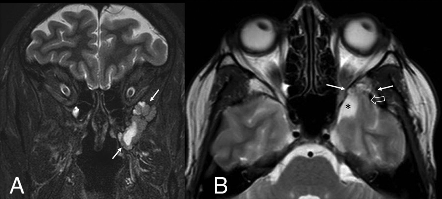
- FIG 1.
Large left middle cranial fossa encephalocele in a patient with left temporal lobe epilepsy. Coronal STIR image (A, arrows) depicts a focally prominent CSF space and distorted temporal lobe parenchyma surrounded by the scalloped bone margins of the encephalocele. Axial T2 TSE image (B) shows a distorted, stretched appearance of a left temporal lobe gyrus (open arrow) extending into the encephalocele (solid arrows) and asymmetric enlargement of the adjacent intracranial subarachnoid space (asterisk).
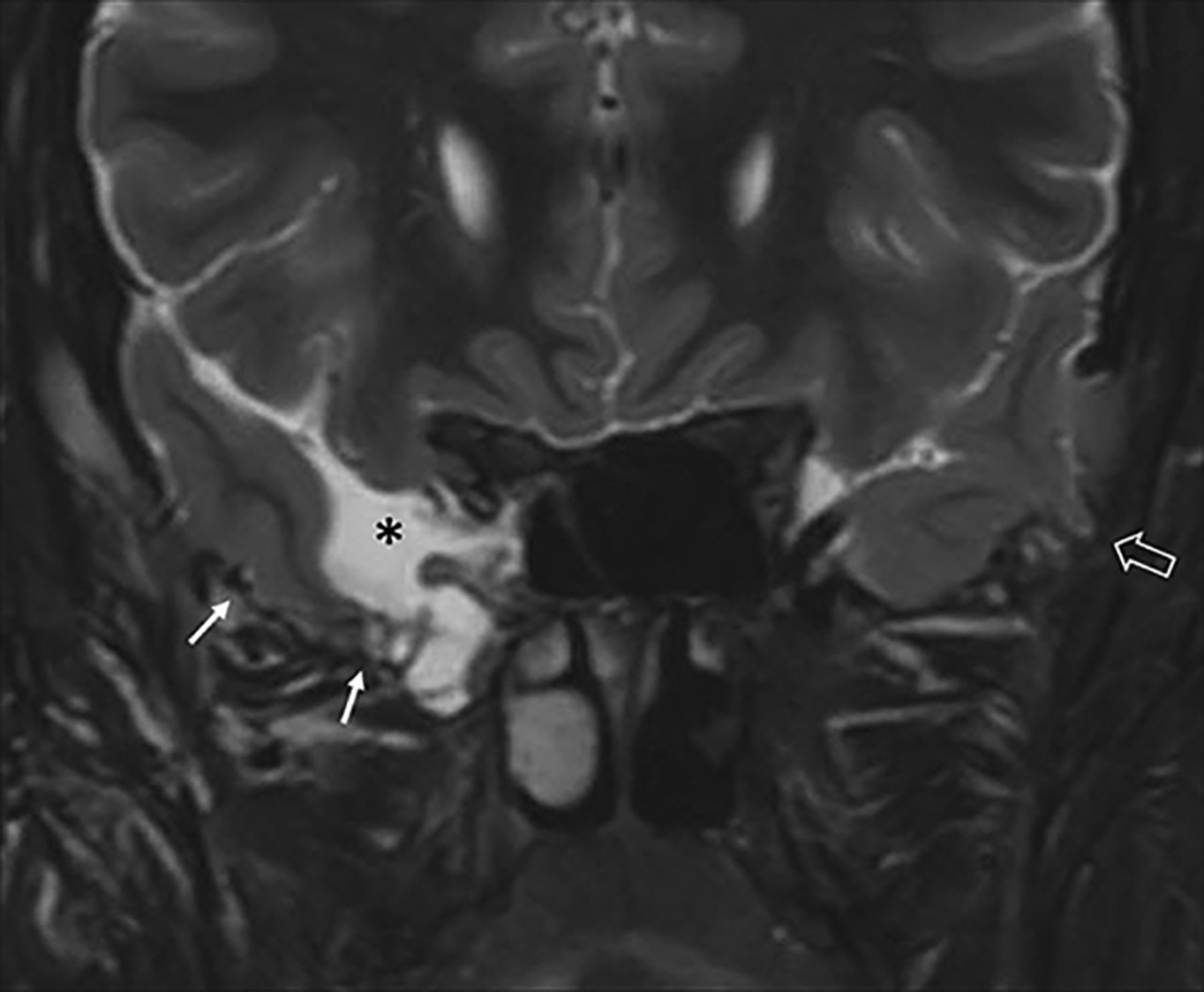
- FIG 2.
Bilateral middle cranial fossa encephaloceles in a patient with no history of seizure. Coronal STIR image depicts distortion and thinning of temporal lobe parenchyma (solid arrows), which appears adherent to the floor of the encephalocele, and asymmetric enlargement of the adjacent intracranial subarachnoid space (asterisk). A small middle cranial fossa encephalocele is present on the left (open arrow), with associated mild parenchymal distortion.


Tables
- Table 1:
MR imaging characteristics of encephaloceles compared between patients with a history of seizure and those with no history of seizurea
Variable Total (n = 77) History of Seizure (n = 35) No History of Seizure (n = 42) P Location of encephaloceles .91 Left 22 (28.6) 10 (28.6) 12 (28.6) Right 16 (20.8) 8 (22.9) 8 (19.1) Bilateral 39 (50.7) 17 (48.6) 22 (52.4) Side of largest encephalocele .80 Left 43 (55.8) 19 (54.3) 24 (57.1) Right 34 (44.2) 16 (45.7) 18 (42.9) Total No. of encephaloceles .12 1 26 (33.8) 15 (42.9) 11 (26.2) >1 51 (66.2) 20 (57.1) 31 (73.8) Median No. of sextantsb containing encephaloceles 3 (2) 2 (2) 3 (3) .21 Largest encephalocele Depth (mm) 5 (3) 5 (3) 5.5 (3) .88 Areal extent (mm2) 117 (142) 119 (144) 117 (154) .93 Volume (mm3) 368 (538) 374 (512) 364 (632) .76 Morphologic distortion of adjacent brain parenchyma .51 Minimal 11 (14.3) 4 (11.4) 7 (16.7) Mild 21 (27.3) 10 (28.6) 11 (26.2) Moderate 32 (41.6) 17 (48.6) 15 (35.7) Severe 13 (16.9) 4 (11.4) 9 (21.4) Longest single dimension of distorted parenchyma adjacent to largest encephalocele (mm) 13 (11) 14 (13) 13 (11) .54 Presence of adjacent encephalomalacia 9 (11.7) 2 (5.7) 7 (16.7) .14 - Table 2:
MR imaging characteristics of middle cranial fossa encephaloceles compared in patients with a history of temporal lobe epilepsy and those with no history of seizurea
Variable Total (n = 62) History of Temporal Lobe Epilepsy (n = 20) No History of Seizure (n = 42) P Encephalocele side .86 Left 17 (27.4) 5 (25) 12 (28.6) Right 30 (21.0) 5 (25) 8 (19.1) Bilateral 32 (51.6) 10 (50) 22 (52.4) Side of largest encephalocele .60 Left 34 (54.8) 10 (50.0) 24 (57.1) Right 28 (45.2) 10 (50.0) 18 (42.9) Total No. of bilateral encephaloceles .14 1 20 (32.3) 9 (45.0) 11 (26.2) >1 42 (67.7) 11 (55.0) 31 (73.8) Median No. of sextantsb containing encephaloceles 3 (2) 3 (2) 3 (3) .23 Largest encephalocele Depth (mm) 5 (3) 4 (2.5) 5.5 (3) .43 Areal extent (mm2) 117 (142) 149 (225.5) 117 (154) .92 Volume (mm3) 368 (538) 423 (513) 364 (632) .74 Morphologic distortion of adjacent brain parenchyma .83c Minimal 9 (14.5) 2 (10.0) 7 (16.7) Mild 17 (27.4) 6 (30.0) 11 (26.2) Moderate 24 (38.7) 9 (45.0) 15 (35.7) Severe 12 (19.4) 3 (15.0) 9 (21.4) Longest single dimension of distorted parenchyma adjacent to largest encephalocele (mm) 13 (11) 15.5 (15) 13 (11) .53 Presence of adjacent encephalomalacia 7 (11.3) 0 (0.0) 7 (16.7) .09c


Jump to section
Related Articles
Cited By...
- No citing articles found.