
Article Figures & Data
Figures
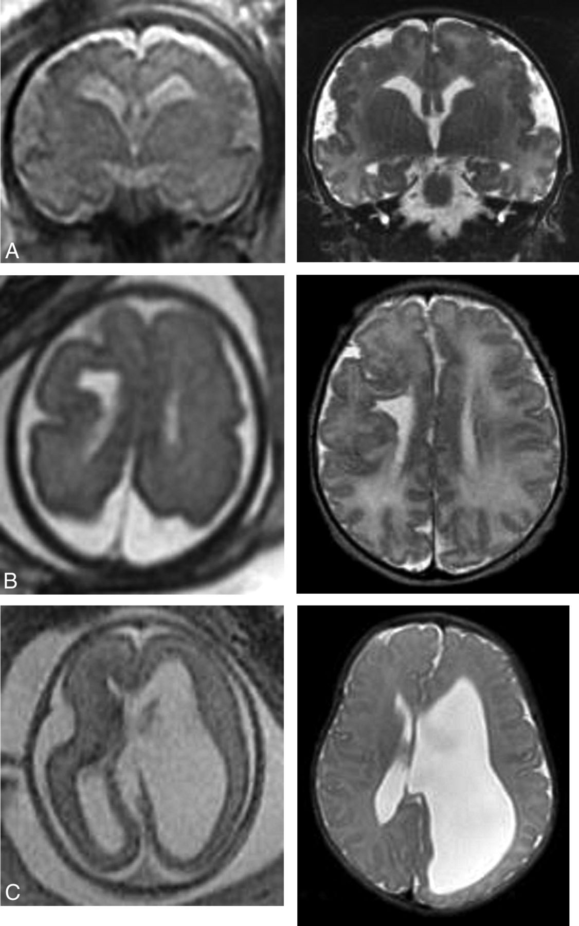
- FIG 1.
Prenatal and postnatal imaging examples of cases in which an indeterminate prognosis was associated with a poor outcome. A, Coronal single-shot fast spin-echo at 33 weeks with extensive bilateral polymicrogyria (peri-Sylvian, frontal, and parietal). Coronal T2 at 2 days of age confirms diffuse polymicrogyria. B, Axial single-shot fast spin-echo at 30 weeks shows a small right cerebral hemisphere with associated abnormal sulcation. There is right-sided polymicrogyria, a left subependymal nodule, and a large middle cystic structure (not shown) inferior to the corpus callosum and extending to the posterior fossa. Axial T2 at 2 days of age confirmed the fetal MR imaging findings. C, Axial FIESTA at 23 weeks demonstrates left unilateral VM. Axial T2 at 3 weeks of age confirms the left VM. The patient later required ventriculoperitoneal shunt due to complications of hydrocephalus and further disconnection surgery without resolution of seizures.
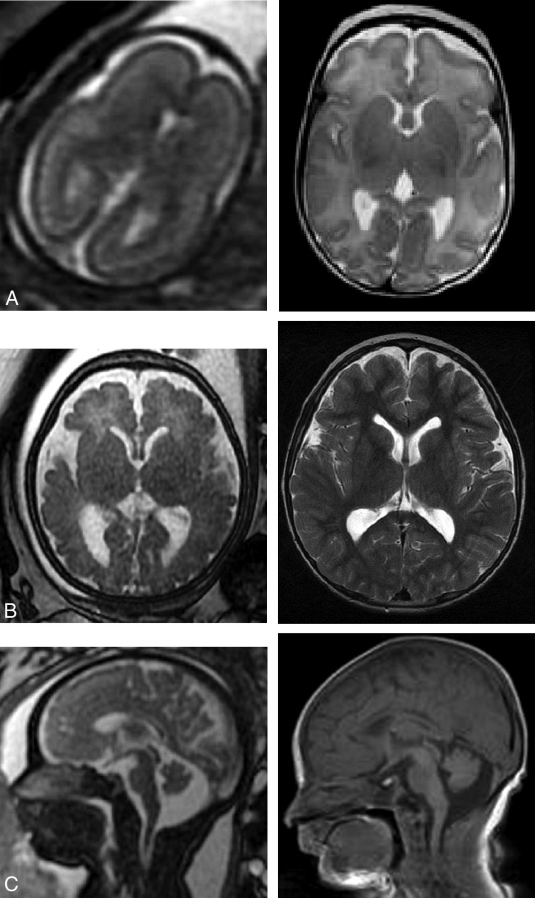
- FIG 2.
Prenatal and postnatal images showing examples of discordant imaging findings. A, Axial T2 feMRI at 27 weeks shows moderate VM, and postnatal axial T2 MRI shows mild VM with subependymal heterotropia (arrows). B, feMRI at 24 weeks shows mild VM and a preserved cerebral mantle, and postnatal MRI shows microcephaly and severe ventriculomegaly with thinning of the cerebral mantle. C, feMRI at 24 weeks shows a thin T2-hypointense cerebral line in the frontal lobes (arrows) that was overlooked. Postnatal MRI confirmed subcortical band heterotopia.
- FIG 3.
Prenatal and postnatal imaging examples of cases in which an indeterminate prognosis was associated with a favorable outcome. A, Axial single-shot fast spin-echo at 23 weeks with primitive sulcation and oligohydramnios. Axial T2 at 2 months of age shows normal brain MRI findings. B, Axial single-shot fast spin-echo at 35 weeks with VM (left 13 and right 12 mm). MRI at 6 years of age shows persistent prominent VM similar to findings on the ultrasound after birth (not shown). The patient has normal neurologic examination findings. C, Sagittal single-shot fast spin-echo at 35 weeks with a large cisterna magna versus vermian hypoplasia. Sagittal T1 at 3 days of life shows a prominent cisterna magna without other abnormalities.


Tables
Characteristic All GA (n = 114) GA <25 Weeks (n = 56) GA ≥25 Weeks (n = 58) GA at fetal MR imaging (range) (median) (IQR) (wk) 19.00–39.43 (25.9 [21.9–32.3]) 19.00–24.43 (21.8 [21.0–23.1]) 25.86–39.43 (32.2 [29.1–34.1]) Maternal age at fetal MR imaging (range) (median) (IQR) (yr) 16.00–41.00 (31.0 [28.0–34.0]) 16.00–41.00 (31.0 [28.8–35.0]) 17.00–41.00 (31.0 [27.2–34.0]) Brain region (No.) (%) Ventriculomegaly 39 (34.2%) 20 (35.7%) 19 (32.8%) Mild 23 (21.9%) 14 (25.0%) 9 (15.5%) Moderate 7 (4.4%) 4 (3.6%) 3 (5.1%) Severe 9 (7.9%) 2 (3.6%) 7 (12.1%) Posterior fossa 26 (22.8%) 12 (21.4%) 14 (24.1%) Corpus callosum 13 (11.4%) 3 (5.4%) 10 (17.2%) Sulcation/migration 15 (13.2%) 9 (16.1%) 6 (10.3%) Normal brain 15 (13.2%) 11 (19.6%) 4 (6.9%) Space-occupying lesion 4 (3.5%) 1 (1.8%) 3 (5.2%) Vascular anomaly 1 (0.9%) 0 (0.0%) 1 (1.7%) Hemorrhage 1 (0.9%) 0 (0.0%) 1 (1.7%) Note:—IQR indicates interquartile range.
No. (%) GA <25 Weeks (No.) (%) GA ≥25 Weeks (No.) (%) Agreement 106 (93.0%) 50 (89.3%) 56 (96.6%) Disagreement 8 (7.0%) 6 (10.7%) 2 (3.4%) GA (wk) Prenatal Diagnosis Postnatal Diagnosis 24 Macrocephaly with moderate asymmetric VM Healthy 22 Vermis and cerebellum slightly small for GA Healthy 27 Moderate VM (Fig 2A) Mild VM and 3 subependymal heterotopias 20 Mild VM and moderate pericardial effusion Cystic encephalomalacia 24 Mild VM (Fig 2B) Microcephaly with severe VM 24 Flattening of inferior surface of cerebellum (Fig 2C) Extensive lissencephaly with band heterotropia 19 Twin A, healthy Congenital CMV 19 Twin B, healthy Congenital CMV Note:—CMV indicates cytomegalovirus.
- Table 4:
Characteristics of participants included in the assessment of concordance between prenatal prognosis and postnatal neurodevelopmental outcome
Characteristic (n = 104) GA <25 Weeks (n = 50) GA ≥25 Weeks (n = 54) GA at fetal MR imaging (range) (median) (IQR) (wk) 19.00–39.43 (26.1 [21.8, 31.9]) 19.00–24.43 (21.7 [21.0, 23.0]) 25.00–39.43 (31.9 [29.6–34.0]) Maternal age at fetal MR imaging (range) (median) (IQR) (yr) 16.00–41.00 (31.0 [28.0–33.0]) 16.00–41.00 (30.0 [28.0–32.0]) 17.00–41.00 (31.0 [28.0–34.0]) Age at postnatal visit (range) (median) (IQR) (yr) 1.00–10.42 (3.0 [1.9–5.9]) 1.00–8.92 (2.8 [1.7–5.1]) 1.00–10.42 (3.8 [2.0–6.0]) Brain region (No.) (%) Ventriculomegaly 32 (30.8%) 17 (34.0%) 15 (27.8%) Mild 20 (19.2%) 12 (24.0%) 8 (14.8%) Moderate 5 (4.8%) 3 (6.0%) 2 (3.7%) Severe 7 (6.7%) 2 (4.0%) 5 (9.3%) Posterior fossa 19 (18.3%) 7 (14.0%) 12 (22.2%) Corpus callosum 11 (10.6%) 2 (4.0%) 9 (16.7%) Sulcation/migration 8 (7.7%) 5 (10.0%) 3 (5.6%) Normal 28 (26.9%) 18 (36.0%) 10 (18.5%) Space 4 (3.8%) 1 (2.0%) 3 (5.6%) Vascular anomaly 1 (1.0%) 0 (0.0%) 1 (1.9%) Hemorrhage 1 (1.0%) 0 (0.0%) 1 (1.9%) Prenatal Prognosis (No.) (%) Postnatal Outcome, Favorable (n = 77) Postnatal Outcome, Intermediate (n = 12) Postnatal Outcome, Poor (n = 15) Favorable (n = 46) 43 (93.5%) 2 (4.3%) 1 (2.2%) Indeterminate (n = 52) 33 (63.5%) 10 (19.2%) 9 (17.3%) Poor (n = 6) 1 (16.7%) 0 (0.0%) 5 (83.3%) Prenatal Prognosis (No.) (%) Absence of Epilepsy (n = 84) Medically Controlled Epilepsy (n = 7) Intractable Epilepsy (n = 5) Favorable (n = 46) 45 (97.8%) 0 (0.0%) 1 (2.2%) Indeterminate (n = 47) 37 (78.7%) 7 (14.9%) 3 (6.4%) Poor (n = 3) 2 (66.7%) 0 (0.0%) 1 (33.3%) Prenatal Prognosis (No.) (%) Postnatal Outcome, Favorable (n = 36) Postnatal Outcome, Intermediate (n = 7) Postnatal Outcome, Poor (n = 7) Favorable (n = 25) 23 (92.0%) 1 (4.0%) 1 (4.0%) Indeterminate (n = 25) 13 (52.0%) 6 (24.0%) 6 (24.0%) Poor (n = 0) 0 (0.0%) 0 (0.0%) 0 (100.0%) Prenatal Prognosis (No.) (%) Postnatal Outcome, Favorable (n = 41) Postnatal Outcome, Intermediate (n = 5) Postnatal Outcome, Poor (n = 8) Favorable (n = 21) 20 (95.2%) 1 (4.8%) 0 (0.0%) Indeterminate (n = 27) 20 (74.1%) 4 (14.8%) 3 (11.1%) Poor (n = 6) 1 (16.7%) 0 (0.0%) 5 (83.3%)


