Advertisement

Main menu

  • Home
  • Content
    • Current Issue
    • Accepted Manuscripts
    • Article Preview
    • Past Issue Archive
    • Video Articles
    • AJNR Case Collection
    • Case of the Week Archive
    • Case of the Month Archive
    • Classic Case Archive
  • Special Collections
    • AJNR Awards
    • Low-Field MRI
    • Alzheimer Disease
    • ASNR Foundation Special Collection
    • Photon-Counting CT
    • View All
  • Multimedia
    • AJNR Podcasts
    • AJNR SCANtastic
    • Trainee Corner
    • MRI Safety Corner
    • Imaging Protocols
  • For Authors
    • Submit a Manuscript
    • Submit a Video Article
    • Submit an eLetter to the Editor/Response
    • Manuscript Submission Guidelines
    • Statistical Tips
    • Fast Publishing of Accepted Manuscripts
    • Graphical Abstract Preparation
    • Imaging Protocol Submission
    • Author Policies
  • About Us
    • About AJNR
    • Editorial Board
    • Editorial Board Alumni
  • More
    • Become a Reviewer/Academy of Reviewers
    • Subscribers
    • Permissions
    • Alerts
    • Feedback
    • Advertisers
    • ASNR Home

User menu

  • Alerts
  • Log in

Search

  • Advanced search
American Journal of Neuroradiology

American Journal of Neuroradiology

ASHNR American Society of Functional Neuroradiology ASHNR American Society of Pediatric Neuroradiology ASSR
  • Alerts
  • Log in

Advanced Search

  • Home
  • Content
    • Current Issue
    • Accepted Manuscripts
    • Article Preview
    • Past Issue Archive
    • Video Articles
    • AJNR Case Collection
    • Case of the Week Archive
    • Case of the Month Archive
    • Classic Case Archive
  • Special Collections
    • AJNR Awards
    • Low-Field MRI
    • Alzheimer Disease
    • ASNR Foundation Special Collection
    • Photon-Counting CT
    • View All
  • Multimedia
    • AJNR Podcasts
    • AJNR SCANtastic
    • Trainee Corner
    • MRI Safety Corner
    • Imaging Protocols
  • For Authors
    • Submit a Manuscript
    • Submit a Video Article
    • Submit an eLetter to the Editor/Response
    • Manuscript Submission Guidelines
    • Statistical Tips
    • Fast Publishing of Accepted Manuscripts
    • Graphical Abstract Preparation
    • Imaging Protocol Submission
    • Author Policies
  • About Us
    • About AJNR
    • Editorial Board
    • Editorial Board Alumni
  • More
    • Become a Reviewer/Academy of Reviewers
    • Subscribers
    • Permissions
    • Alerts
    • Feedback
    • Advertisers
    • ASNR Home
  • Follow AJNR on Twitter
  • Visit AJNR on Facebook
  • Follow AJNR on Instagram
  • Join AJNR on LinkedIn
  • RSS Feeds

AJNR Awards, New Junior Editors, and more. Read the latest AJNR updates

Research ArticleHead and Neck Imaging
Open Access

Non-EPI-DWI for Detection, Disease Monitoring, and Clinical Decision-Making in Thyroid Eye Disease

C. Feeney, R.K. Lingam, V. Lee, F. Rahman and S. Nagendran
American Journal of Neuroradiology August 2020, 41 (8) 1466-1472; DOI: https://doi.org/10.3174/ajnr.A6664
C. Feeney
aFrom the Eye Department (C.F., V.L., F.R., S.N.), Central Middlesex Hospital, London, UK
bImperial Centre for Endocrinology (C.F.), North West Thames, UK
  • Find this author on Google Scholar
  • Find this author on PubMed
  • Search for this author on this site
  • ORCID record for C. Feeney
R.K. Lingam
cDepartment of Radiology (R.K.L.), Northwick Park & Central Middlesex Hospital, London Northwest University Healthcare National Health Service Trust, London, UK.
  • Find this author on Google Scholar
  • Find this author on PubMed
  • Search for this author on this site
  • ORCID record for R.K. Lingam
V. Lee
aFrom the Eye Department (C.F., V.L., F.R., S.N.), Central Middlesex Hospital, London, UK
  • Find this author on Google Scholar
  • Find this author on PubMed
  • Search for this author on this site
  • ORCID record for V. Lee
F. Rahman
aFrom the Eye Department (C.F., V.L., F.R., S.N.), Central Middlesex Hospital, London, UK
  • Find this author on Google Scholar
  • Find this author on PubMed
  • Search for this author on this site
  • ORCID record for F. Rahman
S. Nagendran
aFrom the Eye Department (C.F., V.L., F.R., S.N.), Central Middlesex Hospital, London, UK
  • Find this author on Google Scholar
  • Find this author on PubMed
  • Search for this author on this site
  • ORCID record for S. Nagendran
  • Article
  • Figures & Data
  • Info & Metrics
  • Responses
  • References
  • PDF
Loading

Article Figures & Data

Figures

  • Tables
  • Fig 1.
    • Download figure
    • Open in new tab
    • Download powerpoint
    Fig 1.

    Correlation between CAS and ADC and clinically meaningful group-wise comparisons of ADC and CAS in the overall cohort. Box plots demonstrate a positive Spearman rank correlation coefficient between CAS and ADC of individual EOMs (n = 368) (A) and significantly greater ADC values in CAS ≥ 3 (n = 70) (B) compared with CAS < 3 (n = 298) groups. Two asterisks indicate P < .001, 2-tailed. C, Significantly greater ADC values of all EOMs in cohort 3 compared with those in cohort 1 (P < .001) and cohort 2 (P = .03). For cohort 2, ADC values were significantly higher than those in cohort 1 (P < .001) but lower than those in cohort 3 (P = .03). Single asterisk indicates P < .05.

  • Fig 2.
    • Download figure
    • Open in new tab
    • Download powerpoint
    Fig 2.

    Patient flow chart for clinical decisions influenced by the reported activity of non-EPI-DWI scans in conjunction with CAS and clinical assessment. Whole numbers denote the number of patients. The dashed line represents the opportunity for a non-EPI-DWI-informed clinical decision. MDT indicates multidisciplinary team.
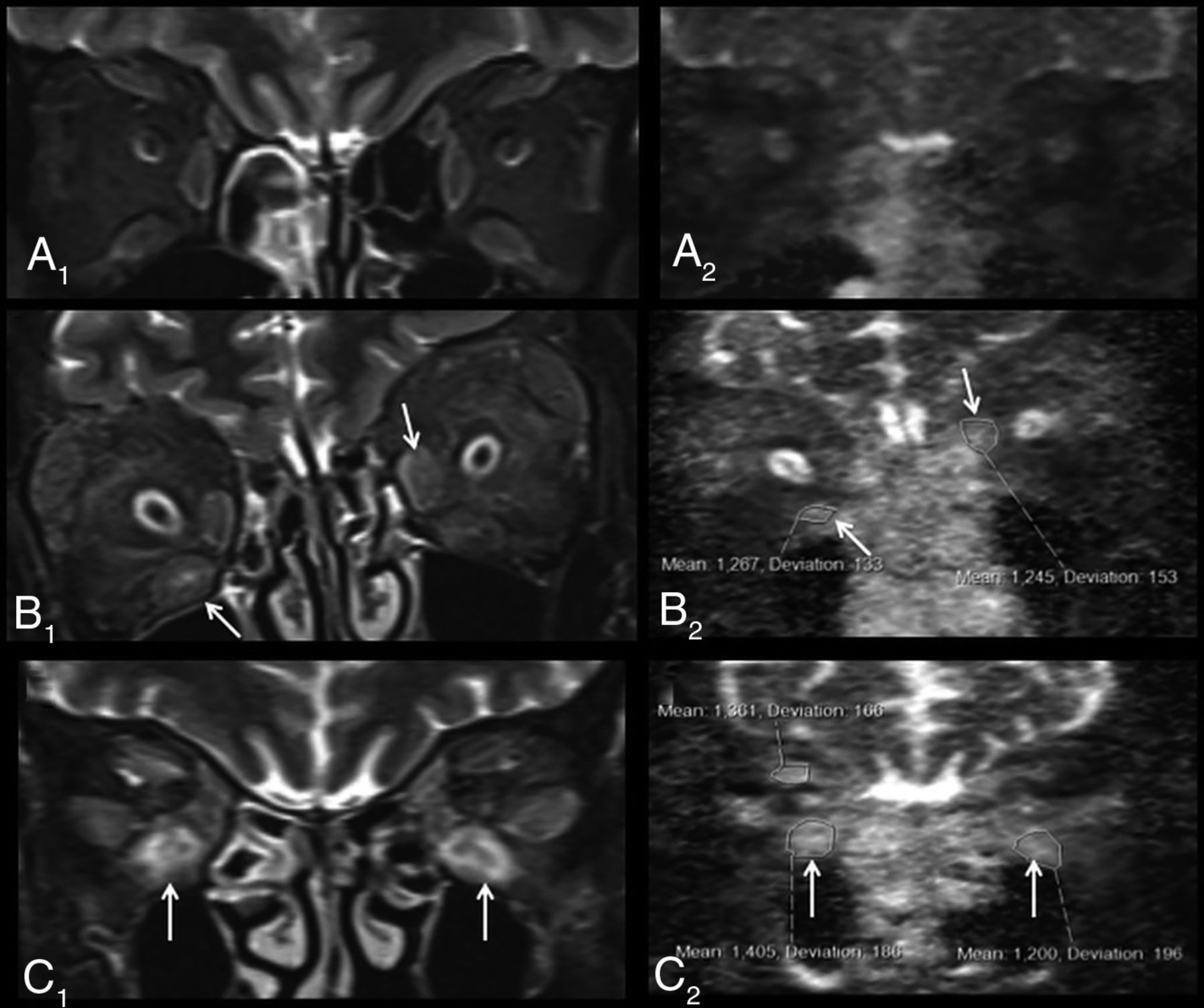
  • Fig 3.
    • Download figure
    • Open in new tab
    • Download powerpoint
    Fig 3.

    Representative examples of non-EPI-DWI of orbital EOMs in patients from each of cohorts 1, 2, and 3 alongside STIR MR imaging. A, Coronal orbital/EOM MR imaging STIR image (A1) and a non-EPI-DWI ADC image (A2) show mild enlargement of the extraocular muscles with mild increased signal and ADC values (right inferior rectus muscle = 590; left inferior rectus muscle = 540), labeled an inactive scan (cohort 1). STIR image (B1) and ADC image (B2) show moderate-to-marked enlargement of the extraocular muscles and increased signal and ADC values, notably at the right inferior and left medial recti muscles (arrows) (cohort 2). STIR image (C1) and ADC image (C2) show moderate-to-marked enlargement of the extraocular muscles and markedly increased signal and ADC values notably at the inferior recti muscles (arrows) (cohort 3).

  • Fig 4.
    • Download figure
    • Open in new tab
    • Download powerpoint
    Fig 4.

    ROCs for ADC values obtained in subjects with no/possible disease (cohort 1) and moderate-severe disease (cohort 3) (area under the curve = 0.737; 95% CI, 0.68–0.80; P < .001). Diagonal line represents a line of no discrimination between disease states. Arrowed number represents the ADC value.

Tables

  • Figures
    • View popup
    Table 1:

    Patient characteristics (cohorts 1–3)

     No.Cohort 1No.Cohort 2No.Cohort 3
    Age at initial scan (yrs)1243.9847.01153.7
    SD + range 15.4 (20.2–74.4) 12.9 (33.9–71.9) 20.8 (22.6–79.8)
    n% female129 (75.0%)85 (62.5%)118 (72.7%)
    n% Afro-Caribbean 11a1 (9.1%)83 (37.5%)115 (45.5%)
    n% current smokers124 (33.3%)83 (37.5%)113 (27.3%)
    n% positive family history124 (33.3%)83 (37.5%)111 (9.1%)
    n% positive autoantibodyb9c5 (55.5%)84 (50.0%)10d10 (100.0%)
    n% antithyroid medicatione126 (50.0%)86 (75.0%)1111 (100.0%)
    n% euthyroidf126 (50.0%)82 (25.0%)110 (0.0%)
    n% DON120 (0.0%)81 (12.5%)114 (36.3%)
    n% IV methylprednisolone120 (0.0%)82 (25.0%)1111 (100.0%)
    n% second-line immunosuppressiong120 (0.0%)80 (0.0%)113 (27.3%)
    n% orbital radiotherapy120 (0.0%)80 (0.0%)117 (63.6%)
    No. of scans12182.5112.5
    SD + range 0 (1.0–1.0) 0.9 (2.0–4.0) 0.8 (2.0–4.0)
    Initial CAS120.581.6113.6
    SD + range 0.5 (0.0–1.0) 0.5 (1.0–2.0) 1.1 (3.0–6.0)
    CAS: 1st Follow-upNANA80.5112.1
    SD + range  0.8 (0–2) 1.9 (0–6)
    Baseline ADC all EOMs966785481165691
    SD + range 171 (340–1141) 256 (311–1426) 208 (240–1088)
    ADC all EOMs:1st Follow-upNANA5277063873
    SD + range  236 (340–1321) 319 (169–1585)
    Time between 1st & 2nd scan (Months)NANA810.5119.5
    SD + range  3.1 (5.5–16.2) 7.8 (2.0–29.0)
    Final CAS12NA80.4111.6
    SD + range  0.1 (0.0–1.0) 2.0 (0.0–6.0)
    Total follow-up period (Months)12NA839.31148
        27.7 (16.3–97.4) 49.3 (8.3–163.0)
    • Note:—NA indicates not applicable.

    • ↵a Data unrecorded (n = 1).

    • ↵b Either thyroid peroxidase or thyroid-stimulating hormone receptor antibody.

    • ↵c Data missing (n  = 3).

    • ↵d Data missing (n = 1).

    • ↵e Either carbimazole, propylthiouracil, or both.

    • ↵f Normal thyroid function and no history of thyroid abnormalities.

    • ↵g Mycophenolate or hydroxychloroquine. Cohort 1 = mild/possibly active (CAS 0 or 1), cohort 2 = mild-to-moderate and active (CAS 1–3), cohort 3 = moderate-to-severe and active (CAS ≥ 3).

PreviousNext
Back to top

In this issue

American Journal of Neuroradiology: 41 (8)
American Journal of Neuroradiology
Vol. 41, Issue 8
1 Aug 2020
  • Table of Contents
  • Index by author
  • Complete Issue (PDF)
Advertisement
Print
Download PDF
Email Article

Thank you for your interest in spreading the word on American Journal of Neuroradiology.

NOTE: We only request your email address so that the person you are recommending the page to knows that you wanted them to see it, and that it is not junk mail. We do not capture any email address.

Enter multiple addresses on separate lines or separate them with commas.
Non-EPI-DWI for Detection, Disease Monitoring, and Clinical Decision-Making in Thyroid Eye Disease
(Your Name) has sent you a message from American Journal of Neuroradiology
(Your Name) thought you would like to see the American Journal of Neuroradiology web site.
CAPTCHA
This question is for testing whether or not you are a human visitor and to prevent automated spam submissions.
Cite this article
C. Feeney, R.K. Lingam, V. Lee, F. Rahman, S. Nagendran
Non-EPI-DWI for Detection, Disease Monitoring, and Clinical Decision-Making in Thyroid Eye Disease
American Journal of Neuroradiology Aug 2020, 41 (8) 1466-1472; DOI: 10.3174/ajnr.A6664

Citation Manager Formats

  • BibTeX
  • Bookends
  • EasyBib
  • EndNote (tagged)
  • EndNote 8 (xml)
  • Medlars
  • Mendeley
  • Papers
  • RefWorks Tagged
  • Ref Manager
  • RIS
  • Zotero
0 Responses
Respond to this article
Share
Bookmark this article
Non-EPI-DWI for Detection, Disease Monitoring, and Clinical Decision-Making in Thyroid Eye Disease
C. Feeney, R.K. Lingam, V. Lee, F. Rahman, S. Nagendran
American Journal of Neuroradiology Aug 2020, 41 (8) 1466-1472; DOI: 10.3174/ajnr.A6664
del.icio.us logo Twitter logo Facebook logo Mendeley logo
  • Tweet Widget
  • Facebook Like
  • Google Plus One
Purchase

Jump to section

  • Article
    • Abstract
    • ABBREVIATIONS:
    • MATERIALS AND METHODS
    • RESULTS
    • DISCUSSION
    • CONCLUSIONS
    • Acknowledgments
    • Footnotes
    • References
  • Figures & Data
  • Info & Metrics
  • Responses
  • References
  • PDF

Related Articles

  • No related articles found.
  • PubMed
  • Google Scholar

Cited By...

  • No citing articles found.
  • Crossref
  • Google Scholar

This article has not yet been cited by articles in journals that are participating in Crossref Cited-by Linking.

More in this TOC Section

Head and Neck Imaging

  • Hydrops Herniation into the Semicircular Canals
  • ASL Sensitivity for Head and Neck Paraganglioma
  • Post SRS Peritumoral Hyperintense Signal of VSs
Show more Head and Neck Imaging

Functional

  • Kurtosis and Epileptogenic Tubers: A Pilot Study
  • Glutaric Aciduria Type 1: DK vs. Conventional MRI
  • Multiparametric MRI in PEDS Pontine Glioma
Show more Functional

Similar Articles

Advertisement

Indexed Content

  • Current Issue
  • Accepted Manuscripts
  • Article Preview
  • Past Issues
  • Editorials
  • Editor's Choice
  • Fellows' Journal Club
  • Letters to the Editor
  • Video Articles

Cases

  • Case Collection
  • Archive - Case of the Week
  • Archive - Case of the Month
  • Archive - Classic Case

More from AJNR

  • Trainee Corner
  • Imaging Protocols
  • MRI Safety Corner
  • Book Reviews

Multimedia

  • AJNR Podcasts
  • AJNR Scantastics

Resources

  • Turnaround Time
  • Submit a Manuscript
  • Submit a Video Article
  • Submit an eLetter to the Editor/Response
  • Manuscript Submission Guidelines
  • Statistical Tips
  • Fast Publishing of Accepted Manuscripts
  • Graphical Abstract Preparation
  • Imaging Protocol Submission
  • Evidence-Based Medicine Level Guide
  • Publishing Checklists
  • Author Policies
  • Become a Reviewer/Academy of Reviewers
  • News and Updates

About Us

  • About AJNR
  • Editorial Board
  • Editorial Board Alumni
  • Alerts
  • Permissions
  • Not an AJNR Subscriber? Join Now
  • Advertise with Us
  • Librarian Resources
  • Feedback
  • Terms and Conditions
  • AJNR Editorial Board Alumni

American Society of Neuroradiology

  • Not an ASNR Member? Join Now

© 2025 by the American Society of Neuroradiology All rights, including for text and data mining, AI training, and similar technologies, are reserved.
Print ISSN: 0195-6108 Online ISSN: 1936-959X

Powered by HighWire