
Article Figures & Data
Figures
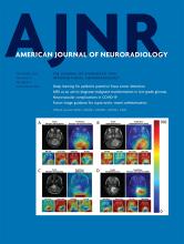
- FIG 1.
Example of DTI planes and ROI drawings from 1 patient. A, T2-weighted structural image with the locations of the C2 level and AC levels. B, ROI drawing at C2. C–E, ROI drawings at the AC levels. D, The MC level. Four DTI metrics were measured, including FA, AD, RD, and MD.
- FIG 2.
ROC analysis of the predictive value of DTI metrics for the postoperative mJOA recovery rate. Results for the average of all AC levels, MC level, and the C2 level, respectively. The area under the curve values for different metrics are shown in Table 11.


Tables
Publications Correlation Method Significance DTI Metrics Prediction Indexes Vertebral Levels Surgery Method Jones et al, 20139 Spearman No FA mJOA MC level, C2–C3 Not specified Wen et al, 201415 Spearman Yes FA mJOA recovery rate Average of C3 to C7 Not specified Vedantam et al, 20175 Pearson Yes FA ΔmJOA MC level Not specified Maki et al, 201716 Spearman Yes FA ΔmJOA, mJOA recovery rate MC level Posterior and anterior Kitamura et al, 202017 Spearman Yes FA ΔmJOA, mJOA recovery rate MC level Posterior Shabani et al, 201918 Intraclass correlation coefficient Yes (–) FA ΔmJOA MC level Not specified Iwasaki et al, 201914 Pearson No FA mJOA MC level Posterior and anterior Note:—(–) indicates negative correlation; ΔmJOA, difference between pre- and postoperative mJOA.
- Table 2:
Comparison of DTI metrics between patients before surgery and the control group for the MC level
Groups/Statistics MD (10−3mm2/s) AD (10−3mm2/s) RD (10−3mm2/s) FA Patients Controls Patients Controls Patients Controls Patients Controls Group 1a 0.97 ± 0.16 0.72 ± 0.04 1.40 ± 0.11 1.35 ± 0.06 0.75 ± 0.21 0.40 ± 0.04 0.41 ± 0.14 0.66 ± 0.04 t/t' value t' = −4.32 t' = −1.24 t' = −4.65 t' = 4.84 P value P = .003d P = .25 P = .002d P = .002d Group 2b 0.97 ± 0.17 0.73 ± 0.04 1.51 ± 0.16 1.32 ± 0.08 0.70 ± 0.21 0.44 ± 0.04 0.48 ± 0.14 0.61 ± 0.04 t/t' value t' = −6.40 t' = −4.78 t' = −5.59 t' = 4.311 P value P < .001d P < .001d P < .001d P < .001d Group 3c 0.99 ± 0.20 0.75 ± 0.05 1.47 ± 0.15 1.26 ± 0.10 0.76 ± 0.26 0.49 ± 0.04 0.43 ± 0.16 0.55 ± 0.05 t/t' value t' = −5.89 t = −5.42 t' = −5.01 t' = 3.51 P value P < .001d P < .001d P < .001d P = .001d - Table 3:
Comparison of DTI metrics between patients before surgery and the control group for the average of AC levels
Groups/Statistics MD (10−3mm2/s) AD (10−3mm2/s) RD (10−3mm2/s) FA Patients (n = 55) 0.87 ± 0.12 1.40 ± 0.12 0.61 ± 0.14 0.50 ± 0.10 Controls (n = 20) 0.73 ± 0.03 1.31 ± 0.07 0.44 ± 0.03 0.61 ± 0.03 t/t' value t' = −8.19 t = −3.15 t' = −8.36 t' = 7.10 P value P < .001a P < .001a P < .001a P < .001a ↵a Significant correlation.
- Table 4:
Comparison of DTI metrics between the patients before surgery and the control group for the C2 level
Groups/Statistics MD (10−3mm2/s) AD (10−3mm2/s) RD (10−3mm2/s) FA Patients (n = 52) 0.74 (0.71–0.77)a 1.35 (1.33–1.42) 0.42 (0.39–0.47) 0.64 (0.59–0.67) Controls (n = 20) 0.75 (0.72–0.79) 1.43 (1.33–1.48) 0.41 (0.37–0.45) 0.68 (0.64–0.70) Z value Z = −0.33 Z = −1.62 Z = −1.04 Z = −2.44 P value P = .74 P = .10 P = .30 P = .02b - Table 5:
Spearman correlations between the DTI metrics at different levels and mJOA before surgery
Levels/DTI Metrics Correlation Coefficient P Value MC level (n = 55) MD –0.13 .34 AD –0.13 .34 RD –0.07 .62 FA –0.004 .98 Average of AC levels (n = 55) MD –0.15 .27 AD –0.12 .39 RD –0.12 .38 FA 0.07 .61 C2 level (n = 52) MD –0.09 .55 AD 0.36 .008a RD –0.29 .04a FA 0.35 .01a ↵a Significant correlation.
- Table 6:
Spearman correlations between the DTI metrics at the MC level and mJOA at 2 follow-up stages after surgery
Follow-Up Stages/DTI Metrics Correlation Coefficient P Value 3-Month follow-up (n = 44) MD –0.12 .41 AD –0.09 .54 RD –0.10 .51 FA 0.10 .51 6-Month follow-up (n = 37) MD –0.31 .04a AD –0.14 .36 RD –0.32 .03a FA 0.33 .03a ↵a Significant correlation.
- Table 7:
Spearman correlations between the DTI metrics for C2 level and mJOA at 2 follow-up stages after surgery
Follow-Up Stages/DTI Metrics Correlation Coefficient P Value 3-Month follow-up (n = 44) MD –0.20 .21 AD 0.16 .33 RD –0.35 .03a FA 0.42 .005a 6-Month follow-up (n = 37) MD –0.44 .01a AD 0.15 .39 RD –0.65 <.001a FA 0.77 <.001a ↵a Significant correlation.
- Table 8:
Spearman correlations between the preoperative DTI metrics of different levels and the postoperative mJOA recovery rate at the final follow-up (1 year)
Levels/DTI Metrics Correlation Coefficient P Value MC level (n = 55) MD –0.10 .34 AD 0.16 .34 RD –0.12 .62 FA 0.19 .98 Average of AC levels (n = 55) MD –0.19 .16 AD 0.08 .59 RD -0.24 .08 FA 0.26 .06 C2 level (n = 52) MD –0.15 .31 AD 0.40 .003a RD –0.42 .002a FA 0.51 <.001a ↵a Significant correlation.
- Table 9:
Results of linear regression to examine the correlation between DTI, T1-weighted, and T2-weighted features and preoperative mJOA
Level/Incorporated DTI Metrics Significant Features Slope (β) P Value MC level (n = 55) MD/AD/RD/FA Axial spinal cord area 0.05 .01 Average of AC levels (n = 55) MD/AD/RD/FA Axial spinal cord area 0.05 .01 C2 level (n = 52) MD MD –10.28 .03 Spinal cord flattened rate 9.00 .005 AD AD 5.58 .04 Spinal cord flattened rate 6.38 .04 RD RD –8.93 .002 Spinal cord flattened rate 8.32 .006 FA FA 9.43 .002 Spinal cord flattened rate 7.12 .02 - Table 10:
Results of linear regression to examine the correlation between DTI, T1-weighted, and T2-weighted features and the mJOA recovery rate
Level Incorporated DTI Metrics Significant Features Slope (β) P Value MC level (n = 55) MD/AD/RD/FA Axial spinal cord area 0.70 .02 Average of AC levels (n = 55) MD/AD/RD Axial spinal cord area 0.70 .02 FA FA 108.67 .01 Spinal cord flattened rate 105.64 .03 C2 level (n = 52) MD MD –157.98 .04 Spinal cord flattened rate 141.57 .01 AD AD 123.60 .01 RD RD –145.13 .004 Spinal cord flattened rate 131.88 .01 FA FA 170.92 .001 Spinal cord flattened rate 111.82 .02 - Table 11:
Prediction capability of the preoperative DTI metrics at different levels for the postoperative mJOA recovery rate using ROC analysis
Level/DTI Metrics AUC P Value Sensitivity Specificity MC level (n = 55) MD 0.46 .66 0.63 0.47 AD 0.64 .10 0.37 0.94 RD 0.43 .40 0.21 0.88 FA 0.62 .15 0.63 0.65 Average of AC levels (n = 55) MD 0.40 .26 0.16 0.88 AD 0.57 .43 0.63 0.59 RD 0.36 .09 0.95 0.06 FA 0.65 .07 0.34 0.94 C2 level (n = 52) MD 0.43 .39 0.97 0.06 AD 0.64 .12 0.39 0.88 RD 0.34 .07 0.97 0.06 FA 0.68 .04a 0.56a 0.81 Note:—AUC indicates area under the curve.
↵a Significant correlation.


Jump to section
Related Articles
Cited By...
- No citing articles found.