Advertisement

Main menu

  • Home
  • Content
    • Current Issue
    • Accepted Manuscripts
    • Article Preview
    • Past Issue Archive
    • AJNR Case Collection
    • Case of the Week Archive
    • Classic Case Archive
    • Case of the Month Archive
  • Special Collections
    • Spinal CSF Leak Articles (Jan 2020-June 2024)
    • 2024 AJNR Journal Awards
    • Most Impactful AJNR Articles
  • Multimedia
    • AJNR Podcast
    • AJNR Scantastics
    • Video Articles
  • For Authors
    • Submit a Manuscript
    • Author Policies
    • Fast publishing of Accepted Manuscripts
    • Graphical Abstract Preparation
    • Manuscript Submission Guidelines
    • Imaging Protocol Submission
    • Submit a Case for the Case Collection
  • About Us
    • About AJNR
    • Editorial Board
  • More
    • Become a Reviewer/Academy of Reviewers
    • Subscribers
    • Permissions
    • Alerts
    • Feedback
    • Advertisers
    • ASNR Home
  • Other Publications
    • ajnr

User menu

  • Alerts
  • Log in

Search

  • Advanced search
American Journal of Neuroradiology

American Journal of Neuroradiology

ASHNR American Society of Functional Neuroradiology ASHNR American Society of Pediatric Neuroradiology ASSR
  • Alerts
  • Log in

Advanced Search

  • Home
  • Content
    • Current Issue
    • Accepted Manuscripts
    • Article Preview
    • Past Issue Archive
    • AJNR Case Collection
    • Case of the Week Archive
    • Classic Case Archive
    • Case of the Month Archive
  • Special Collections
    • Spinal CSF Leak Articles (Jan 2020-June 2024)
    • 2024 AJNR Journal Awards
    • Most Impactful AJNR Articles
  • Multimedia
    • AJNR Podcast
    • AJNR Scantastics
    • Video Articles
  • For Authors
    • Submit a Manuscript
    • Author Policies
    • Fast publishing of Accepted Manuscripts
    • Graphical Abstract Preparation
    • Manuscript Submission Guidelines
    • Imaging Protocol Submission
    • Submit a Case for the Case Collection
  • About Us
    • About AJNR
    • Editorial Board
  • More
    • Become a Reviewer/Academy of Reviewers
    • Subscribers
    • Permissions
    • Alerts
    • Feedback
    • Advertisers
    • ASNR Home
  • Follow AJNR on Twitter
  • Visit AJNR on Facebook
  • Follow AJNR on Instagram
  • Join AJNR on LinkedIn
  • RSS Feeds

Welcome to the new AJNR, Updated Hall of Fame, and more. Read the full announcements.


AJNR is seeking candidates for the position of Associate Section Editor, AJNR Case Collection. Read the full announcement.


Research ArticleAdult Brain

High-Resolution Gadolinium-Enhanced MR Cisternography Using Compressed-Sensing T1 SPACE Technique for Detection of Intracranial CSF Leaks

I.E. Duman, T. Demerath, A. Stadler, S. Elsheikh, E. Raithel, C. Forman, T. Hildenbrand, M. Shah, J. Grauvogel, C. Scheiwe, H. Urbach and S. Meckel
American Journal of Neuroradiology January 2021, 42 (1) 116-118; DOI: https://doi.org/10.3174/ajnr.A6852
I.E. Duman
aFrom the Departments of Neuroradiology (I.E.D., T.D., A.S., S.E., H.U., S.M.)
  • Find this author on Google Scholar
  • Find this author on PubMed
  • Search for this author on this site
  • ORCID record for I.E. Duman
T. Demerath
aFrom the Departments of Neuroradiology (I.E.D., T.D., A.S., S.E., H.U., S.M.)
  • Find this author on Google Scholar
  • Find this author on PubMed
  • Search for this author on this site
  • ORCID record for T. Demerath
A. Stadler
aFrom the Departments of Neuroradiology (I.E.D., T.D., A.S., S.E., H.U., S.M.)
  • Find this author on Google Scholar
  • Find this author on PubMed
  • Search for this author on this site
  • ORCID record for A. Stadler
S. Elsheikh
aFrom the Departments of Neuroradiology (I.E.D., T.D., A.S., S.E., H.U., S.M.)
  • Find this author on Google Scholar
  • Find this author on PubMed
  • Search for this author on this site
  • ORCID record for S. Elsheikh
E. Raithel
dSiemens Healthcare GmbH (E.R., C.F.), Erlangen, Germany
  • Find this author on Google Scholar
  • Find this author on PubMed
  • Search for this author on this site
  • ORCID record for E. Raithel
C. Forman
dSiemens Healthcare GmbH (E.R., C.F.), Erlangen, Germany
  • Find this author on Google Scholar
  • Find this author on PubMed
  • Search for this author on this site
  • ORCID record for C. Forman
T. Hildenbrand
bOtorhinolaryngology (T.H.)
  • Find this author on Google Scholar
  • Find this author on PubMed
  • Search for this author on this site
  • ORCID record for T. Hildenbrand
M. Shah
cNeurosurgery (M.S., J.G., C.S.), University of Freiburg Medical Center, Faculty of Medicine, University of Freiburg, Freiberg, Germany
  • Find this author on Google Scholar
  • Find this author on PubMed
  • Search for this author on this site
  • ORCID record for M. Shah
J. Grauvogel
cNeurosurgery (M.S., J.G., C.S.), University of Freiburg Medical Center, Faculty of Medicine, University of Freiburg, Freiberg, Germany
  • Find this author on Google Scholar
  • Find this author on PubMed
  • Search for this author on this site
  • ORCID record for J. Grauvogel
C. Scheiwe
cNeurosurgery (M.S., J.G., C.S.), University of Freiburg Medical Center, Faculty of Medicine, University of Freiburg, Freiberg, Germany
  • Find this author on Google Scholar
  • Find this author on PubMed
  • Search for this author on this site
  • ORCID record for C. Scheiwe
H. Urbach
aFrom the Departments of Neuroradiology (I.E.D., T.D., A.S., S.E., H.U., S.M.)
  • Find this author on Google Scholar
  • Find this author on PubMed
  • Search for this author on this site
  • ORCID record for H. Urbach
S. Meckel
aFrom the Departments of Neuroradiology (I.E.D., T.D., A.S., S.E., H.U., S.M.)
  • Find this author on Google Scholar
  • Find this author on PubMed
  • Search for this author on this site
  • ORCID record for S. Meckel
  • Article
  • Figures & Data
  • Supplemental
  • Info & Metrics
  • Responses
  • References
  • PDF
Loading

Abstract

SUMMARY: In patients with CSF rhinorrhea, accurate identification of the CSF leakage site is crucial for surgical planning. We describe the application of a novel gadolinium-enhanced high-resolution 3D compressed-sensing T1 SPACE technique for MR cisternography and compare findings with CT cisternography and intraoperative results. In our pilot experience with 7 patients, precise detection of CSF leaks was feasible using compressed-sensing T1 SPACE, which appeared to be superior to CT cisternography.

ABBREVIATIONS:

CS
compressed-sensing
CTC
CT cisternography
MRC
MR cisternography
SPACE
sampling perfection with application-optimized contrasts by using different flip angle evolution

CSF rhinorrhea develops after traumatic or iatrogenic injury to the dura mater, though spontaneous cases have also been reported. Traumatic CSF leaks often close spontaneously and may be treated conservatively. However, if persistent, the CSF leakage site may be a port of entry for bacteria, causing meningitis or other complications, and may need surgical repair. Identification of the precise preoperative leakage site is crucial for surgery and increases success rate while reducing the risk of complications. Although high-resolution CT is commonly the initial noninvasive study of choice, it may not be sufficient to accurately determine the location of a CSF leak.1,2 Intrathecal gadolinium-enhanced MR cisternography (MRC) has been recently introduced with studies reporting its safety and success in locating cranial CSF fistulas.1,2

Recently, a highly accelerated 3D compressed-sensing T1 sampling perfection with application-optimized contrasts by using different flip angle evolution (CS T1 SPACE) sequence (Siemens) has been introduced in MR neuroimaging, mainly as a tool for the assessment of intracranial vessel walls because of its excellent black-blood imaging capabilities.3 The use of this technique for MRC after intrathecal gadolinium application may be advantageous because it offers a global 3D view of the entire skull base, meninges, and brain with high spatial detail and high SNR and contrast-to-noise ratio. Moreover, fusion images combining 3D CS T1 SPACE with high-resolution CT may add important information on bony landmarks for surgical planning and intraoperative navigation.

This article illustrates our pilot experience using high-resolution CS T1 SPACE MRC for presurgical localization of CSF leaks in patients with CSF rhinorrhea. We compared imaging findings with CT cisternography (CTC) and intraoperative results.

TECHNIQUES

After introduction of a novel CS T1 SPACE MRC technique, we perform combined CTC and MRC in all patients with CSF rhinorrhea confirmed by positive B2-transferrin test result. The indication for cisternography was given by a neurosurgeon or otorhinolaryngologist based on the patient’s clinical findings suspicious of a cranial CSF leak. Written informed consent for the intrathecal gadolinium injection as an off-label application and for the MR imaging scan was obtained from all patients.

After intrathecal injection of iodine contrast agent (15  mL of Solutrast 250 M, Bracco Altana Pharma), saline (4 mL) mixed with 0.5 mL of gadoteridol (ProHance, Bracco Diagnostics) is injected into the subarachnoid space. Thereafter, patients are positioned prone in the 30°–40° Trendelenburg position for 10 minutes. CTC images are acquired first in a prone position. During transfer to the CT suite (10–20 min), the patient is kept in bed in a prone position. Later (between 1.5–3 hours after contrast media injection), MRC is obtained in a supine position. Between both examinations, the patient is kept lying in bed in an observation room.

CTC is performed on a 64-section CT scanner (Somatom CT Definition AS, Siemens): section acquisition, 2  ×  0.6  ×  64, by means of a z-flying focal spot; gantry rotation, 0.5 seconds; tube voltage, 80  kV; tube current–time product, 236 mAs/pitch; sharp kernel (B60) reconstructed images (field of view, 100  mm; section thickness, 0.6  mm; increment, 0.4  mm). Bone and soft tissue algorithms were applied to enhance bone and contrast media details.

For MRC, a highly accelerated CS T1 SPACE sequence was applied on a 3T whole-body MR scanner (Magnetom Prisma, Siemens) using a 64-channel head and neck coil. The sequence uses a Poisson-disc variable-density acquisition with elliptical k-space coverage. Image reconstruction was done by combination of CS with L1 norm-based regularization in the Wavelet domain and parallel imaging (number of iterations, 20; regularization parameter, 0.0013). A whole-head sagittal T1-weighted CS SPACE protocol (TR/TE, 800/5.1 ms; field of view, 210 × 210 mm2; 256 slices; section thickness, 0.60 mm; matrix, 384 × 384; fat saturation; pixel bandwidth, 450 Hz/px; echo-train length, 0; total scan time, 6:50 min) with a k-space undersampling factor of 0.22 was implemented, which yields an approximate 5-fold acceleration compared with full k-space sampling. The CS SPACE T1 sequence had an almost isotropic 3D resolution (0.5 × 0.5 × 0.6 mm3).3

Syngo.via software (Siemens) was used to overlay 3D CS T1 SPACE and CTC images to delineate areas of leaks.

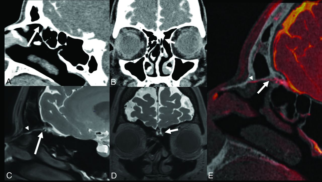
Between November 2019 and March 2020, 7 patients (mean age, 53.1 years; 5 females, 2 males) with CSF rhinorrhea underwent 8 combined CTC/MRC studies (On-line Table). All CSF leaks were precisely depicted on CS T1 SPACE images (Fig 1). In 5 CTC studies, leaks were missed (patient 1, Fig 1), falsely located (patient 2, study 1; On-line Fig 1A–C), or only suspected (patient 2, study 2; On-line Figs 1D–F, patients 3 and 4). All CSF leaks detected on MRC correlated with findings at surgical repair (On-line Videos 1 and 2).

FIG 1.
  • Download figure
  • Open in new tab
  • Download powerpoint
FIG 1.

A 55-year-old woman (patient 1) who was readmitted for recurrent spontaneous left-sided CSF rhinorrhea and persistent slight headaches while sitting or standing after previous endoscopic sinus surgery. One year earlier, sealing of cribriform plate with TachoSil (Takeda Pharma) for suspected CSF leak had been performed. CT cisternography (soft tissue window level, W: 350; L: 50) is unremarkable, even in retrospect (A and B arrows). CS SPACE MR cisternography demonstrates a subtle CSF leak originating from the anterior rim of the left cribriform plate near foramen cecum (C and D, arrows) with a thin CSF collection extending to the anterior nasal cavity (arrowheads in C and E). The site correlated with the patient’s intraoperative findings. After surgical repair, the patient was free of CSF rhinorrhea.

DISCUSSION

In patients with CSF rhinorrhea, precise presurgical localization of CSF leaks is crucial for the success rate of surgery. High-resolution CT and CTC after intrathecal contrast application are both limited in the ability to demonstrate the site of a CSF leak, particularly in patients with multiple osseous defects or small or inactive leaks during imaging.1,2 Anatomically accurate delineation of even a very small dural dehiscence may also be very difficult to diagnose on CTC images in the absence of a larger bony defect.1,2

Standard gadolinium-enhanced MRC is a promising technique that may permit direct, sensitive visualization of the site of CSF leakage.1,2,4-6 Although intrathecal gadolinium injection is not approved by the US Food and Drug Administration, several studies have reported its safe off-label use with long-term follow-up after 4.2 years.1,2 Gadolinium-enhanced MRC has certain advantages over CTC. Although gadolinium distributes readily in the subarachnoid space, the high viscosity of the iodinated contrast agent may impair its free distribution and passage into slowly flowing CSF fistulas. Moreover, CTC requires high radiation dose and may not be able to differentiate CSF leakage from adjacent bone structures. In our series, all patients had confirmation of CSF leakage by surgery. In 5 of 8 CTC studies, the exact site of leakage could not be identified or was only suspected, and in 1 case, the suspected bony defect did not correlate with the site of leakage (patient 4). Related to different patient positioning in MR imaging (supine) and CT (prone), whereas more posterior dependent accumulation of leaked fluid was visible on MRC, CT showed anterior accumulation toward the nasal cavity (patient 4).

The application of 3D CS T1 SPACE for MRC appears particularly attractive because it covers the entire skull base and sinonasal and brain structures. Precise visualization of CSF leaks is enhanced related to its almost isotropic submillimeter 3D resolution, high SNR and contrast-to-noise ratio, and increased contrast between bony or aerated air cell structures, CSF, and soft tissues.3 The conspicuity of contrast medium leakage may also be enhanced because of saturation of medullary bone fat in the cranial base. Compared with earlier reports of gadolinium-enhanced MRC techniques that used spin-echo T1-weighted imaging at 1.5 T,4,5 the CS SPACE T1 technique appears advantageous for visualization of small CSF leaks because of 3D capability, increased spatial resolution, and SNR at 3T.

However, delineation of osseous anatomy at the skull base remains indispensable for surgical planning and intraoperative navigation. Thus, MRC–CTC fusion images combine the advantages of both techniques and allowed, in our series, accurate localization of the CSF leaks. Such fusion images may be alternatively created by combining MRC with low-dose bone-window CT images, thereby skipping the necessity for contrast-enhanced CTC.

Shortcomings of this study relate to the small number of retrospective cases, representing our very early clinical experience with the novel MRC method, which has to our knowledge not yet been described before. Technical limitations relate to the relatively long CS data processing times and relative vulnerability of the sequence to motion.

CONCLUSIONS

High-resolution gadolinium-enhanced CS T1 SPACE MRC is a promising method for detection of CSF leaks in patients with CSF rhinorrhea. In our pilot experience, this technique appears superior to standard CTC. Future prospective comparative studies are necessary to define the diagnostic accuracy of this technique.

Footnotes

  • Paper previously presented at Virtual Conference of the German Society of Neuroradiology, October 7–9, 2020; Frankfurt, Germany.

  • Disclosures: Samer Elsheikh—UNRELATED: Grants/Grants Pending: Bracco Imaging*; Payment for Lectures Including Service on Speakers Bureaus: Daiichi Medical. Esther Raithel—UNRELATED: Employment: Siemens Healthcare GmbH, Comments: I am an employee. Christoph Forman—RELATED: Other: Employee of Siemens Healthcare GmbH, Comments: Salary; UNRELATED: Employment: Siemens Healthcare GmbH; Stock/Stock Options: Siemens Healthineers AG. Horst Urbach—UNRELATED: Board Membership: Co-editor Clin Neuroradiol; Payment for Lectures Including Service on Speakers Bureaus: Bayer, Bracco, Eisai, Stryker, UCB pharma. Stephan Meckel—UNRELATED: Grants/Grants Pending: Bracco S.p.A., Italy.* *Money paid to institution.

References

  1. 1.↵
    1. Reddy M,
    2. Baugnon K
    . Imaging of cerebrospinal fluid rhinorrhea and otorrhea. Radiol Clin North Am 2017;55:167–87 doi:10.1016/j.rcl.2016.08.005 pmid:27890184
    CrossRefPubMed
  2. 2.↵
    1. Eljazzar R,
    2. Loewenstern J,
    3. Dai JB, et al
    . Detection of cerebrospinal fluid leaks: is there a radiologic standard of care? A systematic review. World Neurosurg 2019;127:307–15 doi:10.1016/j.wneu.2019.01.299 pmid:30797912
    CrossRefPubMed
  3. 3.↵
    1. Guggenberger K,
    2. Krafft AJ,
    3. Ludwig U, et al
    . High-resolution compressed-sensing T1 black-blood MRI: a new multipurpose sequence in vascular neuroimaging? Clin Neuroradiol 2019 Dec 18 [Epub ahead of print] doi:10.1007/s00062-019-00867-0 pmid:31853612
    CrossRefPubMed
  4. 4.↵
    1. Aydin K,
    2. Terzibasioglu E,
    3. Sencer S, et al
    . Localization of cerebrospinal fluid leaks by gadolinium-enhanced magnetic resonance cisternography: a 5-year single-center experience. Neurosurgery 2008;62:584–89 doi:10.1227/01.neu.0000317306.39203.24 pmid:18425007
    CrossRefPubMed
  5. 5.↵
    1. Selcuk H,
    2. Albayram S,
    3. Ozer H, et al
    . Intrathecal gadolinium-enhanced MR cisternography in the evaluation of CSF leakage. AJNR Am J Neuroradiol 2010;31:71–75 doi:10.3174/ajnr.A1788pmid:19762462
    Abstract/FREE Full Text
  6. 6.↵
    1. Jinkins JR,
    2. Rudwan M,
    3. Krumina G, et al
    . Intrathecal gadolinium-enhanced MR cisternography in the evaluation of clinically suspected cerebrospinal fluid rhinorrhea in humans: early experience. Radiology 2002;222:555–59 doi:10.1148/radiol.2222010249 pmid:11818627
    CrossRefPubMed
  • Received May 19, 2020.
  • Accepted after revision August 16, 2020.
  • © 2021 by American Journal of Neuroradiology
PreviousNext
Back to top

In this issue

American Journal of Neuroradiology: 42 (1)
American Journal of Neuroradiology
Vol. 42, Issue 1
1 Jan 2021
  • Table of Contents
  • Index by author
  • Complete Issue (PDF)
Advertisement
Print
Download PDF
Email Article

Thank you for your interest in spreading the word on American Journal of Neuroradiology.

NOTE: We only request your email address so that the person you are recommending the page to knows that you wanted them to see it, and that it is not junk mail. We do not capture any email address.

Enter multiple addresses on separate lines or separate them with commas.
High-Resolution Gadolinium-Enhanced MR Cisternography Using Compressed-Sensing T1 SPACE Technique for Detection of Intracranial CSF Leaks
(Your Name) has sent you a message from American Journal of Neuroradiology
(Your Name) thought you would like to see the American Journal of Neuroradiology web site.
CAPTCHA
This question is for testing whether or not you are a human visitor and to prevent automated spam submissions.
Cite this article
I.E. Duman, T. Demerath, A. Stadler, S. Elsheikh, E. Raithel, C. Forman, T. Hildenbrand, M. Shah, J. Grauvogel, C. Scheiwe, H. Urbach, S. Meckel
High-Resolution Gadolinium-Enhanced MR Cisternography Using Compressed-Sensing T1 SPACE Technique for Detection of Intracranial CSF Leaks
American Journal of Neuroradiology Jan 2021, 42 (1) 116-118; DOI: 10.3174/ajnr.A6852

Citation Manager Formats

  • BibTeX
  • Bookends
  • EasyBib
  • EndNote (tagged)
  • EndNote 8 (xml)
  • Medlars
  • Mendeley
  • Papers
  • RefWorks Tagged
  • Ref Manager
  • RIS
  • Zotero
0 Responses
Respond to this article
Share
Bookmark this article
High-Resolution Gadolinium-Enhanced MR Cisternography Using Compressed-Sensing T1 SPACE Technique for Detection of Intracranial CSF Leaks
I.E. Duman, T. Demerath, A. Stadler, S. Elsheikh, E. Raithel, C. Forman, T. Hildenbrand, M. Shah, J. Grauvogel, C. Scheiwe, H. Urbach, S. Meckel
American Journal of Neuroradiology Jan 2021, 42 (1) 116-118; DOI: 10.3174/ajnr.A6852
del.icio.us logo Twitter logo Facebook logo Mendeley logo
  • Tweet Widget
  • Facebook Like
  • Google Plus One
Purchase

Jump to section

  • Article
    • Abstract
    • ABBREVIATIONS:
    • TECHNIQUES
    • DISCUSSION
    • CONCLUSIONS
    • Footnotes
    • References
  • Figures & Data
  • Supplemental
  • Info & Metrics
  • Responses
  • References
  • PDF

Related Articles

  • PubMed
  • Google Scholar

Cited By...

  • Different Glymphatic Kinetics in Spontaneous Intracranial Hypotension
  • Crossref (9)
  • Google Scholar

This article has been cited by the following articles in journals that are participating in Crossref Cited-by Linking.

  • Quantifying cerebrospinal fluid dynamics: A review of human neuroimaging contributions to CSF physiology and neurodegenerative disease
    Neel H. Mehta, Richard A. Suss, Jonathan P. Dyke, Neil D. Theise, Gloria C. Chiang, Sara Strauss, Leslie Saint-Louis, Yi Li, Silky Pahlajani, Vivek Babaria, Lidia Glodzik, Roxana O. Carare, Mony J. de Leon
    Neurobiology of Disease 2022 170
  • Imaging of Acquired Skull Base Cerebrospinal Fluid Leaks
    Daniel J. Scoffings
    Neuroimaging Clinics of North America 2021 31 4
  • Vorgehen bei frontobasalen Frakturen
    F. Sommer, M. Brand, M. O. Scheithauer, T. K. Hoffmann, M.-N. Theodoraki, R. Weber
    HNO 2023 71 1
  • Surgical Repair of Skull Base CSF Leaks after Cisternography Diagnosis: Analysis of Validity and Surgical Outcome and Impact on Future Treatment Strategies
    Christine Steiert, Luisa M. Kraus, Roland Roelz, Horst Urbach, Juergen Beck, Stephan Meckel, Juergen Grauvogel, Ayhan Comert
    BioMed Research International 2022 2022 1
  • Different Glymphatic Kinetics in Spontaneous Intracranial Hypotension
    H. Urbach, I.E. Duman Kavus, C. Zander, L. Becker, E. Kellner, M. Reisert, A. El Rahal, F. Volz, K. Wolf, J. Beck, A. Rau, N. Lützen
    American Journal of Neuroradiology 2024 45 10
  • Reliability of High-resolution Gadolinium-enhanced MR Cisternography and Gasket-seal Technique for Management of Anterior Skull Base Defects
    Mukesch Johannes Shah, Jürgen Beck, Stephan Meckel, Horst Urbach, Ikram Eda Duman, Manuel Christoph Ketterer, Tanja Hildenbrand
    Clinical Neuroradiology 2024 34 1
  • Special MRI Techniques to Suss out Spontaneous Cerebrospinal Fluid Leaks
    Ernest Yushvayev, Bradley N. Delman, Claudia F.E. Kirsch
    Topics in Magnetic Resonance Imaging 2021 30 3
  • Intrathecal Contrast-enhanced Computed Tomography and MR Cisternography for Skull Base Cerebrospinal Fluid Leaks and Other Intracranial Applications
    Daniel J. Scoffings, Tarik F. Massoud
    Neuroimaging Clinics of North America 2025 35 1
  • Radiopaedia.org
    Dalia Ibrahim, Lam Le, Frank Gaillard
    2016

More in this TOC Section

  • Diagnostic Neuroradiology of Monoclonal Antibodies
  • Segmentation of Brain Metastases with BLAST
  • NCCT vs. MRI for Brain Atrophy in Acute Stroke
Show more Adult Brain

Similar Articles

Advertisement

Indexed Content

  • Current Issue
  • Accepted Manuscripts
  • Article Preview
  • Past Issues
  • Editorials
  • Editors Choice
  • Fellow Journal Club
  • Letters to the Editor

Cases

  • Case Collection
  • Archive - Case of the Week
  • Archive - Case of the Month
  • Archive - Classic Case

Special Collections

  • Special Collections

Resources

  • News and Updates
  • Turn around Times
  • Submit a Manuscript
  • Author Policies
  • Manuscript Submission Guidelines
  • Evidence-Based Medicine Level Guide
  • Publishing Checklists
  • Graphical Abstract Preparation
  • Imaging Protocol Submission
  • Submit a Case
  • Become a Reviewer/Academy of Reviewers
  • Get Peer Review Credit from Publons

Multimedia

  • AJNR Podcast
  • AJNR SCANtastic
  • Video Articles

About Us

  • About AJNR
  • Editorial Board
  • Not an AJNR Subscriber? Join Now
  • Alerts
  • Feedback
  • Advertise with us
  • Librarian Resources
  • Permissions
  • Terms and Conditions

American Society of Neuroradiology

  • Not an ASNR Member? Join Now

© 2025 by the American Society of Neuroradiology All rights, including for text and data mining, AI training, and similar technologies, are reserved.
Print ISSN: 0195-6108 Online ISSN: 1936-959X

Powered by HighWire