Advertisement

Main menu

  • Home
  • Content
    • Current Issue
    • Accepted Manuscripts
    • Article Preview
    • Past Issue Archive
    • Video Articles
    • AJNR Case Collection
    • Case of the Week Archive
    • Case of the Month Archive
    • Classic Case Archive
  • Special Collections
    • AJNR Awards
    • Low-Field MRI
    • Alzheimer Disease
    • ASNR Foundation Special Collection
    • Photon-Counting CT
    • View All
  • Multimedia
    • AJNR Podcasts
    • AJNR SCANtastic
    • Trainee Corner
    • MRI Safety Corner
    • Imaging Protocols
  • For Authors
    • Submit a Manuscript
    • Submit a Video Article
    • Submit an eLetter to the Editor/Response
    • Manuscript Submission Guidelines
    • Statistical Tips
    • Fast Publishing of Accepted Manuscripts
    • Graphical Abstract Preparation
    • Imaging Protocol Submission
    • Author Policies
  • About Us
    • About AJNR
    • Editorial Board
    • Editorial Board Alumni
  • More
    • Become a Reviewer/Academy of Reviewers
    • Subscribers
    • Permissions
    • Alerts
    • Feedback
    • Advertisers
    • ASNR Home

User menu

  • Alerts
  • Log in

Search

  • Advanced search
American Journal of Neuroradiology

American Journal of Neuroradiology

ASHNR American Society of Functional Neuroradiology ASHNR American Society of Pediatric Neuroradiology ASSR
  • Alerts
  • Log in

Advanced Search

  • Home
  • Content
    • Current Issue
    • Accepted Manuscripts
    • Article Preview
    • Past Issue Archive
    • Video Articles
    • AJNR Case Collection
    • Case of the Week Archive
    • Case of the Month Archive
    • Classic Case Archive
  • Special Collections
    • AJNR Awards
    • Low-Field MRI
    • Alzheimer Disease
    • ASNR Foundation Special Collection
    • Photon-Counting CT
    • View All
  • Multimedia
    • AJNR Podcasts
    • AJNR SCANtastic
    • Trainee Corner
    • MRI Safety Corner
    • Imaging Protocols
  • For Authors
    • Submit a Manuscript
    • Submit a Video Article
    • Submit an eLetter to the Editor/Response
    • Manuscript Submission Guidelines
    • Statistical Tips
    • Fast Publishing of Accepted Manuscripts
    • Graphical Abstract Preparation
    • Imaging Protocol Submission
    • Author Policies
  • About Us
    • About AJNR
    • Editorial Board
    • Editorial Board Alumni
  • More
    • Become a Reviewer/Academy of Reviewers
    • Subscribers
    • Permissions
    • Alerts
    • Feedback
    • Advertisers
    • ASNR Home
  • Follow AJNR on Twitter
  • Visit AJNR on Facebook
  • Follow AJNR on Instagram
  • Join AJNR on LinkedIn
  • RSS Feeds

AJNR Awards, New Junior Editors, and more. Read the latest AJNR updates

Research ArticleSpine Imaging and Spine Image-Guided Interventions
Open Access

Filtered Diffusion-Weighted MRI of the Human Cervical Spinal Cord: Feasibility and Application to Traumatic Spinal Cord Injury

S.A. Murphy, R. Furger, S.N. Kurpad, V.E. Arpinar, A. Nencka, K. Koch and M.D. Budde
American Journal of Neuroradiology November 2021, 42 (11) 2101-2106; DOI: https://doi.org/10.3174/ajnr.A7295
S.A. Murphy
aFrom the Department of Neurosurgery (S.A.M., R.F., S.N.K., M.D.B.)
  • Find this author on Google Scholar
  • Find this author on PubMed
  • Search for this author on this site
  • ORCID record for S.A. Murphy
R. Furger
aFrom the Department of Neurosurgery (S.A.M., R.F., S.N.K., M.D.B.)
bCenter for Neurotrauma Research (R.F., S.N.K., M.D.B.)
  • Find this author on Google Scholar
  • Find this author on PubMed
  • Search for this author on this site
  • ORCID record for R. Furger
S.N. Kurpad
aFrom the Department of Neurosurgery (S.A.M., R.F., S.N.K., M.D.B.)
bCenter for Neurotrauma Research (R.F., S.N.K., M.D.B.)
  • Find this author on Google Scholar
  • Find this author on PubMed
  • Search for this author on this site
  • ORCID record for S.N. Kurpad
V.E. Arpinar
cCenter for Imaging Research (V.E.A., A.N., K.K.), Medical College of Wisconsin, Milwaukee, Wisconsin
  • Find this author on Google Scholar
  • Find this author on PubMed
  • Search for this author on this site
  • ORCID record for V.E. Arpinar
A. Nencka
cCenter for Imaging Research (V.E.A., A.N., K.K.), Medical College of Wisconsin, Milwaukee, Wisconsin
  • Find this author on Google Scholar
  • Find this author on PubMed
  • Search for this author on this site
  • ORCID record for A. Nencka
K. Koch
cCenter for Imaging Research (V.E.A., A.N., K.K.), Medical College of Wisconsin, Milwaukee, Wisconsin
  • Find this author on Google Scholar
  • Find this author on PubMed
  • Search for this author on this site
  • ORCID record for K. Koch
M.D. Budde
aFrom the Department of Neurosurgery (S.A.M., R.F., S.N.K., M.D.B.)
bCenter for Neurotrauma Research (R.F., S.N.K., M.D.B.)
  • Find this author on Google Scholar
  • Find this author on PubMed
  • Search for this author on this site
  • ORCID record for M.D. Budde
  • Article
  • Figures & Data
  • Supplemental
  • Info & Metrics
  • Responses
  • References
  • PDF
Loading

Article Figures & Data

Figures

  • FIG 1.
    • Download figure
    • Open in new tab
    • Download powerpoint
    FIG 1.

    Schematic representation of the fDWI/DDE MR imaging technique for spinal cord injury. Traumatic injury to the spinal cord (A) results in microscopic damage to axons, illustrated here as beading and end-bulbs that reflect the underlying acute pathology (B). A prominent edema response (light blue) is typical surrounding the injury site. Traditional DWI/DTI derives measures reflecting the bulk sum of all features. With fDWI/DDE, a high-strength diffusion-weighting perpendicular to the spinal cord suppresses extracellular edema (and CSF) to estimate tissue-specific diffusivity metrics less confounded by edema.

  • FIG 2.
    • Download figure
    • Open in new tab
    • Download powerpoint
    FIG 2.

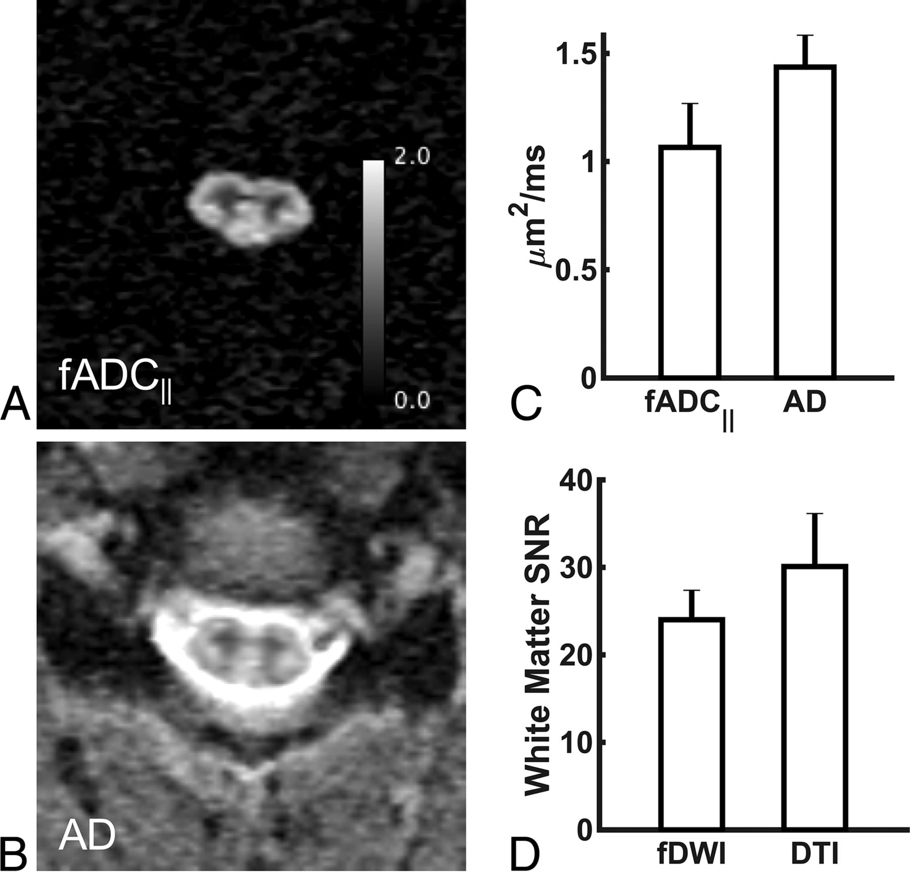
    fADC‖ and AD maps for the healthy spinal cord. Single-subject fADC‖ (A) and AD maps (B) at C4 for a healthy individual on a 3T system. Comparison of the mean white matter fADC‖ or AD values (C) and SNR (D).

  • FIG 3.
    • Download figure
    • Open in new tab
    • Download powerpoint
    FIG 3.

    fADC‖ and AD maps for an individual (subject 3) with an acute spinal cord injury. A T2-weighted image for an individual with an acute spinal cord injury on a 1.5T system. Single slices above, at, and below the injury site (as labeled in the T2 image) for fADC‖ and AD maps. Ax GRE indicates axial gradient recalled-echo.

  • FIG 4.
    • Download figure
    • Open in new tab
    • Download powerpoint
    FIG 4.

    fADC‖ and AD compared at each individual section for acute spinal cord injury (n = 8). There is a large, unidirectional decrease in fADC‖ at the injury site compared with a lesser, multidirectional decrease in AD values. The asterisk indicates significance compared with the first section (P < .05).

  • FIG 5.
    • Download figure
    • Open in new tab
    • Download powerpoint
    FIG 5.

    Correlations of fADC‖ and AD at each individual section for the intact spinal cord and acute spinal cord injury. Correlations are significant for the intact spinal cord; however, a lower correlation and nonrandom residuals for the acute SCI setting indicate that fADC‖ and AD do not have a simple linear relationship, suggesting that they provide differing information.

PreviousNext
Back to top

In this issue

American Journal of Neuroradiology: 42 (11)
American Journal of Neuroradiology
Vol. 42, Issue 11
1 Nov 2021
  • Table of Contents
  • Index by author
  • Complete Issue (PDF)
Advertisement
Print
Download PDF
Email Article

Thank you for your interest in spreading the word on American Journal of Neuroradiology.

NOTE: We only request your email address so that the person you are recommending the page to knows that you wanted them to see it, and that it is not junk mail. We do not capture any email address.

Enter multiple addresses on separate lines or separate them with commas.
Filtered Diffusion-Weighted MRI of the Human Cervical Spinal Cord: Feasibility and Application to Traumatic Spinal Cord Injury
(Your Name) has sent you a message from American Journal of Neuroradiology
(Your Name) thought you would like to see the American Journal of Neuroradiology web site.
CAPTCHA
This question is for testing whether or not you are a human visitor and to prevent automated spam submissions.
Cite this article
S.A. Murphy, R. Furger, S.N. Kurpad, V.E. Arpinar, A. Nencka, K. Koch, M.D. Budde
Filtered Diffusion-Weighted MRI of the Human Cervical Spinal Cord: Feasibility and Application to Traumatic Spinal Cord Injury
American Journal of Neuroradiology Nov 2021, 42 (11) 2101-2106; DOI: 10.3174/ajnr.A7295

Citation Manager Formats

  • BibTeX
  • Bookends
  • EasyBib
  • EndNote (tagged)
  • EndNote 8 (xml)
  • Medlars
  • Mendeley
  • Papers
  • RefWorks Tagged
  • Ref Manager
  • RIS
  • Zotero
0 Responses
Respond to this article
Share
Bookmark this article
Filtered Diffusion-Weighted MRI of the Human Cervical Spinal Cord: Feasibility and Application to Traumatic Spinal Cord Injury
S.A. Murphy, R. Furger, S.N. Kurpad, V.E. Arpinar, A. Nencka, K. Koch, M.D. Budde
American Journal of Neuroradiology Nov 2021, 42 (11) 2101-2106; DOI: 10.3174/ajnr.A7295
del.icio.us logo Twitter logo Facebook logo Mendeley logo
  • Tweet Widget
  • Facebook Like
  • Google Plus One
Purchase

Jump to section

  • Article
    • Abstract
    • ABBREVIATIONS:
    • MATERIALS AND METHODS
    • RESULTS
    • DISCUSSION
    • CONCLUSIONS
    • Acknowledgments
    • Footnotes
    • References
  • Figures & Data
  • Supplemental
  • Info & Metrics
  • Responses
  • References
  • PDF

Related Articles

  • PubMed
  • Google Scholar

Cited By...

  • Contrast-enhanced ultrasound for assessing tissue perfusion and predicting functional recovery following acute traumatic spinal injury: translation from rat model to humans
  • Influence of preprocessing, distortion correction and cardiac triggering on the quality of diffusion MR images of spinal cord
  • Crossref
  • Google Scholar

This article has not yet been cited by articles in journals that are participating in Crossref Cited-by Linking.

More in this TOC Section

  • Management Outcomes For VO Spine Biopsy
  • Characteristics of SIH Type I Culprit Lesions
  • Advanced Imaging of Type 2 Spinal CSF Leaks
Show more Spine Imaging and Spine Image-Guided Interventions

Similar Articles

Advertisement

Indexed Content

  • Current Issue
  • Accepted Manuscripts
  • Article Preview
  • Past Issues
  • Editorials
  • Editor's Choice
  • Fellows' Journal Club
  • Letters to the Editor
  • Video Articles

Cases

  • Case Collection
  • Archive - Case of the Week
  • Archive - Case of the Month
  • Archive - Classic Case

More from AJNR

  • Trainee Corner
  • Imaging Protocols
  • MRI Safety Corner
  • Book Reviews

Multimedia

  • AJNR Podcasts
  • AJNR Scantastics

Resources

  • Turnaround Time
  • Submit a Manuscript
  • Submit a Video Article
  • Submit an eLetter to the Editor/Response
  • Manuscript Submission Guidelines
  • Statistical Tips
  • Fast Publishing of Accepted Manuscripts
  • Graphical Abstract Preparation
  • Imaging Protocol Submission
  • Evidence-Based Medicine Level Guide
  • Publishing Checklists
  • Author Policies
  • Become a Reviewer/Academy of Reviewers
  • News and Updates

About Us

  • About AJNR
  • Editorial Board
  • Editorial Board Alumni
  • Alerts
  • Permissions
  • Not an AJNR Subscriber? Join Now
  • Advertise with Us
  • Librarian Resources
  • Feedback
  • Terms and Conditions
  • AJNR Editorial Board Alumni

American Society of Neuroradiology

  • Not an ASNR Member? Join Now

© 2025 by the American Society of Neuroradiology All rights, including for text and data mining, AI training, and similar technologies, are reserved.
Print ISSN: 0195-6108 Online ISSN: 1936-959X

Powered by HighWire