
Article Figures & Data
Figures
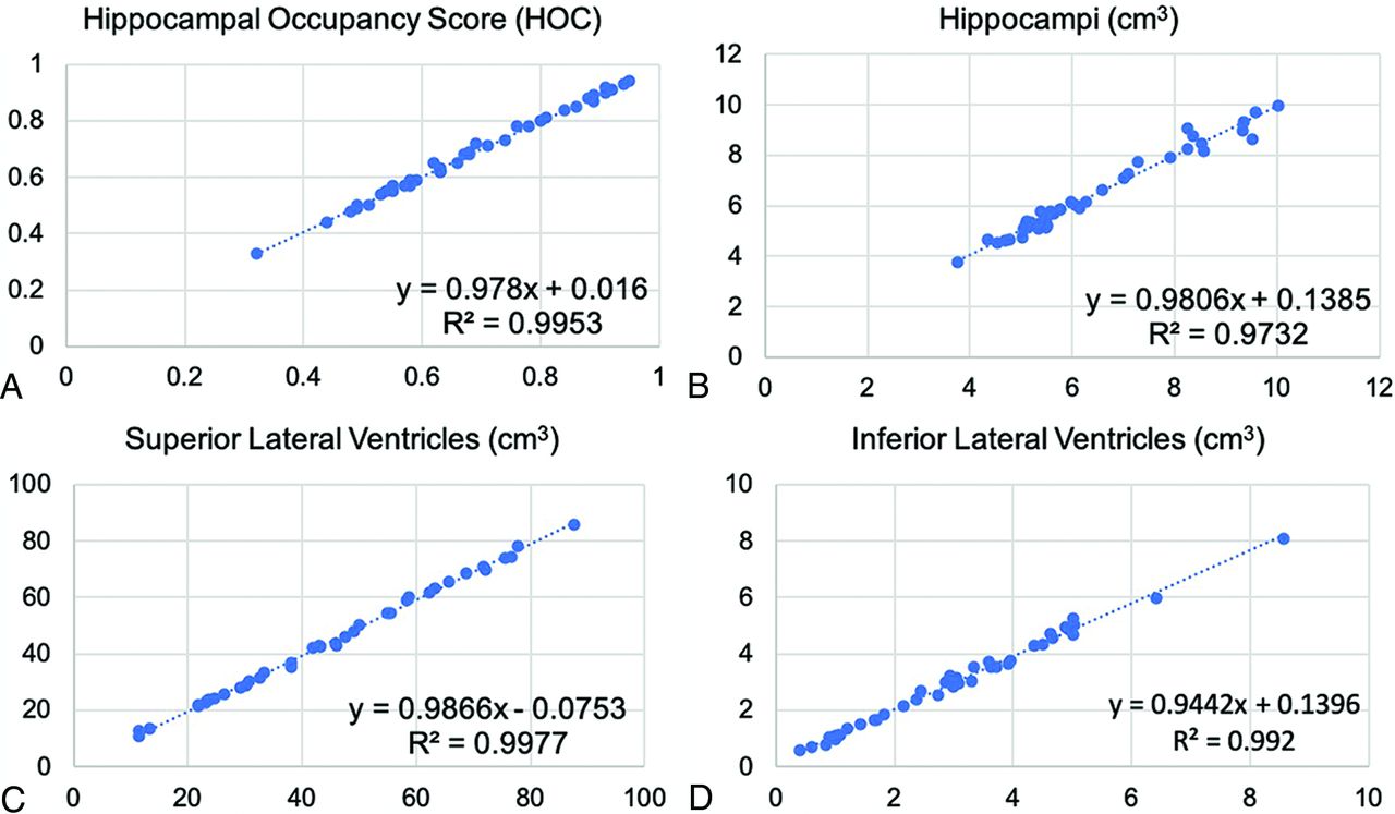
- FIG 1.
Linear regression results for SOC versus FAST-DL. The plot graphs demonstrate linear distribution without scatter, indicating consistent concordance between SOC (x-axis) and FAST-DL (y-axis) in quantitative assessment of HOC (A), HV (B), SLV volume (C), and ILV volume (A).
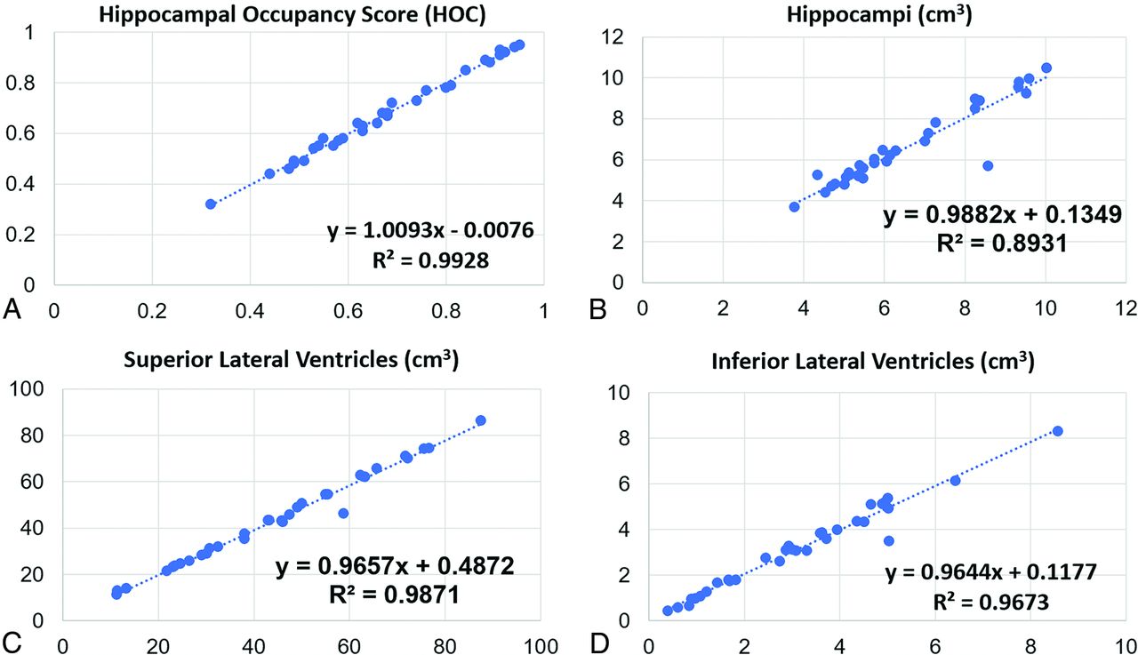
- FIG 2.
Linear regression results for SOC versus FAST. The plot graphs demonstrate a modestly linear distribution though some scatter is present, indicating less optimal concordance of the cross-correlation factor between SOC (x-axis) and FAST (y-axis) (compared with SOC versus FAST-DL) in a quantitative assessment of HOC (A), HV (B), SLV volume (C), and ILV volume (A).
- FIG 3.
Bland-Altman results for SOC versus FAST-DL. The plot graphs demonstrate a linear distribution without significant scatter, indicating consistent concordance between SOC and FAST-DL in the quantitative assessment of HOC, HV, SLV volume, and ILV volume.
- FIG 4.
Bland-Altman results for SOC versus FAST. The plot graphs demonstrate a modestly linear distribution though some scatter is present, indicating less optimal concordance of the cross-correlation factor between SOC versus FAST (compared with SOC versus FAST-DL) in the quantitative assessment of HOC, HV, SLV volume, and ILV volume.
- FIG 5.
Representative axial 3D T1-weighted images on a 3T scanner. Left to right, SOC (scan time, 4 minutes, 55 seconds), FAST (scan time, 2 minutes, 10 seconds), FAST-DL (scan time, 2 minutes, 10 seconds).
- FIG 6.
Representative 3D T1-weighted multiplanar images with volumetric segmentation on a 3T scanner. Left to right, Axial, coronal, sagittal T1-weighted images with SOC (scan time, 5 minutes, 01 second) on the upper row (A) and FAST-DL (scan time, 2 minutes, 37 seconds) on lower row (B).


Tables
Feature SOC vs FAST FAST-DL vs SOC FAST-DL vs FAST Mean P Value Mean P Value Mean P Value Perceived SNR 4.1 (SD, 0.8) <.001 3.5 (SD, 1.3) <.001 4.3 (SD, 1.0) <.001 Sharpness 4.5 (SD, 0.7) <.001 3.5 (SD, 1.5) .005 4.7 (SD, 0.9) <.001 Artifacts 3.9 (SD, 0.8) <.001 3.5 (SD, 1.1) <.001 4.1 (SD, 0.9) <.001 Anatomic/lesion conspicuity 4.2 (SD, 0.7) <.001 3.3 (SD, 1.1) .006 4.3 (SD, 0.7) <.001 Image contrast 4.0 (SD, 0.7) <.001 3.4 (SD, 1.1) .004 4.1 (SD, 0.8) <.001 GM/WM differentiation 4.3 (SD, 0.7) <.001 3.4 (SD, 1.2) .009 4.5 (SD, 0.8) <.001 ↵a SOC is superior to FAST for all criteria (P values <.001). Numbers higher than 3 represent preference for the first of the 2 sequences listed in the upper row. FAST-DL is superior to SOC for all criteria (P values < .008), except for GM/WM differentiation. While this metric trended to be superior for FAST-DL versus SOC, it did not reach statistical significance after Bonferroni correction (P = .009). FAST-DL is superior to FAST for all criteria (P values <.001).
SOC FAST-DL Paired t Test HOC (mean) 0.68 (SD, 0.16) 0.68 (SD, 0.16) 0.58 HV (mean) (cm3) 6.45 (SD, 1.70) 6.47 (SD, 1.69) 0.77 SLV volume (mean) (cm3) 44.30 (SD, 20.60) 43.63 (SD, 20.35) <0.05 ILV volume (mean) (cm3) 3.07 (SD, 1.78) 3.04 (SD, 1.69) 0.27 ↵a There is excellent agreement between SOC and FAST-DL for quantitative assessment of HOC, HV, SLV volume, and ILV volume.
SOC FAST Paired t Test HOC (mean) 0.68 (SD, 0.16) 0.68 (SD, 0.17) 0.63 HV (mean) (cm3) 6.45 (SD, 1.70) 6.56 (SD, 1.88) 0.60 SLV volume (mean) (cm3) 44.30 (SD, 20.60) 43.44 (SD, 20.01) <0.05 ILV volume (mean) (cm3) 3.07 (SD, 1.78) 3.17 (SD, 1.85) 0.93 ↵a There is less optimal agreement between SOC versus FAST (compared with SOC versus FAST-DL) for quantitative assessment of HOC, HV, SLV volume, and ILV volume.


Jump to section
Related Articles
Cited By...
- Alzheimer Disease Anti-Amyloid Immunotherapies: Imaging Recommendations and Practice Considerations for Monitoring of Amyloid-Related Imaging Abnormalities
- The Future of Artificial Intelligence in Clinical Radiology: Savior or False Hope?
- Real-World Adoption of Artificial Intelligence in Radiology: Opportunities and Barriers
- Compressed Sensitivity Encoding Artificial Intelligence Accelerates Brain Metastasis Imaging by Optimizing Image Quality and Reducing Scan Time
- Deep Learning-Generated Synthetic MR Imaging STIR Spine Images Are Superior in Image Quality and Diagnostically Equivalent to Conventional STIR: A Multicenter, Multireader Trial
- Accelerated Synthetic MRI with Deep Learning-Based Reconstruction for Pediatric Neuroimaging
- Validation of a Denoising Method Using Deep Learning-Based Reconstruction to Quantify Multiple Sclerosis Lesion Load on Fast FLAIR Imaging