
Article Figures & Data
Figures
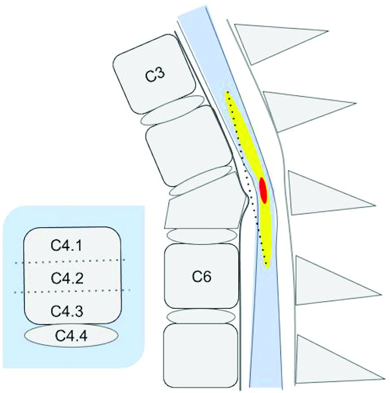
- FIG 1.
Graphic of a SCI on a sagittal T2-weighted image showing the anatomic location designations of the impact zone (center), rostral and caudal limits of spinal cord edema (yellow), and hemorrhage (red). By convention, each vertebral body is arbitrarily subdivided into 3 equal parts (designated as level.1, level.2, and level.3) with the intervertebral disc below the body as the fourth subpart (level.4). On this diagram, the rostral limit of edema is at C3.4, and the caudal extent is at C6.2. Hemorrhage (red) is demarcated by C5. 1 and C5.2. Lesion center is at C5.2. The dotted line represents actual continuous measurement of length of edema demarcated by the upper and lower boundaries on a T2-weighted image that a reviewer would create with electronic calipers.
- FIG 2.
Graphic representation of the BASIC score CDE. The score is based on the extent of the cross-sectional T2-weighted abnormality. A score of 0 is normal. A score of 1 represents signal change in the central GM. A score of 2 represents signal change that extends beyond the central GM but does not involve the entire cross-sectional area. A score of 3 involves the entire cross-section of the spinal cord. A score of 4 features a grade III injury as well as hypointense foci in the central GM indicative of hemorrhage.
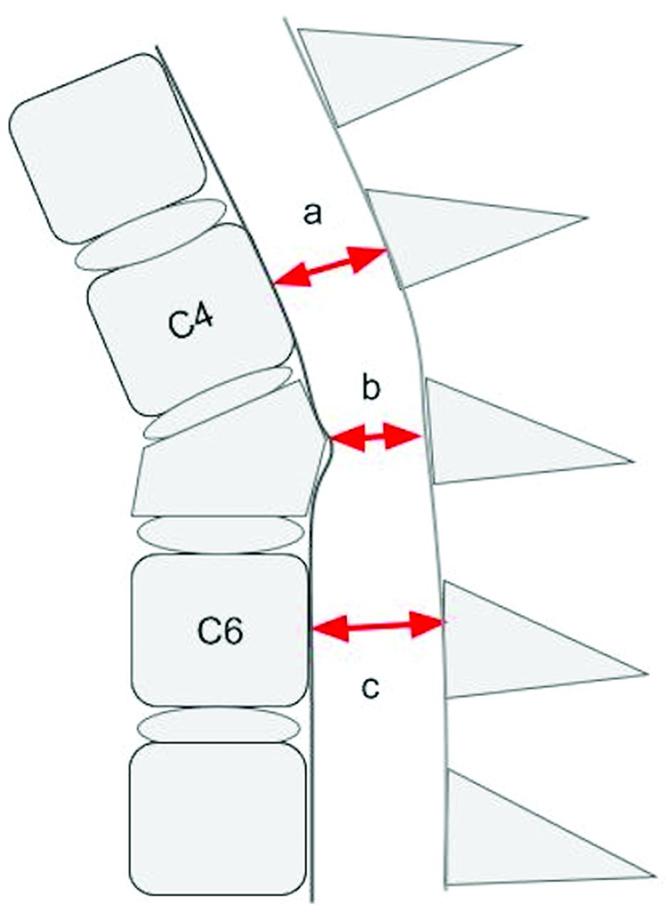
- FIG 3.
Graphic representation of a T2-weighted sagittal MR imaging illustrating an example of 3 key absolute measurements of the sagittal diameter of the spinal canal at the level of injury (b), above the level of injury (a), and below the level of injury (c). Reviewers were instructed to obtain the measurements from the dural boundaries instead of the cortical margins. Measurements obtained rostral and caudal to the injury level are made at the midbody level of the first normal-appearing body above and below the injury level, respectively.
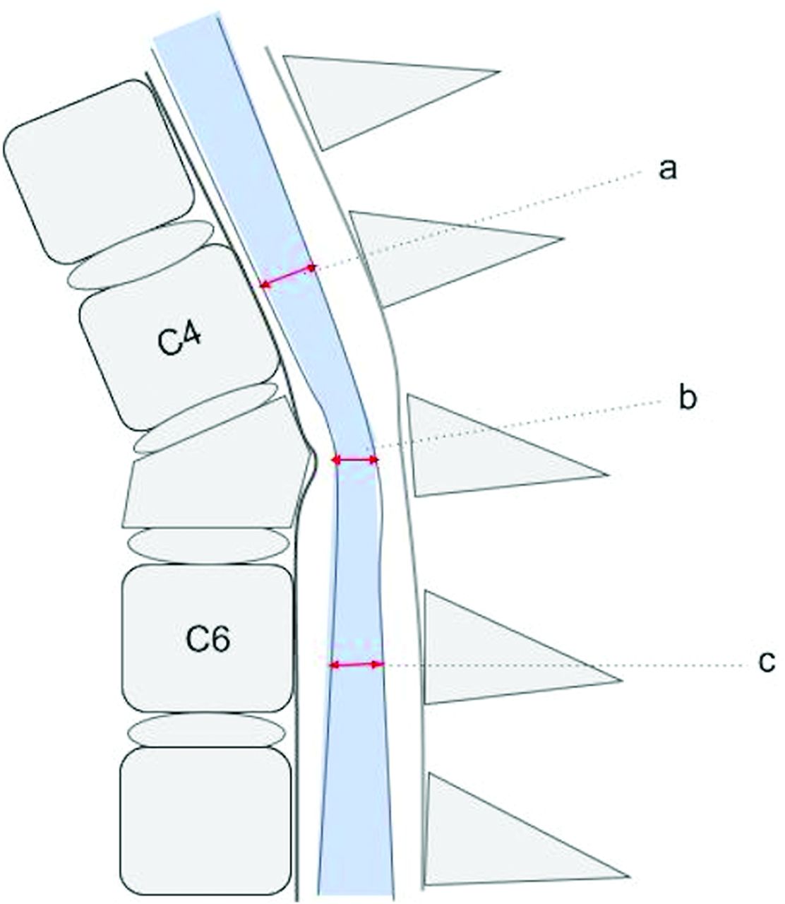
- FIG 4.
Graphic representation of a T2-weighted sagittal MR imaging illustrating an example of 3 key absolute measurements of the sagittal diameter of the spinal cord at the level of injury (b), above the level of injury (a), and below the level of injury (c). Measurements obtained rostral and caudal to the injury level are made at the midbody level of the first normal-appearing vertebral body above and below the injury level, respectively.
- FIG 5.
Graphic representation of an axial MR image of the spine illustrating the methodology for generating absolute measurements of the transverse and anteroposterior dimensions of the dural envelope (arrows). These were measured at 2 locations, the point of maximal compression on the axial dataset and at the first normal rostral midvertebral body level. Reviewers were instructed to estimate the margins from the dural envelope and not the bony cortical boundaries.


Tables
- Table 1:
Inter- and intrarater agreement (ICC) and confidence intervals for CDE features related to SCI using 5 raters, 35 cases, and 2 rounds of evaluationsa
Interrater ICC: Round 1 Interrater ICC: Round 2 Intrarater ICC Center of injury level 0.99 (0.99, 1.00) 0.99 (0.99, 1.00) 1.00 (1.00, 1.00) Rostral extent of spinal cord edema 0.99 (0.98, 0.99) 0.98 (0.98, 0.99) 1.00 (1.00, 1.00) Caudal extent of spinal cord edema 0.97 (0.95, 0.98) 0.97 (0.95, 0.98) 0.98 (0.98, 0.99) Rostral extent of spinal cord hemorrhage 0.68 (0.56, 0.81) 0.70 (0.58, 0.82) 0.96 (0.95, 0.98) Caudal extent of spinal cord hemorrhage 0.69 (0.56, 0.81) 0.69 (0.57, 0.82) 0.95 (0.93, 0.97) Hemorrhage length (mm)b 0.59 (0.44, 0.74) 0.54 (0.39, 0.70) 0.78 (0.72, 0.84) Edema length (mm)b 0.57 (0.42, 0.72) 0.60 (0.46, 0.75) 0.83 (0.78, 0.88) - Table 2:
Inter- and intrarater agreement (ICC) and confidence intervals for direct measures of spinal canal and spinal cord diameters derived from sagittal and axial MR images using the methodology featured in Figs 3–5 using 5 raters, 35 patients, and 2 rounds of evaluations
Interrater ICC: Round 1 Interrater ICC: Round 2 Intrarater ICC Sagittal cord diameter at level of injury (mm)a 0.83 (0.75, 0.91) 0.82 (0.74, 0.90) 0.84 (0.80, 0.89) Sagittal cord diameter at first rostral segment above injury (mm)a 0.37 (0.20, 0.53) 0.27 (0.11, 0.43) 0.79 (0.73, 0.85) Sagittal cord diameter at first caudal segment below injury (mm)a 0.43 (0.27, 0.60) 0.51 (0.36, 0.67) 0.69 (0.61, 0.77) Sagittal canal measurement at level of injury (mm)a 0.80 (0.71, 0.89) 0.72 (0.60, 0.83) 0.85 (0.81, 0.90) Sagittal canal measurement rostral to injury (mm)a 0.23 (0.07, 0.38) 0.31 (0.15, 0.48) 0.86 (0.82, 0.91) Sagittal canal measurement caudal to injury (mm)a 0.56 (0.41, 0.71) 0.38 (0.21, 0.54) 0.90 (0.86, 0.93) Anteroposterior diameter (mm) of the spinal cord at the point of maximal compression or epicenter of injury from axial imagea 0.76 (0.65, 0.86) 0.69 (0.57, 0.82) 0.86 (0.82, 0.90) Transverse diameter (mm) of the spinal cord at the point of maximal compression or epicenter of injury from axial imagea 0.41 (0.25, 0.58) 0.33 (0.17, 0.50) 0.78 (0.72, 0.85) Anteroposterior diameter (mm) of the spinal cord on axial image at the nearest adjacent rostral normal levela 0.37 (0.20, 0.53) 0.27 (0.11, 0.43) 0.82 (0.77, 0.87) ↵b Continuous data.


Jump to section
Related Articles
Cited By...
- No citing articles found.